
Mapa mental
Kafli 6 : Vision
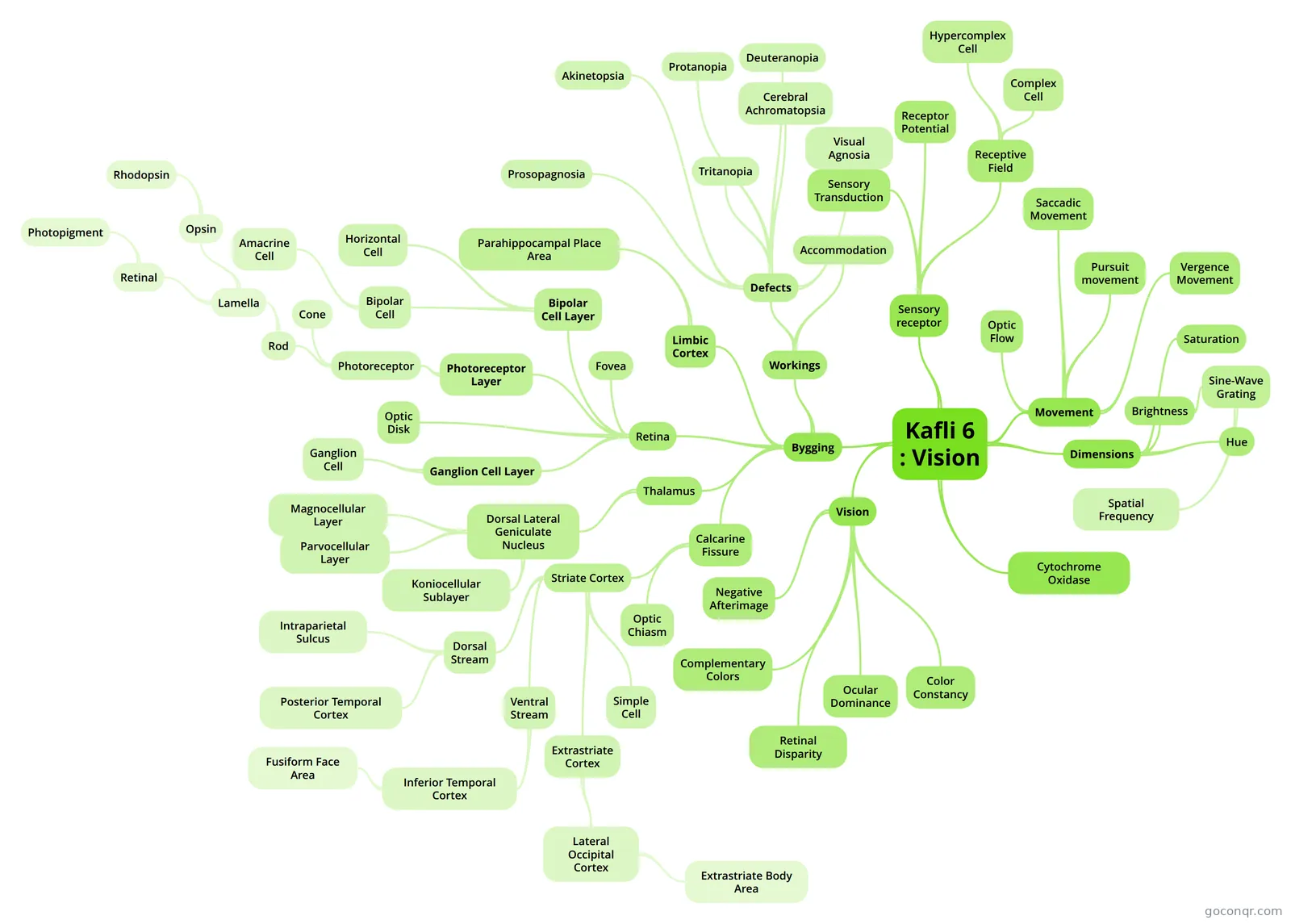
Mind map: Vision
Contenido del mapa
Kafli 6 : Vision
Sensory receptor
- A specialized neuron that detects a particular category of physical events.
- Sensory Transduction
- The process by which sensory stimuli are transduced into slow, graded receptr potentials.
- Receptor Potential
- A slow, graded electrical potential produced by a receptor to a physical stimulus.
- Receptive Field
- That portion of the visual field in which the presentation of visual stimuli will produce an alteration in the firing rate of a particular neuron.
- Complex Cell
- A neuron in the visual cortex that responds to the presence of a line segment with a particular orientation located within its receptive field, especially when the line moves perpendicularly to its orientation.
- Hypercomplex Cell
- A neuron in the visual cortex that responds to the presence of a line segment with a particular orientation that enda at a particular point within the cell's receptive field.
Dimensions
- Hue
- One of the perceptual dimensions of color; the dominant wavelength.
- Brightness
- One of the perceptual dimensions of color; intensity.
- Sine-Wave Grating
- A series of straight parallel bands varying continuously in brightness according to a sine-wave function, along a line perpendicular to their lengths.
- Spatial Frequency
- The relative width of the bands in a sine-wave grating, measured in cycles per degree of visual angle.
- Saturation
- One of the perceptual dimensions of color; purity.
- Hue
Movement
- Vergence Movement
- The cooperative movement of the eyes, which ensures that the image of an object falls on identical portions of both retinas.
- Saccadic Movement
- The rapid, jerky movement of the eyes used in scanning a visual scene.
- Pursuit movement
- The movement that the eyes make to maintain an image of a moving object on the fovea.
- Optic Flow
- The complex motion of points in the visual field caused by relative movement between the observer and environment; provides information about the relative distances of objects from the observer and of the relative direction of movement.
- Vergence Movement
Bygging
- Retina
- The neural tissue and photoreceptive cells located on the inner surface of the posterior portion of the eye.
- Fovea
- The region of the retina that mediates the most acute vision of birds and higher mammals. Color-sensitive cones constitute the only type of photoreceptor found in the fovea.
- Optic Disk
- The location of the exit point from the retina of the fibers of the ganglion cells that form the optic nerve; responsible for the blind spot.
- Bipolar Cell Layer
- Bipolar Cell
- A bipolar neuron located in the middle layer of the retina, conveying information from the photoreceptors to the ganglion cells.
- Amacrine Cell
- A neuron in the retina that interconnects adjacent ganglion cells and the inner processes of the bipolar cells.
- Horizontal Cell
- A neuron in the retina that interconnects adjacent photoreceptors and the outer processes of the bipolar cells.
- Bipolar Cell
- Ganglion Cell Layer
- Ganglion Cell
- A neuron located in the retina that receives visual information from bipolar cells; its axons give rise to the optic nerve.
- Ganglion Cell
- Photoreceptor Layer
- Photoreceptor
- One of the receptor cells of the retina; transduces photic energy into electrical potentials.
- Rod
- One of the receptor cells of the retina; sensitive to light of low intensity.
- Lamella
- A layer of membrane containing photopigments; found in rods and cones of the retina.
- Retinal
- A chemical synthesized from vitamin A; joins with an opsin to form a photopigment.
- Photopigment
- A protein dye bonded to retinal, a substance derived from vitamin A; responsible for transduction of visual information.
- Opsin
- A class of protein that, together with retinal, constitutes the photopigments.
- Rhodopsin
- A particular opsin found in rods.
- Cone
- One of the receptor cells of the retina; maximally sensitive to one of three different wavelengths of light and hence encodes color vision.
- Photoreceptor
- Thalamus
- Dorsal Lateral Geniculate Nucleus
- A group of cell bodies within the lateral geniculate body of the thalamus; receives inputs from the retina and projects to the primary visual cortex.
- Magnocellular Layer
- One of the inner rwo layers of neurons in the dorsal lateral geniculate nucleus; transmits information necessary for the perception of form, movement, depth, and small differences in brightness to the primary visual cortex.
- Parvocellular Layer
- One of the four outer layers of neurons in the dorsal lateral geniculate nucleus; transmits information necessary for perception of color and fine details to the primary visual cortex.
- Koniocellular Sublayer
- One of the sublayers of neurons in the dorsal lateral geniculare nucleus found ventral to each of the magnocellular and parvocellular layers; transmits information from short-wavelength ("blue") cones to the primary visual cortex.
- Dorsal Lateral Geniculate Nucleus
- Calcarine Fissure
- A horizontal fissure on the inner surface of the posterior cerebral cortex; the location of the primary visual cortex.
- Striate Cortex
- The primary visual cortex.
- Simple Cell
- An orientation-sensitive neuron in the striate cortex whose receptive field is organized in an opponent fashion.
- Extrastriate Cortex
- A region of visual association cortex; receives fibers from the striate cortex and from the superior colliculi and projects to the inferior temporal cortex.
- Lateral Occipital Cortex
- A region of the extrastriate cortex, involved in perception of objects other that people's bodies and faces.
- Extrastriate Body Area
- A region of the visual association cortex located in the lateral occipitotemporal cortex; involved in perception of the human body and body parts other than faces.
- Dorsal Stream
- A system of interconnected regions of visual cortex involved in the perception of spatial location, beginning with the striate cortex and ending with the posterior parietal cortex.
- Posterior Temporal Cortex
- The highest level of the dorsal stream of the visual association cortex; involved in perception of movement and spatial location.
- Intraparietal Sulcus
- The end of the dorsal stream of the visual association cortex; involved in perception of location, visual attention and control of eye and hand movements.
- Ventral Stream
- A system of interconnected regions of visual cortex involved in the perception of form, beginning with the striate cortex and ending with the inferior temporal cortex
- Inferior Temporal Cortex
- The highest level of the ventral stream of the visual association cortex; involved in perception of objects, including people's bodies and faces.
- Fusiform Face Area
- A region of the visual association cortex located in the inferior temporal; ivolved in perception of faces and other complex objects that require expertise to recognize.
- Optic Chiasm
- A cross-shaped connection between the optic nerves, located below the base of the brain, just anterior to the pituitary gland.
- Workings
- Accommodation
- Changes in the thickness of the lens of the eye, accomplished by the ciliary muscles, that focus images of near or distant objects on the retina.
- Defects
- Protanopia
- An inherited form of defective color vision in which red and green hues are confused; "red" cones are filled with "green" cone opsin.
- Deuteranopia
- An inherited form of defective color vision in which red and green hues are confused; "green" cones are filled with "red" cone opsin.
- Tritanopia
- An inherited form of defective color vision in which hues with short wavelengths are confused; "blue" cones are either lacking or faulty.
- Cerebral Achromatopsia
- Inability to discriminate among different hues; caused my damage to area V8 of the visual cortex.
- Visual Agnosia
- Deficits in visual perception in the absence of blindness; caused by brain damage.
- Prosopagnosia
- Failure to recognize particular people by the sight of their faces.
- Akinetopsia
- Inability to perceive movement, caused by damage to area V5 of the visual association cortex.
- Protanopia
- Accommodation
- Limbic Cortex
- Parahippocampal Place Area
- A region of limbic cortex on the medial temporal lobe; involved in perception of particular places.
- Parahippocampal Place Area
- Retina
Vision
- Negative Afterimage
- The image seen after a portion of the retina is exposed to an intense visual stimulus; consists of colors complementary to those of the physical stimulus.
- Complementary Colors
- Colors that make white or gray when mixed together.
- Retinal Disparity
- The fact that points on objects located at different distances from the observer will fall on slightly different locations on the two retinas; provides the basis for stereopsis.
- Ocular Dominance
- The extent to which a particular neuron recieves more input from ove eye than from the other.
- Color Constancy
- The relatively constant appearance of the colors of objects viewed under varying light conditions.
- Negative Afterimage
Cytochrome Oxidase
- The central region of a module if the primary visual cortex, revealed by a stain for cytochrome oxidase; contains wavelength-sensitive neurons; part of the parvocellular system.
Información
- Descripción
- (Hugtök) Physiology of Behavior Mapa Mental sobre Kafli 6 : Vision, creado por bykkja el 22/04/2013.
- Creado por
- bykkja
- Visitas
- 36
- Etiquetas
- Valoración