
Upper Limb Radiography
Diapositivas del conjunto (23)
-
- AP: Patient turns away from the table and extends the affected arm out behind them over the table. The posterior aspect of thumb is in contact with image receptor and fingers are moved and supported
- PA: Medial aspect of hand is in contact with IR. It is externally rotated 90 degrees and the fingers are extended and superimposed vertically. Long axis should be parallel to IR
- Lateral: Thumb should lie on laterally with its phalangeal condyles superimposed. Long axis should be parallel to IR; radiolucent pad is used under palmar to aid immobilisation or could use fist
- Centring: Over the 1st metacarpophalangeal joint
- Collimation: All phalanges, 1st metacarpal, trapezium, soft tissue
- Criteria: All of the collimated points are included, joint spaces, symmetry of the phalangeal condyles, sharp image of soft tissue margins of the thumb, thenar eminence, bony cortex and trabeculae
-
- Positioning: Patient is sat with affected side adjacent to the table; lead rubber is applied to the waist. The affected hand is pronated and the finger is placed on the cassette. Fingers are extended to show the interphalangeal joint spaces andslightly separated
- Centring point: Over the proximal interphalangeal joint
- Collimation: All phalanges of the finger under examination, the metacarpophalangeal joint and adjacent finger/s to facilitate correct finger identification
- Criteria: Adjacent fingers and soft tissue outline; symmetry of the phalangeal condyles, spaces between the joints, sharp mage showing bone cortex and trabeculae
-
- Index: From DP, internally rotate at 90 degrees, the finger will be seperated and extended. The remaining fingers are flexed. Long axis of the index finger is parallel to the long axis of the image receptor
- Middle finger: From DP, externally rotate at 90 degrees, the finger will be seperated and extended. The remaining fingers are flexed. Long axis of the index finger is parallel to the long axis of the image receptor
- Ring and little finger: From DP, externally rotate through 90 degrees, the index and middle are flexed and held by the thumb. Medial aspect is in contact with the IR
- Centring point: Over the proximal interphalangeal joint of the finger under examination
- Collimation: All phalanges, soft tissue outlines and metacarpophalangeal joint
- Criteria: All phalanges and metacarpophalangeal joint with outline of adjacent fingers joints and phalangeal joints are shown and superimposed
-
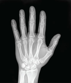
- DP: Hand is pronated, palmar aspect is placed in contact with IR, fingers and thumb are extended and slightly separated
- DP oblique: Hand is externally rotated until palmer/anterior aspect of the hand makes an angle of 45 degrees wih the IR
- Centring: Over the head of the 3rd metacarpal
- Collimate: Include whole hand, wrist joint and distal radius and ulna
- To see the extent of any displacement of fracture fragments (take all if a fracture is present) & the position of a foreign body (take DP then lateral)
- Lateral: Hand is externally rotated so the medial aspect is in contact with IR. Thumb is extended & abducted away from the hand. Metacarpals should be superimposed
- Centring: Over the head of the 2nd metacarpal
- Collimate: all of the hand, wrist joint and distal radius and ulna
- Criteria: Fingers are separated, joints are clear, symmetrical appearance of the heads of metacarpals 2 to 4, obliquity of thumb and heads of metacarpals 1 & 5, sharp image
-
- Head or neck of 5th metacarpal "Boxer's fracture" displaces anteriorly
- Bennett's fracture: an oblique fracture of the base of the 1st metacarpal; the proximal fragment is triangular in shape and remains in normal alignment with the trapezium; the rest of the thumb displaces proximally and posteriorly
-
- PA: Elbow is flexed, wrist and forearm placed onto the cassette. Fingers are slightly flexed to raise the hand and keep the wrist in contact with the cassette. Styloid processes are equidistant from the cassette
- Centring Point: Midway between the styloid processes
- Collimation: The heads of the metacarpals and the distal third of the radius and ulna
- Criteria: Clear demonstration of distal radioulnar joint, radial & ulnar styloid processes seen on the lateral & medial margins of these bones, sharp image demonstrating the soft tissue margins of the area, bony cortex and trabeculae, adequate penetration to demonstrate hook of hamate.
- Lateral: Hand is rotated so the palmer aspect is at 90 degrees to the cassette. Elbow is flexed. Wrist may be rotated a further 5 degrees posteriorly to superimpose the styloid processes
- Centring Point: Over radial styloid process
- Collimation: The proximal third of the metacarpals and the distal third of the radius and ulna
- Criteria: Penetration of carpus to demonstrate individual carpal bones, same as before
-
- Fracture of distal radius (Colles' fracture) - usually one inch of the distal articular surface; displaced posteriorly & laterally; patient may present with a 'dinner-fork' deformity
- Smiths fracture - one inch of the distal radius articular surface; falling onto a palmar flexed hand, distal fragment displaced anteriorly; less common than a Colles' fracture
-
- Caused by a fall on an outstretched hand, with the weight landing on the palm
- DP with ulnar deviation: Arm extended over image receptor,anterior aspect of wrist in contact with image receptor, styloid processes equidistant from image receptor, ulnar deviation is applied, there should be no flexion of the wrist.
- DP oblique with ulnar deviation: Arm remains extended over image receptor. From the PA position, the arm is externally rotated so that the anterior aspect of the wrist makes an angle of 45º with the image receptor.
- Centring: Over anatomical snuff box
- Collimation: Include carpal bones, 1st carpometacarpal joint, radiocarpal joint
- Criteria: Separation of carpal joints through ulnar deviation, sharp image demonstrating bony cortex and trabeculae,
- Lateral: Same as lateral wrist
- Collimate: Include carpal bones, 1st carpometacarpal joint, radiocarpal joint, the radial and ulnar styloid processes
- Criteria: Lunate looks like a crescent, proximal end of 3rd metacarpal, distal radius, capitate and lunate should be aligned, scaphoid should superimposed over pisiform
- AP oblique: Arm remains extended over image receptor. From the lateral position, the arm is externally rotated so that the posterior aspect of the wrist makes an angle of 45º with the image receptor.
- Criteria: Scaphoid seen above the radius, partially overlapping the lunate, but clear of the pisiform and triquetral
- Centring: Anatomical snuffbox
- Zitters: Same as 1st scaphoid projection.
- Centring: Over the trapezium with the central ray angled 30º towards the elbow.
-
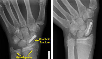
Common injuries in the Scaphoid
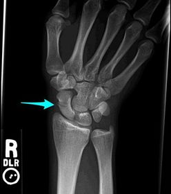
- Pie de imagen: Caused by FOOSH with weight landing on the palmar aspect
-
- PA positioning
- Positioning : Arm extended over image receptor. Posterior aspect of forearm in contact with image receptor. Shoulder, elbow & wrist all on same horizontal plane. Humeral epicondyles & styloid processes equidistant from image receptor
- Centring: Midway between the wrist and elbow joints
- Collimate: Include elbow, wrist, shafts of radius and ulna and soft tissue outline of forearm
- Criteria: Partial superimposition of radius & ulna at proximal & distal ends with separation of shafts, humeral epicondyles equidistant from coronoid & olecranon fossae, radial & ulnar styloid processes seen on lateral aspects of these bones, sharp image demonstrating soft tissue margins of the forearm, bony cortex & trabeculae, overlap of olecranon over distal humerus
- Lateral Positioning
- Positioning: Arm remains extended over image receptor. From the AP position, arm is internally rotated so that medial aspect of forearm is in contact with the film. Elbow is flexed. Shoulder, elbow & wrist all on same horizontal plane. Styloid processes & humeral epicondyles should each be superimposed.
- Centring: Midway between elbow & wrist joints with a Vertical central ray
- Collimate: Include elbow, wrist, shafts of radius and ulna and soft tissue outline of forearm
- Criteria: Partial superimposition of radius & ulna at proximal & distal ends with separation of shafts, humeral epicondyles equidistant from coronoid & olecranon fossae, radial & ulnar styloid processes seen on lateral aspects of these bones, sharp image demonstrating soft tissue margins of the forearm, bony cortex & trabeculae.
-
- Galeazzi fracture dislocation: fracture of the lower third of the radius with dislocation of the head of ulna. Caused by a fall onto the hand with a rotational force
- Monteggia fracture: upper third of the ulna with dislocation of the radial head. Caused by a fall onto the hand with forced pronation of the forearm
-
- AP
- Positioning: Arm extended over image receptor. Posterior aspect of elbow in contact with cassette. Shoulder, elbow & wrist on same horizontal plane. Humeral epicondyles & styloid processes equidistant from cassette.
- Centring: Between the humeral epicondyles with a VCR
- Collimation: Proximal third of radius and ulna, distal third of humerus and soft tissue outline
- Criteria: The proximal radius/ulna, elbow joint, distal shaft of the humerus and soft tissue outlines surrounding the elbow joint demonstrated; partial superimposition of the radius & ulna at the proximal end – radial tuberosity overlap cortex of ulna at most; humeral epicondyles equidistant from the coronoid and olecranon fossae
- Lateral
- Positioning: Arm internally rotated & elbow flexed through 90º. Medial aspect of elbow in contact with image receptor. Shoulder, elbow & wrist on same horizontal plane. Humeral epicondyles & styloid processes superimposed.Centring: Over lateral humeral epicondyle with VCR
- Collimation: Proximal third of radius and ulna, distal third of humerus and soft tissue outline
- Criteria: The proximal radius/ulna, elbow joint, distal shaft of the humerus and soft tissue outlines surrounding the elbow joint are demonstrated; superimposition of surfaces of trochlea & capitulum with posterior portion of radial head shown over coronoid process of ulna; evidence of joint space at elbow seen; shaft of radius seen anterior to that of ulna
-
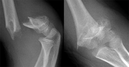
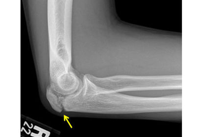
- Pie de imagen: Radial head fracture: occurs in adults; caused by FOOSH which pushes the radial head against the capitulum, it can fracture in a number of ways: vertical split, tilt, shear fracture and crush
- Pie de imagen: Quite difficult to spot on x-ray so we look for fat pad displacement, in a normal elbow the posterior fat pad isn't visible and injury displaces the fat pads away from the bone. A visible posterior FP is always abnormal and a visible anterior FP is normal unless displaced away from the bone
-
- Positioning of AP: Standing with back to receptor. Arm extended, abducted slightly & supinated. Humeral epicondyles equidistant to image receptor.
- Centring: Midway between shoulder & elbow joints with a horizontal central ray (HCR) on the anterior aspect of the humerus
- Collimate: the shoulder joint, shaft of humerus, elbow joint and soft tissue outlines
- Criteria: Shoulder joint, shaft of humerus, elbow joint & soft tissues are demonstrated, humerus is clear of the soft tissue of the trunk, greater tuberosity of humerus is in profile laterally on the head of humerus, humeral epicondyles are equidistant from coronoid and olecranon fossae, sharp image demonstrating soft tissue margins around the elbow, bony cortex & trabeculae, adequate penetration to demonstrate joints whilst maintaining trabecular detail over the humeral shaft
- Positioning of lateral: Standing with back to the receptor. Arm abducted slightly, elbow flexed & hand placed on abdomen.Lateral aspect of humerus in contact with image receptor. Humeral epicondyles superimposed.
- Centring: Midway between shoulder & elbow joints with a HCR on the medial aspect of the humerus
- Collimation: The shoulder joint, shaft of humerus, elbow joint and soft tissue outlines
- Criteria: Shoulder joint, shaft of humerus, elbow joint and sift tissues are demonstrated, humerus is clear of soft tissue of trunk and humeral head is cleared from the image of the scapula, greater tuberosity of humerus is seen over the middle of the head, some superimposition of trochlea and capitulum.
-
Common injuries to the Humerus
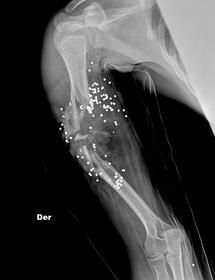
- Fracture mid-shaft humerus: caused by direct impact or rotation; mainly spiral or oblique, can have a "butterfly" fragment
- Surgical neck of humerus fracture: FOOSH, common in the elderly (osteoporotic fracture)
- Anterior dislocation: create a weakness so future dislocations are more likely (compression fracture) very likely
- Posterior dislocation: light bulb sign, often dismissed as a muscle spasm; rare around 5% - mechanism: electric shock, fitting
-
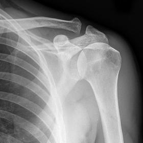
- 20 - 30 degrees for trauma cases
- Positioning criteria: Posterior aspect of affected shoulder in contact with image receptor. Arm is extended, abducted slightly and supinated. Patient turns 20 - 30° towards the affected side. Head should be turned towards other shoulder.
- Centring: To the coracoid process with a HCR.
- Other info: 100cm SID. Provides a general view of the shoulder girdle.
- Collimation: Clavicle, inferior angle of the scapula, soft tissues and sternoclavicular joint
- 45 degrees for non-traumatic cases
- Positioning criteria: Posterior aspect of affected shoulder in contact with IR. Arm is extended, abducted slightly and supinated. Patient turns 45° towards the affected side. Head should be turned towards other shoulder.
- Centring: To the coracoid process with a HCR.
- Other info: 100cm SID Provides a view of gleno-humeral joint.
- Axial Shoulder
- Patient seated with affected side next to table.Positioning: Image receptor placed on the table top. Patient leans towards table so that the arm is abducted over the image receptor. Elbow flexed for support; hand pronated (placed palm down)
- Centre: over the head of humerus with a VCR.
- Collimate: to include the head and proximal third of humerus, glenoid cavity, coracoid and acromion processes and surrounding soft tissues 100cm SID
-
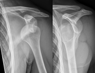
- Pie de imagen: Arthiritis, trauma (dislocation, fractured neck of humerus), frozen shoulder
-
- PA projection
- Positioning: Chin on top of cassette & feet separated slightly for stability. Median saggital plane coincident with longitudinal midline of IR. MSP perpendicular to IR. Sternoclavicular joints equidistant to IR. Shoulders relaxed. Elbows flexed & backs of hands on hips. Elbows rolled forwards to clear scapulae from lung fields. Expose on arrested inspiration & height of IR should be slightly above the shoulders
- Centring: Midline of thorax & cassette. Spinous process of T7 (body of T8) with horizontal central ray
- FFD: 200cm (2 metres)
- Collimation: 1st thoracic vertebra, 1st rib, lateral margins of ribs, 2 - 10 and costophrenic angles
- 35 x 43 IR or erect bucky
- Criteria: 3-5cm apical tissue shown above clavicles, medial ends clavicles equidistant from midline of thoracic vertebrae, 6 anterior or 9 posterior ribs, visible trachea, diaphragms & costophrenic angles shown, sharp image of lungs in contrast to air filled lung tissue and dense like the hilae & mediastinum
- AP Projection
- Positioning: Same as PA but patient has posterior aspect of the chest in contact with the IR
- Centring: Middle of thorax & receptor (approx midway between sternal angle & Xiphisternum) with a horizontal central ray.
- FFD: 180cm or 200cm (2metres)
- Collimate to include the 1st thoracic vertebra, 1st rib, lateral margins of ribs and costophrenic angles
- 35 x 43 cassette
- Lead applied to waist
- Criteria: Same as PA chest, do get magnification of heart, adequate inspiration is harder to achieve, lordosis more likely
- and difficult for infirm patient to lift chin
-
Clinical Indications for a Chest X-Ray
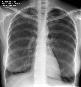
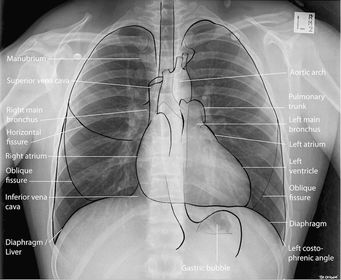
- Pie de imagen: Heart disease, respiratory, infection(SOB, pyrexia), chest pains, cardiac conditions, TB, trauma, suspected malignancy, metastases, post arrest, pre-anaesthetic, perforation, pneumothorax, post insertion/removal of lines & drains, haemoptysis, long term cough (6weeks), sarcoidosis in the eye
-
- Lateral Chest
- Positioning: arms raised & lateral aspect of chest in contact with IR. Height of cassette adjusted until the top is level with top of shoulder. Midline between the anterior & posterior chest walls is coincident with the vertical midline of the IR. Elbows are flexed & hands clasped at the back of head. Humeri are abducted medially until parallel & slight forward tilt of trunk brings thorax into a vertical position. MSP parallel to cassette
- Centre: Middle of cassette
- FFD: 180cm and 90 degrees
- Collimation: shoulder, sternum, spinous processes of thoracic vertebrae, posterior and anterior costophrenic angles
- 35 x 43cm IR vertically
- Criteria: Shoulder & soft tissue of upper arms overlying lung apices only, condyles on posterior aspect of T spine are superimposed, posterior aspect of ribs are SI, intervertebral joint spaces are clear, left diaphragm higher than right, T11 is seen above posterior diaphragm, sharp image of lung markings in contrast with the heart, aorta, air filled trachea & ribs.
- Lateral Sternum
- Positioning: Patient stands or sits with lateral aspect of chest placed in contact with image receptor. Feet separated for stability. If CR adjust cassette so middle is coincident with point midway between sternal notch and xiphisternum. MSP parallel to image receptor. Arms raised above head.
- Centre: Midway between sternal notch and xiphisternum
- Collimate: sternoclavicular joints, manubrium, sternal body, xiphisternum, soft tissues of anterior sternal area & retrosternal lung tissue
- FFD: 100cm
- Criteria: Area of interest included. Arms cleared of ROI. Sternoclavicular joints superimposed. Sharp image
-
Clinical Indication for a Sternum
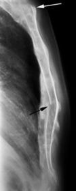
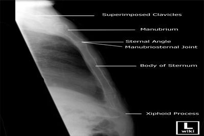
- Pie de imagen: Fractures - check for a step in the cortex of the sternum. Swelling of the surrounding soft tissue can accompany bone fracture.
-
- Infero-superior Positioning
- Positioning criteria: Posterior aspect of shoulder in contact with image receptor
- Centring: To the middle of the clavicle with the tube angle 30°towards the head (cephallically / cranially)
- FFD: 100cm SID
- Collimate to include all of clavicle
- Criteria: Clavicle is projected clear of lung fields. Normally used to assess fracture union.
- Antero-posterior Positioning
- Position: Patient either remains ‘straight’ with median sagittal plane at 90º to the receptor or turns approx 20º towards the affected side (this brings the medial end of the clavicle away from the spine). Patient’s head is turned away from the side under examination.
- Centre over the mid point of the clavicle with a horizontal central ray.
- Collimate to include all of clavicle (including acromioclavicular and sternoclavicular joints)
- 100cm SID.
- Postero-anterior Positioning
- Positioning criteria: Patient rotates affected shoulder towards the image receptor so that anterior aspect of shoulder is in contact with the receptor and the clavicle is parallel to the receptor.
- Centring: To the middle of the image receptor so that the central ray emerges at the mid shaft of the clavicle
- Other info: 100cm SID
- Collimate to include all of the clavicle
- Not used as a first line investigation in trauma. You would do an AP shoulder (rotated 20-30º or ‘straight’)
- Can be used as a follow up projection
-
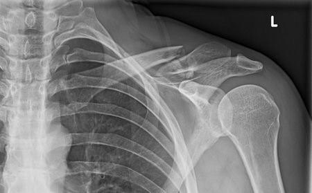
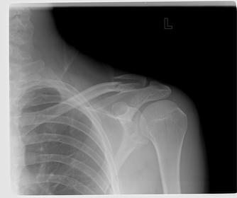
- Pie de imagen: A fracture of the clavicle characteristically leads to inferior displacement of the distal component - weighed down by the whole arm
- Pie de imagen: Transverse/oblique mid shaft fracture, Inferior displacement of the distal clavicle
Información
- Descripción
- 1 (Principles of Radiographic Imaging) Diagnostic Radiography Diapositivas sobre Upper Limb Radiography , creado por Rutendo Chingomb el 02/11/2015.
- Creado por
- Rutendo Chingomb
- Visitas
- 13
- Valoración