12056545
Description
Flashcards by Sara. H S., updated more than 1 year ago
|
|
Created by Sara. H S.
almost 8 years ago
|
|
| Question | Answer |
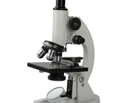
| What is a microscope? | A microscope is an instrument used to magnify a small object. Some types use light to go through the image, some use electrons. |
| Define the following term: Monocular microscope | Monocular microscopes are light microscopes that use one ocular lens (mono- meaning one). |
| What do you call a light microscope with two ocular lenses? | Binocular |
| What is the difference between binocular and monocular telescopes? | Monocular microscopes only have one ocular lens while binocular (bi- means two) have two ocular lenses. |
| Contrast light microscopes and stereo microscopes. | Three differences include: 1. Unlike light microscopes, stereo microscopes make three-dimensional images 2. Light microscopes have a maximum magnification of x1000 while stereo microscopes have it at x100. 3. Light reflects off the specimen in stereo microscopes while light passes through the specimen of light microscopes. |
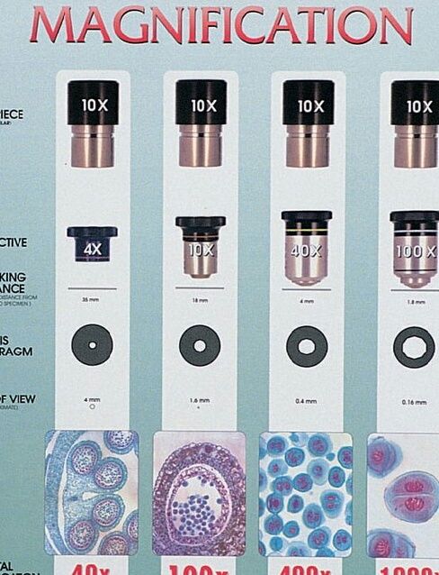
| How do you calculate magnification? | To calculate magnification, multiply the magnification of the ocular lens and the total magnification of the objective lens (e.g. x 10 x 4 = x40). |
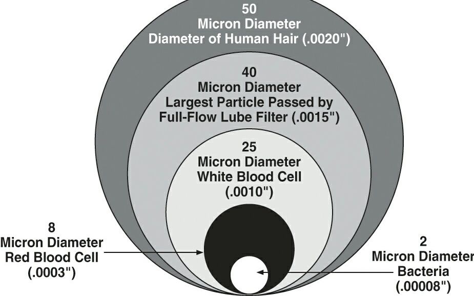
| What is a micrometre? | A metric unit of measurement used by scientists to measure microscopic objects. |
| How does an electron microscope work? | Electron microscopes use beams of elementary particles called electrons in order to magnify images up to x1,000,000. |
| What are the two types of electron microscopes? | The two types of electron microscopes are TEM (transmission electron microscope) and SEM (scanning electron microscope). |
| Give three differences between SEM and TEM. | Three differences between SEM and TEM are: 1. Unlike SEM, specimens have to be sliced with TEM. 2. Electrons reflect off the surface of specimens in SEM while they pass through the specimen in TEM. 3. SEM greatly magnifies objects while TEM shows their inner structure. |
| What happens when an image seen in a monocular microscope when the slide is moved (e.g. to the left)? | The image moves to the opposite direction that you moved the slide. |
| Why must specimens be prepared and cut thin in order to be viewed in a light microscope? | Cutting the specimen into a thin slice allows the light to pass through it easily. |
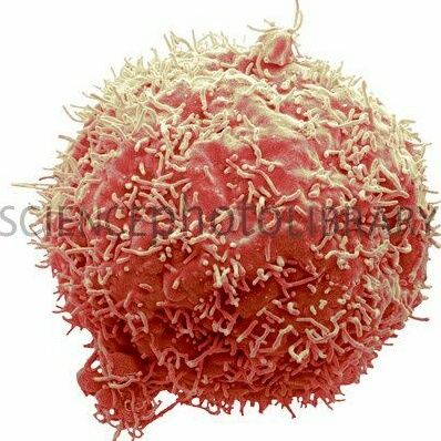
| You are shown a magnified three-dimensional image of a human liver cancer cell. What type of microscope was used to make the image? | A stereo microscope was used to make the image. |
| What is the function if this part of a microscope: the mirror/lamp? | The mirror/lamp directs light up through the stage and specimen. |
| What is the function of this part of a microscope: the handle? | The handle is used to carry the microscope safely. |
Want to create your own Flashcards for free with GoConqr? Learn more.