12596476
Description
Flashcards by Dolu Falowo, updated more than 1 year ago
|
|
Created by Dolu Falowo
almost 8 years ago
|
|
| Question | Answer |
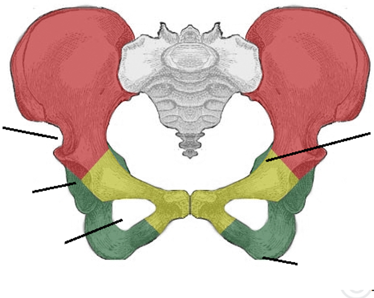
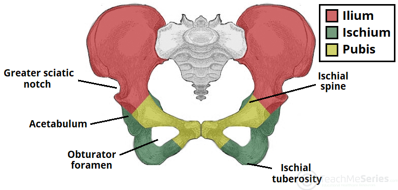
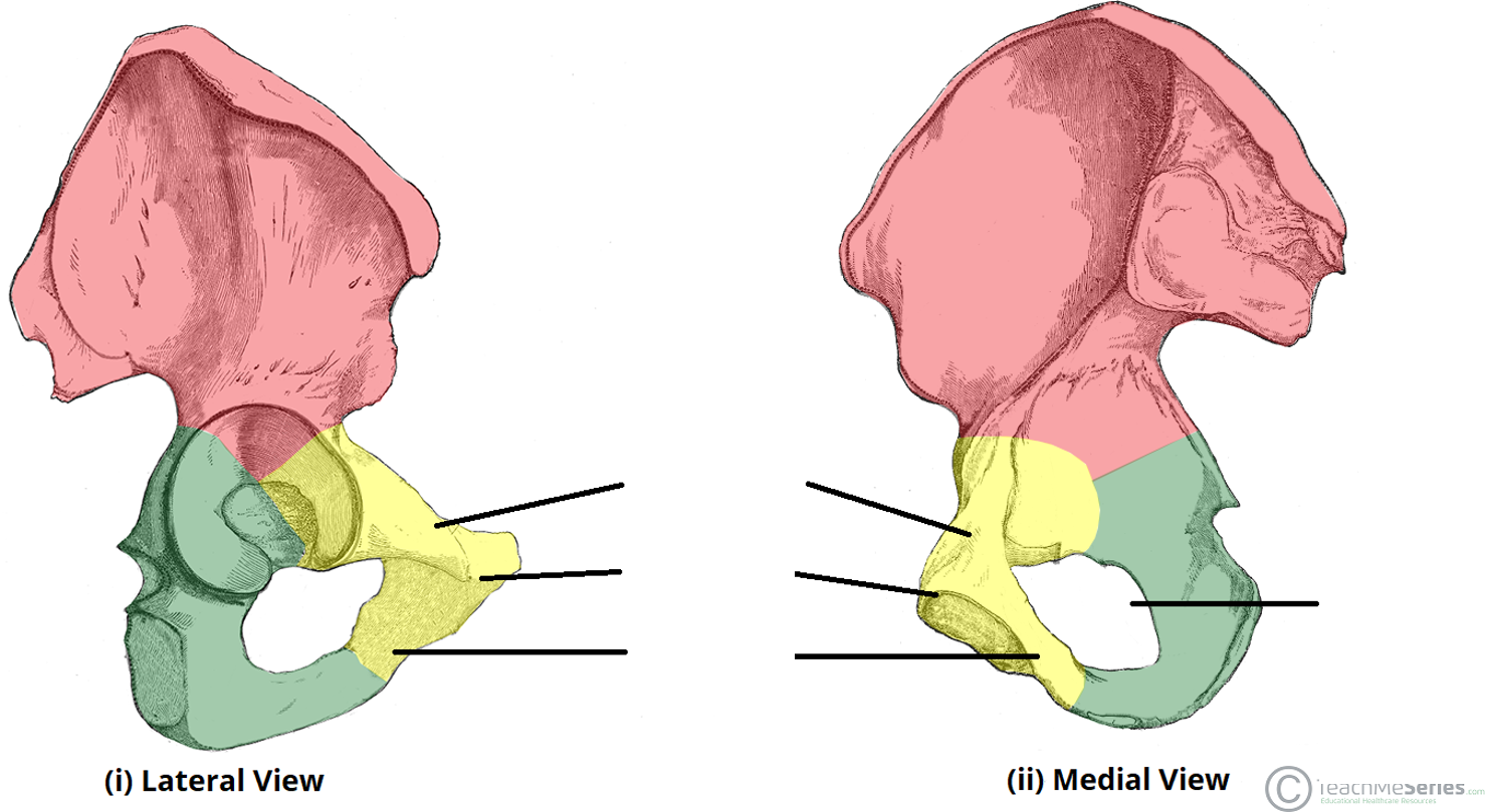
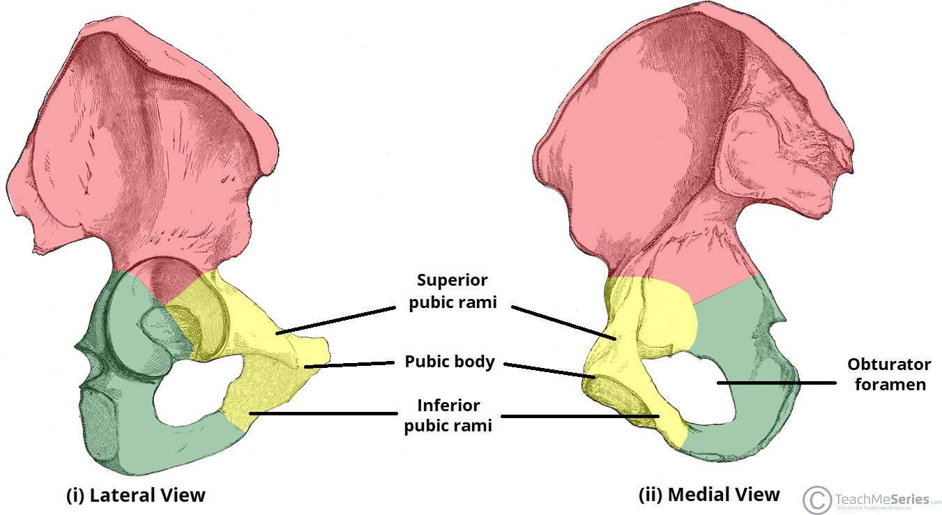
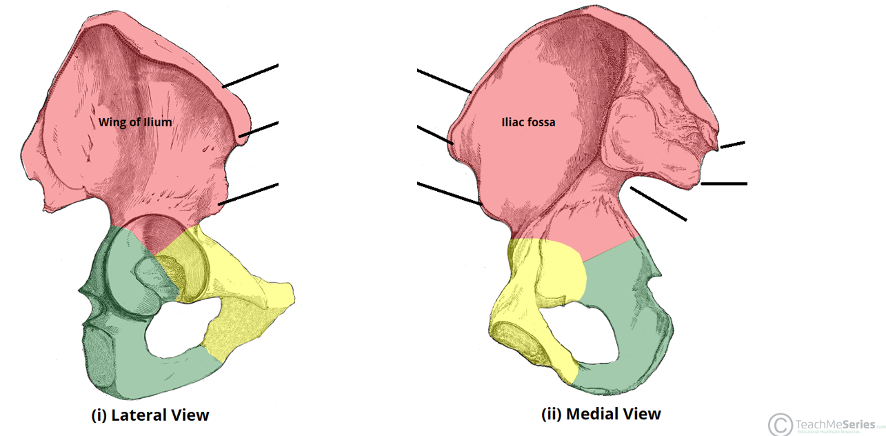
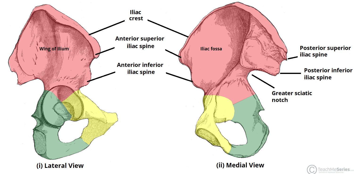
| What connects the pubis, ilium and ischium? | Triradiate cartilage. Fuses at around 15-17 |
| Which ligaments provide stability to the hip joint? | Anterior: ileofemoral and pubofemoral ligament Posterior: ischiofemoral ligament |
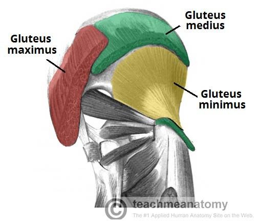
| Describe the gluteus maximus in terms of: -movement -nerve -spinal levels | -Extends and lateral rotation -Inferior gluteal nerve -L5-S2 |
| Describe the gluteus medius in terms of: -movement -nerve -spinal levels | -Abducts and medial rotation -Superior gluteal nerve -L4-S1 |
| Describe the gluteus minimus in terms of: -movement -nerve -spinal levels | -Abducts and medial rotation -Superior gluteal nerve -L4-S1 |
| Describe the tensor fascia lata in terms of: -movement -nerve -spinal levels | -Abduction and medial rotation -Superior gluteal nerve -L4-S1 |
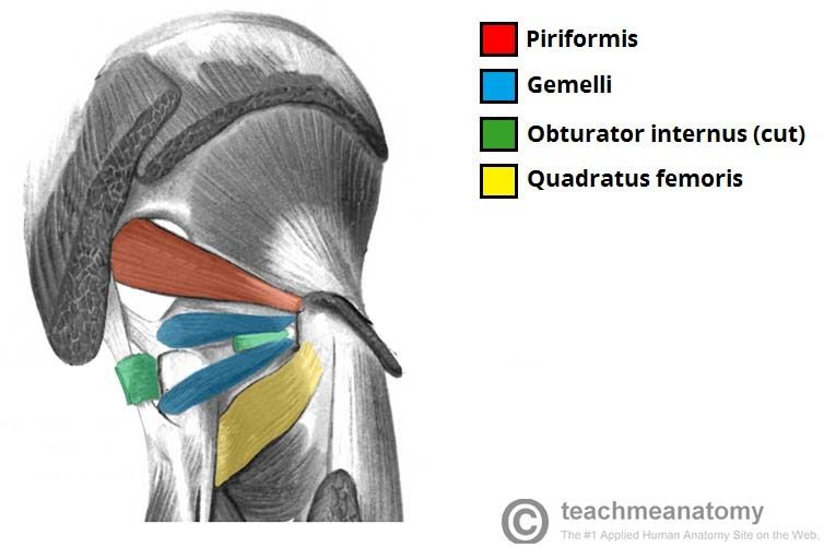
| Describe the piriformis in terms of: -movement -nerve | -Lateral rotation and abduction -Nerve to piriformis |
| Describe the obturator internus in terms of: -movement -nerve | -Lateral rotation and abduction -Nerve to obturator internus |
| Describe the gemelli (superior/inferior) in terms of: -movement -nerve | -Lateral rotation and abduction -Superior: nerve to quadratus internus -Inferior: nerve to quadratus femoris |
| Describe the quadratus femoris in terms of: -movement -nerve | -Lateral rotation -Nerve to quadratus femoris |
| The adductors of the hip are arranged into 2 layers What are they? What muscles are in each? | Superficial layer: adductor longus, gracilis Deep layer: adductor brevis, adductor magnus, obturator externus |
| Describe the psoas major in terms of: -action -nerve | -Flexion of thigh at hip and lateral flexion of vertebral column -Anterior rami of L1-L3 |
| Describe the psoas minor in terms of: -action -nerve | -Flexion of vertebral column -Anterior rami of L3 |
| Describe the iliacus in terms of: -action -nerve | -Flexion of thigh at hip joint -Femoral nerve L2-4 |
| Describe the pectineus in terms of: -action -nerve | -Adduction and flexion at hip joint -Femoral nerve (and also obturator nerve) |
| Describe the sartorius in terms of: -action -nerve | -At hip: flexor, abductor, lateral rotator -At knee: flexor -Femoral nerve |
| What are the various boundaries of the femoral triangle? -superior -lateral -medial -roof -base | Superior: inguinal ligament Lateral: medial border of sartorius muscle Medial: medial border of adductor longus Roof: fascia lata, skin Base: pectineus, ilipsoas, adductor longus |
| Describe the contents of the femoral triangle | -Femoral nerve -Femoral artery -Femoral vein -Femoral canal (contains deep lymph nodes) |
| What are the extensors in the hip? | -Semimembranosus -Semitendinosus -Biceps femoris (long/short head) |
| Describe the semimembranosus and semitendinosus in terms of: -action -nerve | SAME ACTION/INNERVATION -Flexion at knee -Extension of thigh at hip -Medial rotation of thigh at hip/leg at knee -Tibial part of sciatic nerve |
| Describe the biceps femoris long/short head in terms of: -action -nerve | -Flexion at knee -Extends leg at hip -Lateral rotation at hip/knee Long head: tibial part of sciatic nerve Short head: common fibular part of sciatic nerve |
| Why is the femoral artery important to cardiologists? | -A catheter can be inserted and passed up into the heart -Dye is injected (coronary angiogram) -Check for blockages/narrowings |
| At which site is fracture of the hip most likely to occur? | Neck of femur as there is a large amount of cancellous bone |
| What other consequences of a posterior hip dislocation can occur? | -Associated fractures of posterior lip of acetabulum -Damage to acetabular labrum -Sciatic nerve damage (L4-S3) -Avascular necrosis |
| What is a waddling gait/positive Trendelenberg sign? | Weakness/paralysis of abductor muscles of the hip (gluteus medius/minimus) When standing on one leg, pelvis drops of the other side Superior gluteal nerve is damaged |
| What happens in a 'pulled elbow'? Why is it common in children? | Radial head subluxation occurs. Arm is held in pronation, and it is difficult to supinate. Annular ligament is immature so the radial head can be pulled out of place |
| Is an X-ray needed to diagnose a 'pulled elbow'? | Yes -Mechanism of injury is unknown -Check for supracondylar fractures |
| What muscles are involved in pronation? What is their innervation? | -Pronator Teres (median nerve) -Pronator quadratus (median nerve-anterior interosseous branch) -Brachioradialis (radial nerve) |
| What muscles are involved in supination? What is their innervation? | -Supinator (radial nerve-deep branch) -Biceps brachii (musculocutaneous nerve) |
| Describe the procedure used to repair a pulled elbow | -Hold wrist/forearm -Turn hand to supinate it -Put pressure at the radial head with a hand -Slowly flex the elbow -A faint pop will be heard when the joint is back in place -Put arm is sling in flexed position for 2 weeks |
| What is a Pott's fracture? | -Medial ligaments are damaged -Tibia internally rotates, but foot stays planted down -Tibia hits on fibular and the force is transmitted causing a fracture |
| What is a sprain? Signs/symptoms? | Torn/damaged ligament Pain, swelling, bruising, inability to move limb, difficulty using affected extremity |
| Describe how a posterior hip dislocation occurs ie. in a RTC | -Angle of joint is changed when sititing (hip is flexed) -Major force pushes backwards -Posterior side of the hip is a weakness as the iliofemoral, pubofemoral and ischiofemoral ligaments are anterior to the hip |
| Which nerve is vulnerable to damage? Consequences if damaged? | Sciatic nerve (L4-S3) -Posterior compartment of leg via tibial nerve (flexion) -Anterior and lateral compartment of leg via common fibular nerve (extension) -Can result in a foot drop -Lower back referred pain |
| Which bones are vulnerable to injury at the 'temple'? Why is this life-threatening? | -Behind the temple lies the pterion region (frontal, parietal, temporal and sphenoidal bones join) -This is the weakest area of the skull -Anterior division of middle meningeal artery runs underneath the pterion. Rupture can lead to an epidural haematoma |
| Which nerves may be affected by a fractured humerus? | -Ulnar (posterior to medial epicondyle) -Radial (anterior to lateral epicondyle and radial groove) -Median (anterior to medial epicondyle) -Musculocutaneous (anterior to humerus) |
| What is a straddle injury of the pelvis? | -Caused by compression of soft tissues against bony margins of the pelvic outlet -Fractures both the superior and inferior pubic rami |
| What soft tissues are at risk from straddle injuries? | -Injury/compression of genitalia (testes/vulvar tissue) -Urethra -Urinary bladder |
Want to create your own Flashcards for free with GoConqr? Learn more.