13319674
Description
Flashcards by Yasmin Beer, updated more than 1 year ago
|
|
Created by Yasmin Beer
almost 8 years ago
|
|
| Question | Answer |
| What is the heart? Define cardiac output. | *The heart is the pump that moves the blood around the body = its activity is described as the cardiac output Cardiac Output = Heart rate x Stroke volume *Heart rate is driven by waves of electrical activity that induce the cardiac muscles to contract. |
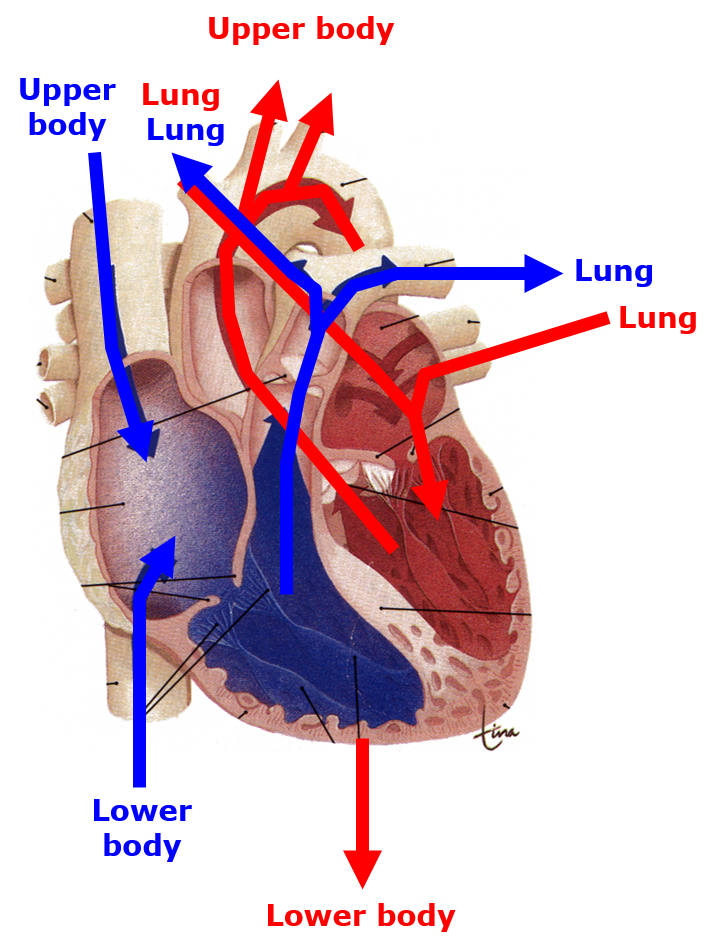
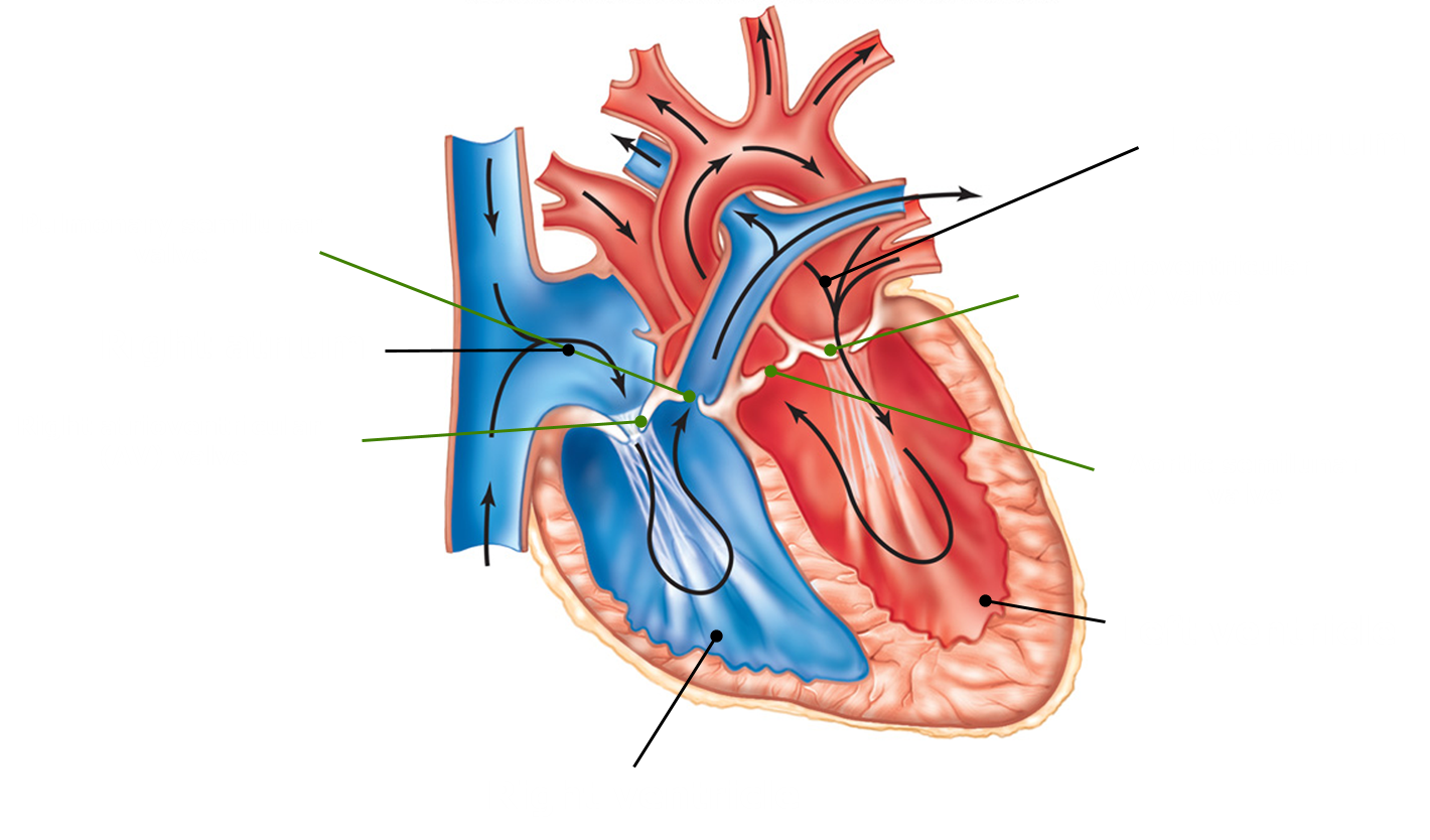
| Describe blood flow in the heart. | |
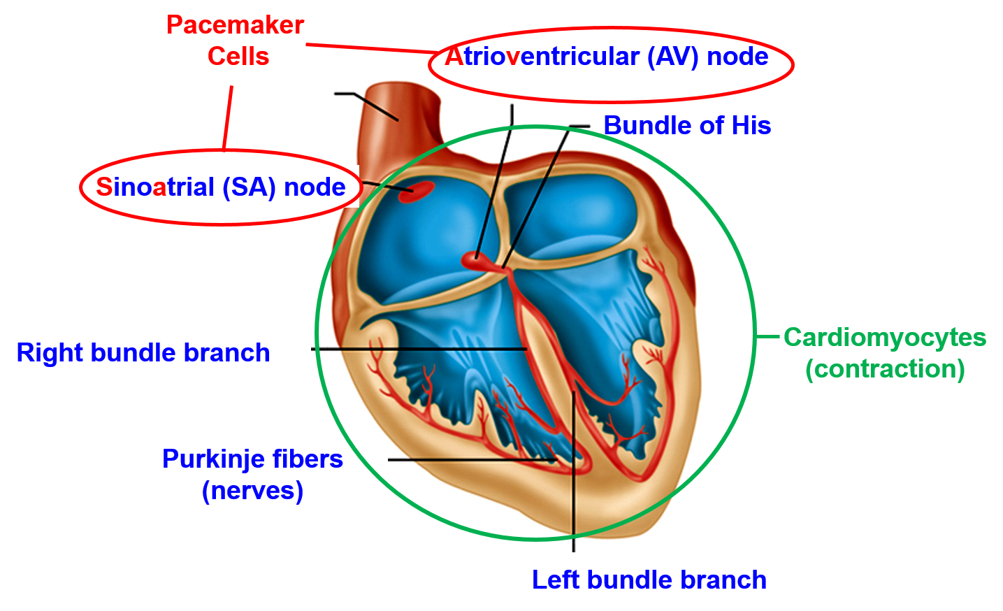
| Outline the components of the conduction system of the heart. | Heartbeat is myogenic. Heartbeat is driven by specialised myocytes composed of the sino-arterial node and the artrioventricular node . |
| Outline the sequence of cardiac excitation. | Spread of action potentials via the purkinje fibres results in coordinated contraction of ventricles. |
| What causes the sino-atrial node to trigger an action potential? | Low resting membrane potential (-60-70v) Na+ leakage =Action potential |
| Outline the mechanism underlying spontaeous action potential generation in pacemaker cells. | *Sodium ions “leaking” in through the F-type channels and calcium ions moving in through the T-type channels cause a threshold graded depolarization. *The rapid opening of voltage-gated calcium L-types channels is responsible for the rapid depolarization phase. *Reopening of potassium channels and closing of calcium channels are responsible for the repolarization phase. |
| Explain the mechanism of contraction of the ventricular cardiomyocytes. | *The rapid opening of voltage-gated sodium channels is responsible for the rapid depolarization phase. *The prolonged “plateau” of depolarization is due to the slow but prolonged opening of voltage-gated calcium channels and closure of potassium channels *Opening of potassium channels results in the repolarization phase. |
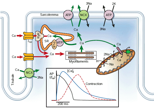
| How does calcium produce contraction of cardiac muscles? | *Excitation-Contraction Coupling in Cardiac Muscle *Calcium ions regulate the contraction of cardiac muscle: The entry of extracellular calcium ions causes the release of calcium from the sarcoplasmic reticulum. |
| What is the refractory period? Explain its purpose. | *Refractory period = time required before it is possible to re-stimulate muscle contraction > Cardiac muscle (250ms) *Cardiac muscle has a prolonged refractory period which allows for ventricles to fill with blood prior to pumping. |
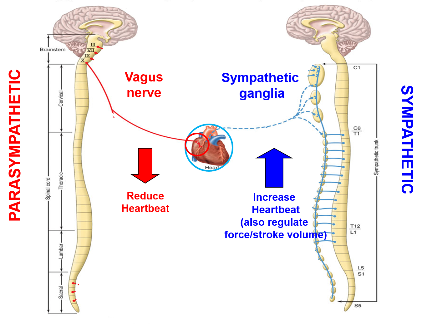
| Outline the role of the Autonomic nervous system in regulation of the sino-atrial node. | |
| Outline regulation of heart beat by para/sympathetic nervous system. | 1. Spontaneous depolarisation of the sino-atrial node is the driver of heart rate 2.Sino-atrial node is regulated by the parasympathetic (reduce) and sympathetic (increase) nerves 3.Electrical activity flows from sino-atrial node – atrioventricular node – purkinje fibres - cardiomyocytes |
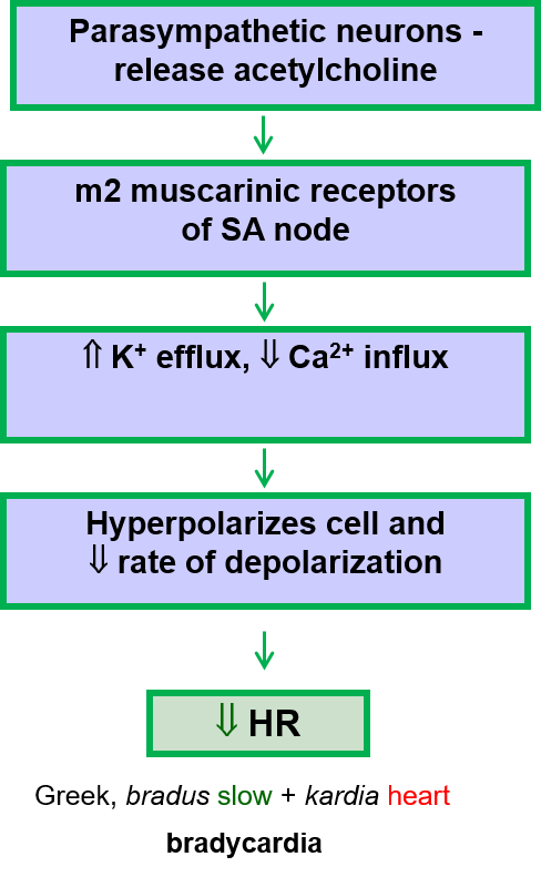
| Describe parasympathetic regulation of the depolarisation of the sino-atrial node? | |
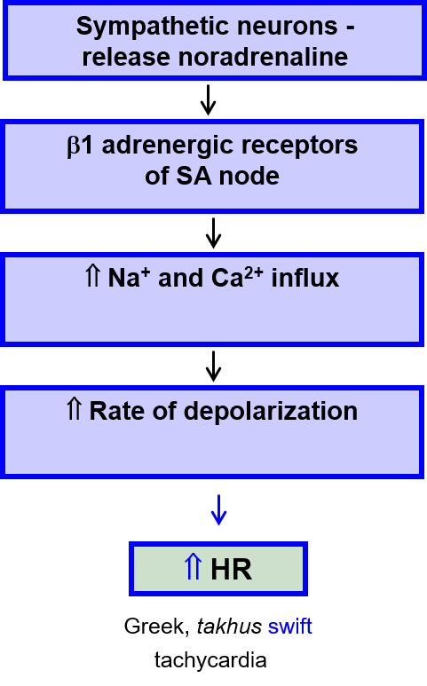
| Describe sympathetic regulation of the depolarisation of the sino-atrial node? | |
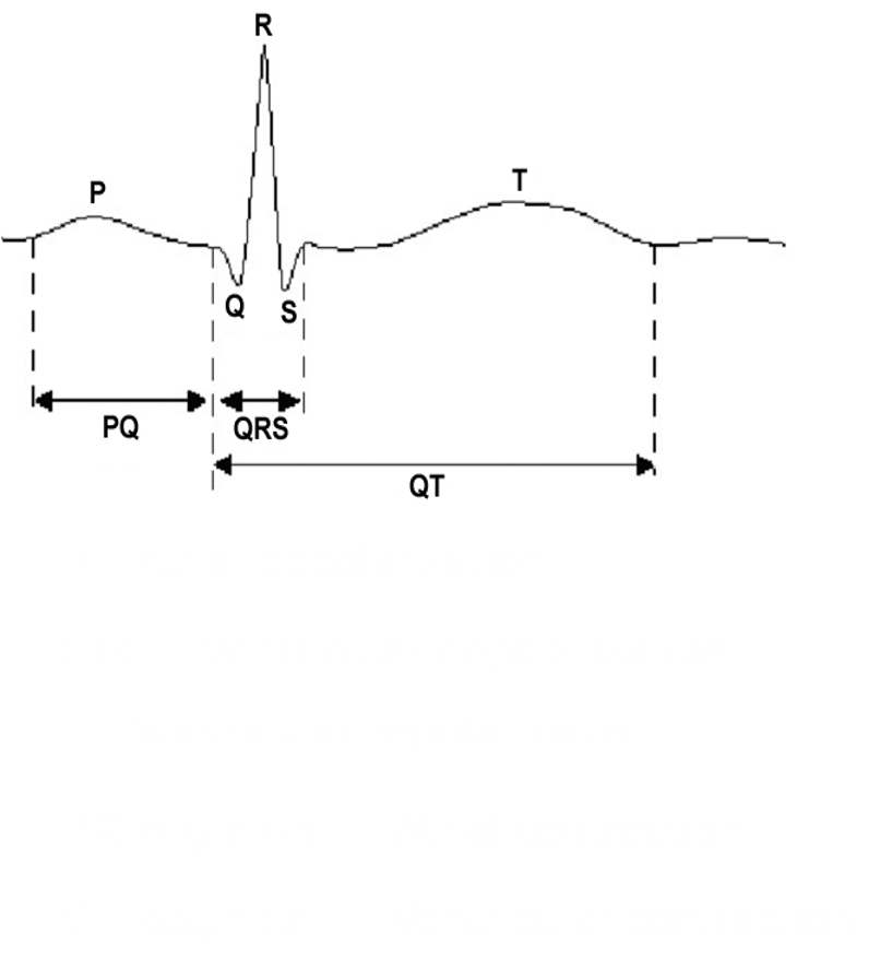
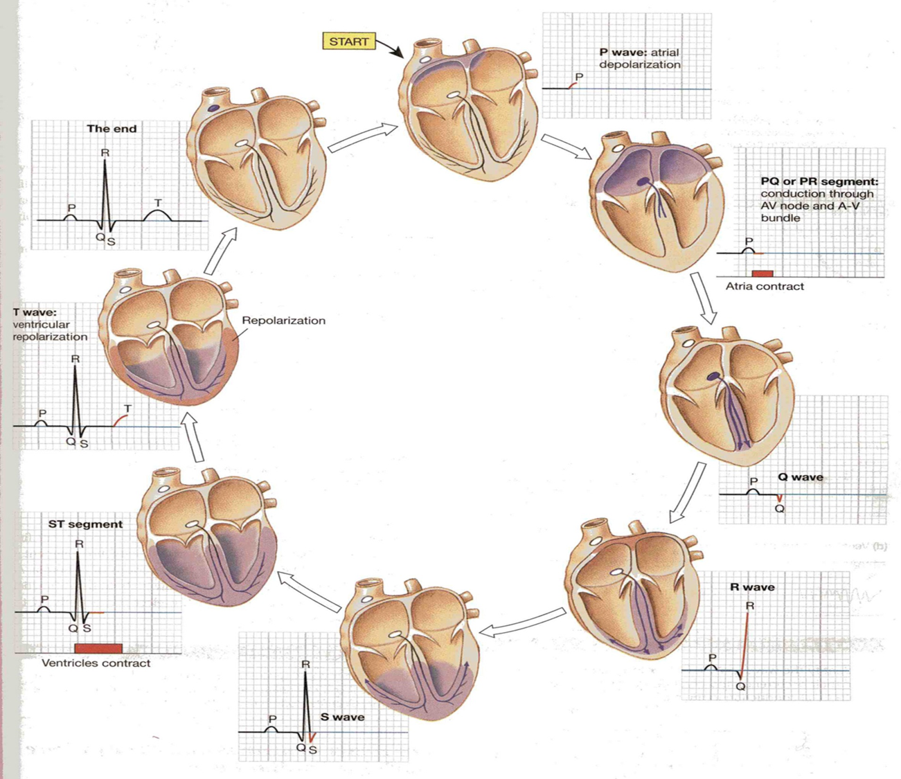
| What is an electrocardiogram? | *ECG is a summation of the spread of action potentials through the various sections of the heart. *PQRSTU assigned to various deflections |
| Label general electrocardiogram. | |
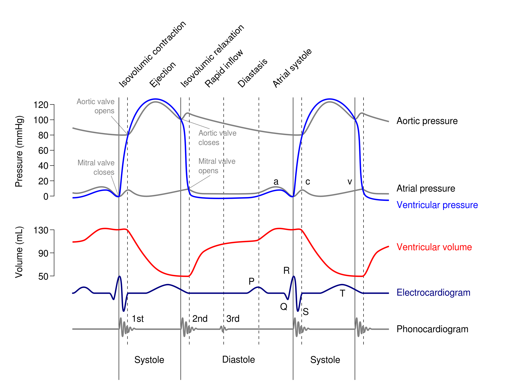
| Outline electrical events of the cardiac cycle. | |
| Explain how the ECG is used to diagnose disease. | *Provides valuable information on the electrical activity of the heart -Heart rate/ Heart rhythm -Disturbances of rhythm and conduction (arrhythmia, pacemaker) -Conduction velocity -Anatomical orientation of the heart -Relative size of chambers -Condition of tissue within the heart -Damage to the myocardium -Influence of certain drugs |
| What is ventricular fibrillation? | RESULTS - Random Firing of heart, Fibrillating ventricles cannot pump blood, Fatal after a few minutes 450,000 deaths/year. CAUSES - Myocardial Infarction (heart attack), Electrical shock, Drug intoxication Impaired cardiac metabolism |
| Describe structure of the heart. | |
| What are the two phases of the cardiac cycle? | SYSTOLE – ventricular contraction and blood ejection DIASTOLE– ventricular relaxation and blood filling |
| Explain pressure changes in different blood vessels. | *Blood pressure is reduced as it passes from arteries to veins since bigger lumen *Lower artiole pressure in pulmonary system since dont want formation of blood pressure in lungs |
| Explain contractions in one heart beat. | *Passive filling of atria *Atria contact semi lunar valves shut *When pressure in atria greater than in ventricles AV valves open and blood forced into ventricles *ventricles begin to contract AV valves shut *Semi lunar valves only open when pressure in ventricles greater than that of the aorta. *Diastole |
| Describe the pressure and volume changes that occur during the cardiac cycle. | |
| Explain the heart sounds as heard by a stethoscope. | IST SOUND – closure of the atrioventricular (AV) valves ‘lub’ - onset of systole (contraction) 2ND SOUND – closure of pulmonary and aortic valves ‘dub’ - onset of diastole (relaxation) |
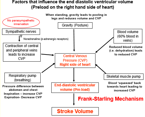
| Outline the factors that influence the end diastolic ventricular volume. | |
| What is the frank starling mechanism? | *Ventricles contract with more force (i.e. stroke volume ↑) if it contains more blood (i.e. end-diastolic ventricular volume ↑) *Frank-Starling mechanism determined by length-tension relationship in the muscle i.e. the greater the stretch the more tensioned developed in the cardiac muscle |
| Explain how the frank-starling mechanism maintains balance between the left and right sides of the heart. | -If increased venous return to right ventricle then increased contraction = more blood to lungs - increased venous return to left ventricle then increased contraction = increased stroke volume *blood doesnt accumulate in lungs |
| Define stroke volume. | *Stroke volume (ventricular ejection) is determined by the venous end-diastolic volume (pre-load), arterial (peripheral) resistance (after-load) and sympathetic stimulation. |
| Describe regulation of stroke volume. | SYMPATHETIC NERVOUS SYSTEM REGULATES STOKE VOLUME. *Sympathetic ganglia *Sympathetic nerves= b1 adrenergic receptors *Increases force and speed of cardiac muscle contraction = Increases ventricular contraction at any given end-diastolic ventricular volume *Arterial blood pressure opposed ejection of blood. |
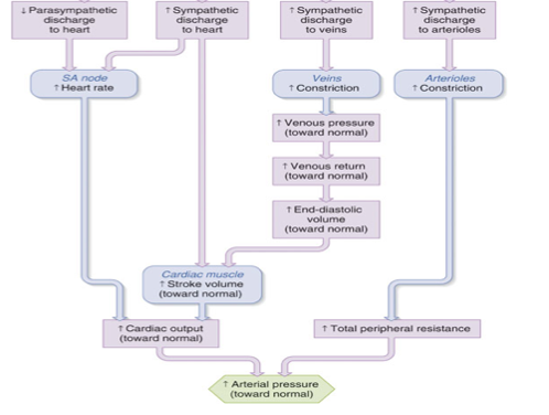
| Give an overview of regulation of cardiac output. | |
| What is congestive heart failure? | *Congestive Heart Failure (CHF) -Reduced Cardiac Output/ Tiredness and shortness of breath/ Fluid Retention to increase cardiac output (venous return) *Chronic Left Ventricular Failure most common -Coronary artery disease leading to ischaemic heart disease and myocardial infarction( systolic dysfunction) -Hypertension (high blood pressure) (diastolic dysfunction) -Cardiomyopathy (viral infection, heavy drinking) |
| Explain what happens during heart failure - systolic dysfunction. | *Heart Attack (myocardial infarction (MI)) *Damage to heart muscle *Ventricular contractility ↓ *Stroke Volume ↓ |
| Explain what happens during heart failure - diastolic dysfunction. | *High Blood Pressure(Hypertension) *Arterial Pressure increased *Cardiac resistance (afterload) *Ventricular Muscle (hypertrophy) *Stiffening (reduce compliance) of ventricular wall *End-diastolic ventricular volume *Stroke Volume ↓ |
Want to create your own Flashcards for free with GoConqr? Learn more.