13811772
Description
Flashcards by zakiya gaisie, updated more than 1 year ago
|
|
Created by zakiya gaisie
almost 6 years ago
|
|
| Question | Answer |
| What is the magnification of a light microscope, TEM and SEM? | Light - X1500 TEM - X500,000 SEM - X100,000 |
| Define Magnification | The amount of times something is enlarged by, compared to the original object |
| What is the resolution of a light microscope TEM & SEM ? | light = 200nm TEM = 0.2nm SEM = 0.2nm |
| Define resolution | The ability to distinguish between two separate points with clarity. |
| How do you calculate magnification? | image size ---------------------- actual size |
| How many micrometres in a millimetre? | 1000um |
| How many nano-metres in a micrometer? | 1000nm |
| For a light microscope how do you prepare a slide (dry mount)? | 1. Take a thin slice of your specimen 2. Using tweezers place your slice in the middle of a clean slide 3. Place a cover slip on top of your slice |
| How do you prepare a slide for a light microscope (wet mount)? | Pipette a small drop of water onto slide 2. Using tweezers place slice, and the cover slip on top of water drop, ensuring there are no air bubbles under the cover slip 3. Add the stain by placing a drop at one edge of the cover slip and a paper towel at the opposite edge to draw stain through |
| Name 4 similarities between Prokaryotic and Eukaryotic cell. | They both have: -Plasma membrane - Cytoplasm - Ribosomes - DNA and RNA |
| Name 4 differences between Prokaryotic and Eukaryotic cells. | - much smaller (2µm in diameter vs eukaryotes that are 10-100 µm) - Free floating, circular DNA (nucleoid) not linear/straight and not contained within a nucleus - Less well-developed cytoskeleton - A cell wall made from peptidoglycan |
| Describe the function of the nucleus | - Houses all of cell’s genetic material - Chromatin consists of DNA and proteins - Has instructions for making proteins |
| Describe the structure of the nucleus | - Largest organelle - Contains chromatin - Surrounded by nuclear envelope and contains nucleolus |
| Describe the structure of the nucleolus | - Dense spherical structure inside nucleus |
| Describe the function of the nucleolus | - Makes RNA and ribosomes |
| Describe the structure of the nuclear envelope | - Surrounds nucleus - 2 membranes with fluid between them - Nuclear pores go through envelope |
| Describe the function of the nuclear envelope | - Pores allow passage of relatively large molecules (e.g. hormones and mRNA) |
| Describe the structure of the RER | - Flattened membrane sacs called cisternae - Continuous with outer nuclear membrane - Studded/littered with ribosomes |
| Describe the function of the RER | - Modifies and transports proteins made on attached ribosomes - Some proteins are secreted from the cell - Some are placed on plasma membrane |
| Describe the structure of the SER | - Flattened membrane sacs called cisternae - No ribosomes |
| Describe the function of the SER | - Involved in lipid production (e.g. cholesterols and steroid hormones) - Involved in lipid absorption from the gut |
| Describe the structure of the golgi apparatus | - Stack of membrane bound, flattened sacs - Vesicles can often be seen around the edges |
| Describe the function of the golgi apparatus | - Modifies proteins (e.g. folding or adding sugar) - Packages modified proteins into vesicles for transportation - Some modified proteins are secreted from surface of the cell |
| Describe the structure of the flagella | - Extension sticking out from cell - Cylinder contains nine microtubules arranged in a circle and has 2 in the middle '9,2 formation' - Long - Usually present alone or in pairs |
| Describe the function of the flagella | - Enables movement |
| Describe the structure of the mitochondria | - Spherical or oval shaped and only 2-5 µm long - Enveloped in two membranes - Highly folded inner membrane forms cristae - Central part called matrix |
| Describe the function of the mitochondria | - Site of ATP (adenosine triphosphate) production and aerobic respiration |
| Describe the structure of a lysosome | - Small spherical sacs - Surrounded by single membrane |
| Describe the function of lysosomes | - Keep damaging enzymes separate from the cell - Can engulf old/worn out organelles and foreign substances, digesting them with their enzymes |
| Describe the structure of chloroplast | - Double membrane - Separated by fluid filled space - Continuous inner membrane with network of flattened membrane sacs called thylakoids - Stack of thylakoids = granum |
| - Site of photosynthesis | |
| Describe the structure of cilia | - Hair-like extensions from the cell surrounded by the plasma membrane - contain microtubules from the cytoskeleton and are formed from centrioles |
| Describe the function of cilia | - Can act as antennae in cell signalling - Or can act in large groups on a cell surface to move material around (e.g. cilia in airways wafting mucus) |
| Describe the structure of the plasma membrane | - Continuous outer membrane - Contains a variety of molecules such as proteins and lipids |
| Describe the function of the plasma membrane | - Controls what goes in and out of cell - Receptors on cell surface allow for endocytosis and exocytosis |
| Describe the structure of the vacuole | - A fluid filled sac within a membrane called a tonoplast |
| Describe the function of the vacuole | - The fluid contains water and a variety of solutes that helps plant cells regulate their water |
| Describe the structure of ribosomes | - No outer membrane - They are tiny organelles - Each consists of 2 sub-units - Some found in cytoplasm, some bound to RER |
| Describe the function of ribosomes | - Site of protein synthesis in the cell - Act as an assembly line where mRNA is used to assemble proteins from amino acids |
| Describe the structure of centrioles | - Made of microtubules - Small tubes of protein fibres |
| Describe the function of centrioles | - Take part in cell division - Form spindle fibres, moving chromosomes in cell division |
| Describe the structure of the cellulose cell wall | - Made from cellulose fibres - Found on the outside of the plasma membrane of plants |
| Describe the function of the cellulose cell wall | - Supports the cell and maintains their shape - contributing to the shape of the whole plant - Prevents cells from bursting when they are swollen with water (turgid) - The cell wall is permeable to allow solutions to pass in and out |
| What are the steps in protein synthesis? | 1. Genes are copied into messenger RNA (mRNA), which leaves the nucleus via nuclear pores and attaches to a ribosome 2. Ribosomes read mRNA and assembles amino acids into a unique sequence 3. Proteins produced by ribosomes on the rough ER are modified (e.g. sugars added and polypeptides folded) within the rough ER. 4. The polypeptide(s) are transported to the Golgi apparatus in a vesicle. 5. Further modified and packaged into a vesicle 6. Vesicle transported to cell surface membrane and secreted (by exocytosis). |
| What is the function of the cytoskeleton? | - Helps maintain the shape of cell - Enables movement - Helps transport vesicles around cell |
| What is the structure of the cytoskeleton? | - A large network of protein fibres |
| What are the two types of protein that the cytoskeleton consists of? | - Actin filaments: Small microfilament strands 7nm in diameter help support the cell and give it mechanical strength. Intermediate filaments 10nm in diameter anchor the nucleus and can extend between some cells for communication. - microtubules: Stacks of protein cylinders, 18-30nm in diameter and made from a protein called tubulin. provides the cell with its shape and support. They form cilia, undulipodia and centrioles. Cytoskeletal motor proteins are present on microtubules. They use ATP to move the cell’s contents along the fibres. |
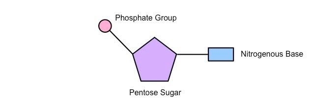
| What is the structure of a nucleotide? | |
| What are the 5 bases? | - Adenine - Thymine - Guanine - Cytosine - Uracil |
| Which bases are purines? | Adenine and Guanine |
| Which bases are pyrimidines? | Thymine Cytosine and Uracil |
| How many rings do purines have? | 2 |
| How many rings do pyrimidines have? | 1 |
| What are phosohorylated nucleotides? | nucleotides with extra phosphate groups attached |
| Give examples of phosphorylated nucleotides | ATP (adenosine triphosphate) - has 3 phosphate groups attached ADP (adenosine diphosphate) - has 2 phosphate groups attatched |
| What kind of reaction bonds nucleotides together? | condensation reaction |
| What are phosphodiester bonds? | Covalent bonds which hold the polynucleotide together (at carbon 5 and carbon 3 ) |
| What does it mean when 2 polynucleotide chains are running in antiparallel? | Parallel but pointing in opposite directions. One strand runs from 3’ to 5’ and the other from 5’ to 3’. |
| What is the process of DNA purification? | 1. Break up the DNA containing tissue (e.g. cheek cells from a swab) to break it up into cells 2. Add a detergent – detergents break apart lipids, like those found in the plasma membrane 3. Shake with ethanol to precipitate the DNA out of solution |
| What are the steps in DNA replication? | 1. DNA double helix unwinds and “unzips”; the hydrogen bonds between base pairs are broken. The unzipping of the hydrogen bonds is catalysed by an enzyme called DNA helicase. 2. The exposed nucleotide bases act as a template for assembly of the new DNA strand. 3. Free nucleotides move towards these exposed bases according to the base pair rule. 4. An enzyme called DNA polymerase binds the nucleotides together with covalent bonds, forming the new sugar-phosphate backbone. (travels from 5’ to 3’) 5. The leading strand is replicated continuously, and the lagging strand is synthesised in fragments (discontinuously) that are then joined by ligase enzymes. 6. This results in 2 daughter DNA strands |
| What does DNA Helicase do? | Catalyses the unzipping of hydrogen bonds |
| Why do we call it semi-conservative relpication? | one of the original/parental strands is conserved on each daughter DNA molecule. |
| What does DNA polymerase do? | What does DNA polymerase do? |
| What does ligase do? | Joins the fragments of the lagging strand together |
| What does degenerate mean? | Several/lots of different triplets can code for the same amino acid |
| What does it universal mean? | the genetic code is the same for all living organisms |
| What does non-overlapping mean? | One triplet is coded for, one after another |
| What is a gene? | A sequence of DNA nucleotides that codes for a protein/ |
| Define transcription | the process of copying RNA from DNA |
| What are the steps in transcription? | 1. DNA strand unwinds and unzips by DNA helicase 2. RNA nucleotides pair up with the exposed bases on one strand of DNA – called the template strand 3. The new nucleotides form the coding strand, which is complementary to the template strand 5. DNA rezips and rewinds after the coding strand of mRNA detaches 6. mRNA migrates/leaves through nuclear pores to the ribosomes for translation into a protein |
| Define translation | the process of creating polypeptides based off mRNA |
| What are the steps in translation? | 1. mRNA attaches to a ribosome on the RER 2. Transfer RNA (tRNA) carries the corresponding amino acid to each codon on the mRNA 3. The anti-codon is a triplet of bases that form part of a tRNA molecule which is complementary to the codons on the mRNA strand 4. The mRNA has a start codon at the beginning and a stop codon (that doesn’t code for an amino acid) at the end 5. One by one amino acids are added as the tRNA anti-codons temporarily pair with the codons on the mRNA strand. 6. Adjacent amino acids are joined together by peptide bonds, creating a polypeptide chain 7. This process continues until the ribosome reaches the stop codon. At this point, the polypeptide breaks loose from the ribosome and is free |
| Is translation an active or passive process? | Active process - requires ATP |
| What are the differences between DNA and RNA? | DNA = Double stranded RNA = Single stranded DNA - Found in Nucleus only RNA - Nucleus + Cytoplasm Only 1 form of DNA 3 forms of RNA (tRNA mRNA rRNA) DNA - Deoxyribose sugar RNA - Ribose sugar DNA - Thymine RNA - No Thymine it has Uracil instead |
| How many hydrogen bonds form between bases A and T? | 2 Hydrogen bonds |
| How many hydrogen bonds form between bases G and C? | 3 Hydrogen bonds |
| How do TEMs work? | Uses electromagnets to focus a beam of electrons which is transmitted through the specimen. |
| Why do same parts of an image from a TEM look darker? | Denser parts of the specimen absorb more electrons making it look darker. |
| how do SEMs work? | Scans a beam of electrons across the specimen. This knocks off electrons from the specimen and are gathered in a cathode ray tube. |
| Do SEMs produce 2D or 3D images? | 3D images |
| Define diffusion | The passive, net movement of particles from an area of high concentration to low concentration (down a concentration gradient) |
| What type of molecules diffuse through cell membranes? | Small non-polar molecules e.g. O2 and CO2. Also water as it is small enough to fit between phospholipids despite it being polar |
| What factors affect the rate of diffusion? | 1. Concentration gradient - higher = fast rate of diffusion 2. Thickness of exchange surface - Thinner = short distance for particles to travel = faster rate of diffusion 3. Surface area - large S.A = fast rate of diffusion 4. Temperature - warmer = particles have more kinetic energy = they move faster = fast rate of diffusion |
| What is facilitated diffusion? | The passive movement of larger molecules down a concentration gradient through carrier or channel proteins. |
| How do carrier proteins work? | 1. Large molecule attaches to a carrier protein in the membrane 2. Protein changes shape 3. Releases molecule on the opposite side of the membrane |
| How do channel proteins work? | They form pores in the membrane for charged particles to diffuse through - down the concentration gradient |
| Define active transport | The active movement of particles against a concentration gradient (from low to high concentration) It involves carrier proteins |
| Define osmosis | The passive movement of water molecules across a partially permeable membrane down a water potential gradient. |
| What molecule has the highest water potential? | Water |
| What is a hypotonic solution? | A solution with a higher water potential than the cell |
| What is an isotonic solution? | A solution with the same water potential as the cell |
| What is a hypertonic solution? | A solution with a lower water potential than the cell |
| What happens to animal cells when placed in a hypotonic solution? | The water potential is higher outside the cell, so water molecules move in the cell causing it to fill up and burst |
| What happens to a plant cell when placed in a hypotonic solution? | The water potential is higher outside the cell, so water moves in the cell causing the vacuole and cytoplasm to push against the cell wall - the cell becomes turgid |
| What happens to an animal cell when placed in an isotonic solution? | The water potential is equal both in and outside the cell - the cell stays the same |
| What happens to a plant cell when placed in an isotonic solution? | The water potential is equal both in and outside the cell - the cell stays the same |
| What happens when an animal cell is placed inside a hypertonic solution? | Water potential is lower outside the cell, so water moves out of the cell causing it to shrink - crenation |
| What happens when a plant cell is placed in a hypertonic solution? | Water potential is lower outside the cell, so water moves out of the cell, it becomes flaccid, the cytoplasm and membrane pull away from the cell wall - plasmolysis |
| Why does endocytosis take place? | Because some molecules are too large to be taken in by carrier proteins |
| How does endocytosis work? | 1. The cell surrounds the substance with part of its membrane 2. membrane pinches off to form a vesicle in the cell containing the ingested substance |
| Why do we need exocytosis? | Some substances produced by the cell need to be released |
| How does exocytosis work? | 1. Vesicles containing the substance pinch off from sacs from the golgi apparatus 2. this moves towards the membrane 3. vesicles fuse with the plasma membrane and release the contents outside the cell |
| Which processes are examples of bulk transport? | exocytosis endocytosis |
| Which processes are passive? | Diffusion Facilitated diffusion osmosis |
| Which processes are active? | Active transport endocytosis and exocytosis (bulk transport) |
| What is cell signalling? | Communication between cells |
| How does cell signalling work? | 1. A cell releases a messenger molecule 2. The molecule travels (e.g in blood) to another cell 3. The messenger molecule is detected by the cell because it binds to a receptor on the cell membrane |
| Give an example of a messenger molecule | Hormones |
| What is the structure of a phospholipid? | It has a hydrophillic head - attracts water, and a hydrophobic tail - repels water |
| What is the role of phospholipids in cell membranes? | They form a barrier to dissolved substance They arrange themselves into a bilayer (heads face out, tails face in). Because the centre is hydrophobic it doesn't let water soluble substances through it |
| What is cholesterol? | A type of lipid which fits between phospholipids in the cell membrane |
| What is the role of cholesterol? | They bind to tails of phospholipids making them pack more closely together, making the membrane less fluid and more rigid. Gives stability to the membrane |
| How do glycoproteins and glycolipids stabilise the membrane? | They form hydrogen bonds with surrounding water molecules. |
| What is the role of glycolipids and glycoproteins? | 1. Site where drugs, hormones and antigens bind 2. Act as receptors for cell signalling 3. They are antigens - involved in the immune response |
| What happens to membrane permeability below 0°C? | Phospholipids have little energy so they can't move around much. They're packed closely together and the membrane is rigid. But channel and carrier proteins deform, increasing permeability. Ice crystals may form and pierce the membrane making it highly permeable. |
| What happens to membrane permeability between 0 and 45°C? | Phospholipids can move around and aren't packed as tightly together, the membrane is partially permeable. As temperature increases, phospholipids move more as they have more energy, which increases membrane permeability |
| What happens to membrane permeability above 45°C? | Bilayer starts to melt/break down and membrane becomes more permeable. Water inside the cell expands putting pressure on the membrane. Channel and carrier proteins deform so they can't control what enters and leaves the cell, increasing permeability. |
| How does changing the solvent increase permeability? | surrounding cells in solvent increases permeability, because solvents dissolve lipids in the cell membrane, causing it to lose its structure |
| Give examples of solvents that affect membrane permeability | - ethanol - methanol |
| What type of bond joins monosaccharides together? | Glycosidic |
| How many proteins does mRNA code for? | 1 protein |
| What is the endocytosis of liquids called? | pinocytosis |
| What is activation energy? | The amount of energy a reaction needs to occur |
| How do enzymes reduce activation energy? | They create an alternative pathway for the reaction, lowering the activation energy, so the reaction can occur at lower temperatures, speeding up the rate of reaction. |
| Why are enzymes described as biological catalysts? | They speed up metabolic reactions. (Takes place in a living things) They aren' t used up in the reaction |
| What do anabolic enzymes do? | They build more complex molecules by combining several/lots of smaller ones |
| What do catabolic enzymes do? | They breakdown bigger molecules into smaller ones |
| Give examples of how enzymes can affect structure | Enzymes can affect structures such as in the production of collagen or keratin |
| Give examples of how enzymes can affect functions | Enzymes can affect functions in a cell such as respiration, photosynthesis and DNA replication. |
| Describe the structure of enzymes | - Globular proteins - Enzymes have an active site with a specific shape that substrate molecules bind to - The tertiary structure dictates the shape of the active site - The shape of the substrate is complementary to the active site |
| Give an example of an enzyme that catalyses intracellular reactions | Catalase - breaks down hydrogen peroxide into water and oxygen. Hydrogen peroxide is a toxic by-product of several metabolic processes. If it builds up it can kill cells and tissues. |
| Give two examples of enzymes that catalyse extracel lular reactions | - Amylase catalyses breakdown of starch into maltose.. its found in saliva, secreted into the mouth by the salivary glands, as the first chemical step of digestion. - Trypsin catalyses the breakdown of peptide bonds. its produced by the pancreas and secreted into the small intestine to help digest proteins. |
| What is activation energy? | the amount of energy a reaction needs to occur/start |
| How do enzymes lower activation energy? | they provide an alternative pathway for a reaction, one with a lower activation energy, |
| What does lowering activation energy mean in terms of temperature? | Reactions can occur at lower temperatures than they would normally, this speeds up the rate of reaction. |
| What are cofactors | They are additional non-protein molecules that are attached to or within enzymes which allows them to function |
| What are prosthetic groups? | They are cofactors that form part of an enzymes structure |
| What is the lock and key model? | The active site of an enzyme is rigid and only works with substrates that are the exact fit • A substrate with the right shape binds to form the enzyme-substrate complex • The reaction occurs and the enzyme-product complex forms • The products separate from the enzyme and the enzyme remains unchanged. • This is what scientists thought when first studying enzymes |
| What is the induced fit model? | • The active site of an enzyme is not rigid, it can change shape • A substrate that can change the shape of the active site in the right way binds • The enzyme-substrate complex is catalysed to an enzyme-product complex by the active site • The products leave the site and the shape of the active site returns to its original shape. • This is the modern, accurate view on enzyme function |
| Give an example of a cofactor for an enzyme | Cl- is a cofactor for the enzyme amylase which is involved in the breakdown of starch |
| Give an example of a prosthetic group for an enzyme | Zn2+ for carbonic anhydrase which turns carbon dioxide into carbonic acid in the blood |
| Give an example of a source of coenzymes | vitamins |
| What are coenzymes? | A type of cofactor that form temporary associations with the enzyme |
| What is the effect of temperature on enzyme activity? | - The rate increases when the temperature increase because there is more kinetic energy in the molecules meaning they move faster. - Enzymes are more likely to collide with substrates and with more force/energy meaning enzyme substrate complexes are more likely to form - If temperature gets too high the reaction stops because the enzymes denature. |
| What is the effect of pH on enzyme activity? | - Enzymes have an optimum pH value that they operate fastest at. The closer pH is to the optimum the faster the rate of reaction - Enzymes will denature at pH values that are significantly different from their optimum pH. |
| What pH does pepsin work best at and why? | pH 2 because it is used in the stomach to digest protein |
| What is the effect of substrate concentration on enzyme activity? | High substrate concentration = fast rate of reaction because there is a greater chance of a collision that form enzyme-substrate complexes. • A saturation point will be reached at which all the enzymes are operating at full capacity – their active sites are full. • As substrate concentration decreases over time (as they get used up), the rate of reaction decreases over time unless more is added. |
| What is the effect of enzyme concentration on enzyme activity? | The higher the enzyme concentration, the greater the chances of a substrate colliding with an enzyme and forming an enzyme-substrate complex, so the rate of reaction will increase. • If the amount of substrate is low then there will be a saturation point where the rate of reaction no longer increases with enzyme concentration. |
| What are competitive inhibitors? | inhibitiors which compete with the substrate to bind with the enzyme. When they bind, no reaction takes place. |
| What are non competitive inhibitors? | Inhibitors which don't compete with the substrate to bind to the enzyme. They bind to the allosteric site of an enzyme causing the active site to change shape so that no substrate can bind to it. The effect of this type of inhibition is much stronger than that of competitive inhibitors. |
| what happens when inhibitors form covalent bonds with the enzyme? | covalent bonds are strong so this makes the reaction irreversible |
| What happens when inhibitors form hydrogen bonds? | Hydrogen bonds are weaker so this makes the reaction reversible |
Want to create your own Flashcards for free with GoConqr? Learn more.