16396078
Description

Flashcards by Alice Hathaway, updated more than 1 year ago
|
|
Created by Alice Hathaway
over 5 years ago
|
|
| Question | Answer |
| Polymer Properties | Geometry electrostatic properties Reactivity of side groups |
| Types of macromolecule and bonding | Polymer of repeating monomer nucleic acids = phosphodiester, protein = peptide, polysaccharide = glycosidic |
| Size of macromolecules | DNA/ glycogen >10MDA RNA 50KDA Protein 5.5KDA-10MDa |
| Formation of bonds | Condensation Endergonic hence requires ATP synthesis |
| Monosaccharides | Trios, hexose, pentose contains C=O as aldose or ketone Central carbon is chiral (except dihydroxyacetone) found as D enatiomers |
| Chiral centres | Different biochemical properties from unique arrangeemnt n chiral centres = 2^n isomers |
| Epimers | Sugars differing in single chiral centre e.g glucose and galactose |
| Intramolecular Cyclisation | Formes pyranose ring New chiral centre at C1 New chiral centre = anomeric carbon Similar occurs to form furganose ring |
| Anomeric CArbon | alpha and beta forms Alpha has OH on opposite side of CH2OH on carbon, determining L or D |
| Different forms | In solution, equilibrium of cyclic and chain forms Chair form more stable as fewer clashes between O Boat form less stable |
| Disaccharide | Condensation forming glycosidic bonds Diverse - many isomers of anomeric carbon and react with different OH groups 2 alpha glucose --> maltose 2 beta glucose --> cellobiose (forms cellulose) |
| Glycogen | Alpha glucose Energy store in liver and muscle 1 -> 4 glycosidic bonds. Branchpoint 10 monomers 1->6 bonds |
| Starch | Alpha glucose Amylopectin - like glycogen but less frequent branching Amylose - helical shape through 1-> 4 linking making curved |
| Reducing vs non-reducing | Reducing if contain reactive carbonyl group Polysaccharide directionality based on reducing and non-reducing end |
| Linear vs branched polysaccharide carbons | Linear = free anomeric carbon at reducing end Branch multiple non-reducing, one reducing ned |
| Chitin | Monosaccharide modification by substitution of hydroxyl with other groups Chitin 1,4 polymer of N-acetylglucosamine |
| Glycoproteins | Carbohydrate attached to proteins e.g. antibodies Secreted in plasma membrane Ogliosaccharides added in Golgi |
| Glucosaminoglycans | Synovial fluid embryonic skeletal cartilage Sulphated GAG resist compression under load |
| Purine vs pyrimidine | Purine = adenine and guanine. Hexose and pentose Pyridine = Thymine, uracil, cytosine |
| Nucleotide | Sugar, base, 3x phosphate |
| Nucleoside | Ribosome and base |
| Polynucleotide | Phosphodiester linkage Free phosphoryl end labelled 5' Free hydroxyl group 3' end Al written 5'-3' by convention Antiparallel keeps bases inside |
| Ribose | Contains extra OH hence can hydrolyse. Means RNA more readily degraded, suiting temporary function |
| Hydrogen bonding | Between bases A- - T C - - - G Strongest when straight line Amino groups of A and C H-bond donors, carbonyl groups of G, T, U H-bond acceptors |
| Double Helix | Antiparallel Right handed helix 1 turn = 10 bases Maximal efficiency of base pair packing Complementary strands |
| Grooves | Major and minor grooves Allow access to bases in helix Specific DNA recognition |
| Protein monomers | Amino acids Differ in R group Some non-polar, some charged Glycine = simplets Proline = cyclic All but glycine chiral L isomers |
| Non ionisable side chains | 3 states + at low pH Zwitterion at pH 7 - at high pH |
| pKA | Centre point of titration Amino acid lose proton 300x more readily than acetic acid |
| Peptide bonds | Charges lost when bond formed Free animo acid end - N terminus, written N-> C by convention 2 resonance hybrid forms - 40% double bond character. Little rotation (rigid and planar) Usually trans - stereospecific |
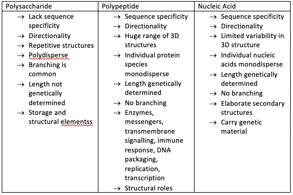
| Compare macromolecules | |
| Primary structures | Unique in each protein Peptide bonds Order of amino acids Determines the function and structure e.g. those binding to DNA contain many + change (lysine and argentine) |
| Sequencing | Sanger 1953 first sequenced PS of insulin EDMAN degredation used for 10 amino acids |
| Rotation Angles | Atoms in peptide bonds fixed in same plane Angle of rotation: N-C(alpha) = phi C(alpha)-C = psi Possible combination os these displaced as Ramchadram plot Can use to describe protein conformation |
| Ramchadran plot | Possible phi and psi combinations Most angles within permitted regions Proline restricted phi angle -60 to -77 dye to ring Glycine more combination as H so smaller Only for L amino acids |
| Criterial for stable secondary structure | Peptide bond planar with favourable bond length/ angle Every carbonyl O and amide N involved in H bonding H bonded in straight line Operation of translation and rotation always the same Side chains project out from structure to minimise steric effect |
| Alpha helix | CO bonded to NH 2 residues ahead. 3.6 amino acids per turn side chains outwards right handed helix amphipathic can show in helical wheel |
| Beta sheet | Strands - parallel or antiparallel Antiparallel more stable Side chains alternatively above and below Reverse B turn changes direction Minimal 4 residues |
| Tertiary structure | H bonds -> hold sheet/ helix Ionic interaction -> amino acids with charge at physiological pH. Varies with distance Hydrophobic interactions -> non-polar side chain associate with each other rather than water (entropy) Van der Waals -> electron distribution fluctuates around atom, inducing complementary fluctuations around atom. only if close. weak Interactions individually weal, but collectively strong Many only occur in water solvent Disulfide between cysteine |
| Supersecondary structure | B hairpin A hairpin 2 alpha helix bundle beta alpha beta motif Greek ket motif |
| Beta hairpin | antiparallel joined by B turn held by H bonds |
| Alpha hairpin | Amphipathic - held by Van Der Waals and hydrophobic interactions |
| 4 Alpha helix bundle | Found in proteins binding to haem |
| Beta alpha beta | In parallel, B sheets cannot be connected by b turn Arrange into Rossman fold e.g. lactate dehydrogenase |
| Greek key motif | Antiparallel Used by beta sandwich Hydrophobic side chain interactions e.g. immunoglobin fold |
| 3D structures | X-ray diffraction NMR spectroscopy Cryo-electron microscopy Atomic force microscopy |
| X-ray diffraction | Ordered structures X rays scattered by electrons and waves combine or cancel depending on phase diffraction pattern recorded and related to furrier form |
| NMR spectroscopy | Small protein in solution Irradiated at radio frequency in strong magnetic field 3d model created using spectra to determine inner proton distance |
| Cryo electron microscopy | Frozen rapidly - preserve and protect Computation reorientation creates 3d image from images at different angles |
| Atomic force microscopy | sharp tip scanned over surface with feedback mechanisms piezoelectric scanners maintain tip at constant force to obtain high information and topography of sample mapped |
| Quaternary structure | Multiple polypeptide chains bound together |
| DNA binding proteins | Dimers - bind to repeating DNA sequences e.g CRO from bacteriophage dimer with 2 alpha helices binding to adjacent major grooves |
| Haemoglobin | 4 myoglobin chains in tetramer. Precise molecular fir of chains held by same interactions as in tertiary structure |
| Sickle cell anaemia | Replace charged glycine with hydrophobic valine Stick together via hydrophobic interactions Polymerise in RBC - distort and rupture Less O2 and block capillaries |
| Protein folding | In vivo, chaperone proteins held fold correctly. Native form most thermodynamically stable Proteins fold in defined pathways |
| Ribonuclease Denaturing | Urea and B-mercaptoethanol denatures. When reducing agent and denaturant removed, ribonuclease spontaneously refolds to catalytic active site in certain conditions |
| Protein misfolding | Can cause disesase Alzheimers - amyloid fibrils form plaques of many amyloid proteins in repetitive beta sheets Prion proteins form BSE - protein is the infection material |
Want to create your own Flashcards for free with GoConqr? Learn more.