21013696
| Question | Answer |
| Cytoskeleton overview | Components: - Actin filaments aka microfilaments -Microtubules -Intermediate filaments Function: 'the dynamic muscles and bones of the cell': Gives cell structure, provides mechanical force for movement and changing shape, internal force for organelle movement, provides mechanical force for cell division |
| Actin filaments aka microfilaments | Structure: - 7nm in diameter - Twisted chains of identical globular actin -Strong yet flexible -Has structural polarity- a barbed end and a pointed end - Treadmilling- ATP binds faster to barbed end, leading to a net growth on barbed end and a net loss on pointed end. |
| Actin filaments aka microfilaments cont Nucleation of microfilaments | Nucleation: the process by which new actin filaments are formed. - 3 actin monomers must associate, which is difficult as the rate of disassociation is high, making it a genetically unfavourable process. However, once the trimers have formed, the rate of disassociation decreases, and the molecule becomes more stable. - ATP hydrolysis acts as a timer for actin filament assembly/disassembly- ATP bound to the filament is hydrolysed into ADP- decreasing stability |
| Different structures formed by actin filaments in cells | - Lamellipodia- branched actin forms the protrusion for cell migration - Filopodia- projections that protrude beyond the lamellipodia in parallel bundles for cell migration - Stress fibres- link with myosin II to form anti-parallel bundles that maintain cell structure - Skeletal muscle cells- actin and myosin filaments work together |
| Actin polymerisation- regulated by actin binding proteins | - Actin monomer binding proteins - Actin nucleators - Actin filament elongation factors - Actin filament capping proteins - Actin filament severing proteins - Actin filament cross-linking proteins |
| Cell migration | - Lamellipodium are positioned on the the surface of the cell membrane, with the barbed end facing the plasma membrane - The actin filaments of the lamellipoduim then extend at the barbed end, pushing cell forward, allowing it to migrate - The retraction of the cell at the rear is then mediated by myosin. |
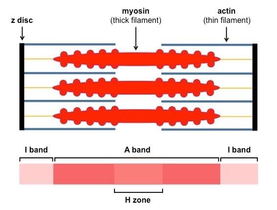
| Actin in human movement | - Myosin II is bound to actin in skeletal muscle cells -Myosin II contains 2 heavy and 2 light chains, filaments are wound together to form thick filaments with myosin heads - Sacrcomere consists of thick and thin filaments - During muscle contraction, myosin II walk towards disks, in direction of the barbed ends |
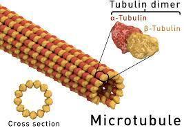
| Microtubules: Structure | - Composed of dimers of a- and b- tubulin, bound to GTP or GDP - Form hollow tubes composed of 13 parallel protofilaments - All tubulin dimers face in the same direction - - Largest 25mn in diameter |
| Microtubules: assembly | - Ennuculate from microtubule organising centres (MTOCS, which are made of y-tubulin rings) - Microtubules positive end grow towards the cell periphery from the centrosome |
| Microtubules: dynamic instability | - Microtubules polymerise and catastrophise quickly - This instability is driven by GTP hydrolysis- microtubules bound with GDP are unstable - "GTP cap" keeps microtubules stable - The function of dynamic instability is to allow microtubules to explore the whole of the cytoplasm |
| Microtubules: function | - Seperation of the chromosomes during mitosis - Form network in cytoplasm for the movement and functioning of organelles - Provide structure and motility to the cilla and flagella - Covers cytoplasm tracks for movement of organelles |
| Microtubules: regulation | - Microtubules are regulated by microtubule associated proteins, which bind to the side of microtubules. |
| Microtubule stabilisation: cancer drugs that act on microtubules | - Some cancer drugs stabilise microtubes, interfereing with dynamic instability EG: - Vinblaste: binds between tubulin dimers - Taxol: binds B tubulin - Colchine & nocodazole: bid to tubulin dimers, inhibiting assembly |
| Microtubules: microtubule motor proteins | Microtubule motor proteins move cargo around the cell- rely on ATP hydrolysis - Kinesins- move cargo to + end - Dynesins- move cargo to - end They are made of 2 globular heads and a tail. |
| Intermediate filaments: structure | - Rope-like/ flexible - Good tensile strength - 10nm diameter - Anchor to adjacent cells - Connected to the extracellular matrix by hemidesosomes - NOT polarised - Coiled from monomers into dimers, then into tetrameters. Then 8 tetrameters join together to form the rope-like structure of the intermediate filament |
| Intermediate filaments: function | - Strengthen cells against mechanical stress by... - Preventing excessive stretching of cells/ epithelial tissues - Reinforcing axons of nerve cells - forming mesh inside nuclear lamina in nucleus |
| Intermediate filaments: types | - Keratins in epithelia - Vimentin in connective tissue, muscle cells and glial cells - Neurofilaments in nerve cells - Nuclear lamins in all animal cells |
| Issues with intermediate filaments resulting in disease | - Keratins- blistering skin, brittle hair/nails - Neurofilaments- neuropathies - Nuclear lamins- cardiomyopathy - Nuscular dystrophy |
Want to create your own Flashcards for free with GoConqr? Learn more.