21115427
| Question | Answer |
| Biological membranes | - Purpose: compartmentalisation (allows chemical reactions to occur quicker, as provides ideal enviroment) and limiting the movement of molecules (selective permeability barriers). - Organelles surrounded by membranes, and the cell itself surrounded by one too. - Phospholipid bilayers- 'islands of proteins in a sea of lipids', with carbohydrates for signalling. Hydrophillic heads on the outside, hydrophobic tails inside. - small uncharged/hydrophobic molecules freely transverse bilayer whilecharged polar proteins require specialist transport proteins. |
| Rates of diffusion: different types of molecules | - Hydrophobic molecules EG O2, benzene, short chain fatty acids. Diffuse freely at a high rate through membrane. - Small uncharged polar molecules EG H2O, CO2, urea, glycerol. Diffuse through bilayer but at slower rate. - Large uncharged polar molecules EG glucose, sucrose; specialist proteins needed for their transport. - Ions EG H+ Na+; lipid bilayer impermeable to them - Charged polar molecules; amino acids, ATP; cannot pass through lipid bilayer To summarise: Permeability of lipid bilayer is higher for molecules that are uncharged, non-polar and small |
| Membrane ion concentrations at rest | Na+ higher conc outside so moves in K+ higher conc inside so moves out Cl- higher conc outside so moves in Ca2+ higher conc outside so moves in At rest, the inside of the cell is negatively charged compared to the outside |
| Transport mechanisms | Simple diffusion- diffusion down conc gradient Facilitated diffusion- diffusion down conc gradient using transmembrane proteins (no energy required) Primary active transport- Energy from ATP hydrolysis used to move substances against conc gradient through proteins Secondary active transport- Energy from electrochemical gradient used to transport substances through proteins against conc gradient |
| Examples of transport mechanisms | Simple diffusion- H2O Facilitated diffusion- Glucose by GLUT transporters Primary active transport- Na+/K+ by ATPase Secondary active transport- Na+/glucose transporter in the intestine |
| Simple diffusion | 'Solute moves from one side of the membrane to another along its conc gradient' until equilibrium is reached. - Depends on: concentration gradient and octanol/water partition coefficient (how well the molecule partitions between octanol (oil) and water), shown as Kow. - The higher the Kow value, the more lipid soluble it is, and thus the faster it can travel through the lipid bilayer. |
| Facilitated diffusion | 'movement of substances across a biological membrane through a concentration gradient by means of a carrier molecule.' - Mineral ions and hydrophilic molecules - Ion channels are usually gated: - voltage-gated -Ligand gated (extracellular ligand) - Ligand gated (intracellular ligand) - Mechanically gated Types of transport mechanisms: - Uniport- transports one solute in one direction - Symport- one protein that allows movement of 2 different types of molecule in the same direction - Antiport- one protein that allows movement of 2 different types of proteins in opposite directions |
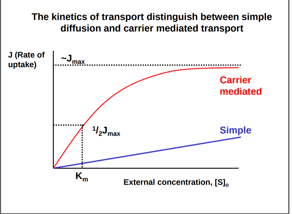
| Distinguishing between simple and carrier mediated transport Km= transporter affinity for substrate- lower the Km, the higher the affinity. | |
| Facilitative glucose transporters: The GLUT transporters | GLUT transporters: Facilitated glucose transport is mediated by a family of distinct transporters, which show sequence homology but tissue specific distribution. GLUT1: ubiquitous locations, abundant in erythrocytes. Mediates basal transport of glucose to a wide range of cells (Km 1.8mM). GLUT2: liver/pancreatic B-cells, low affinity for glucose compared to GLUT 1 (20mM) GLUT3: low Km, highest expression in neurones GLUT4: muscle/adipocytes, regulated by insulin (Km -5mM) GLUT 5: fructose transporter |
| Structure of the human glucose transporter GLUT 1 | |
| Regulation of GLUT4 by insulin | - Receptors for insulin on muscle cells - After a meal glucose travels in bloodstream, reaches receptors on muscle/fat cells, activating them, triggering an intracellular reaction: 1. Phosphorylation cascade, which signals an intracellular pool of GLUT 4 2. This signals GLUT4 transporters to translocate to the membrane of the cell 3. These transporters then take in glucose through the membrane as long as insulin is around 4. When insulin levels fall, GLUT4 is recycled to its pool, away from the cell surface membrane |
| Active transport | - Uses energy to transport solutes against concentration gradients - 2 types: 1. Primary active transport- energy harnessed through the hydrolysis of ATP (through Na+/K+ ATPase) 2. Secondary active transport- energy harnesses using an electrochemical gradient |
| Primary active transport: how energy is harnessed through Na+/K+ ATPase | - To begin with- Na+ levels are higher on the outside, K+ higher on the inside. 1. Na+ binds to intracellular binding site on ATPase in cell membrane 2. This triggers autophosphorylation of the ATP pump (it gains potassium from ATP, which becomes ADP). 3. The small amount of energy released with phosphorylation is enough to cause a conformational change to the ATPase, so it releases Na+ out of the cell, exposing a K+ binding site. 4. K+ can now bind to these sites, triggering dephosphorylation as they do so. 5. This dephosphorylation produces more energy, used to turn the pump back to its original conformation, so K+ is released into the cell - For every cycle, 3Na+ are pumped out and 2K+ are pumped in, helping maintain the negative charge of the inside of the cell. |
| Na+/K+ ATPase as a drug target in the treatment of congestive heart failiure | - In heart muscle, Na+/Ca+ antiporter (NCX) contributes to the removal of Ca2+ from the cytosol, allowing cardiac relaxation to occur. - The drug eg oubain, prevents K+ binding (antagonist), decreasing the rate at which Na+ is released from muscle cells. - This results in increased intracellular Na+, reducing activity of the NCX, thus slowing Ca2+efflux so high levels of Ca2+ are maintained, which maintains cardiac muscle contraction, slowing heart failure. |
| Secondary active transport | - Utilises the electrochemical gradient to produce energy for movement. - Important in: Intestinal epithelial cells for the absorption of dietary glucose and Epithelial cells in the proximal tubules of the kidney for reabsorption of glucose from the primary urine |
| When active transport is disrupted: how cholera toxin acts on lumen in the gut to cause huge electrolyte and fluid losses in the intestine | Mechanisms through which cholera toxin works: 1. Cholera toxin binds to GM1 gangloside receptor on the apical membrane of intestinal cells. 2. It is then internalised by endocytosis and transported through the golgi body to the ER 3. At the ER, subunits of the toxin are split, and the A1 subunit enters the cytosol 4. A1 subunit binds to and activates the GTPase, activating adenylyl cyclase; increasing AMP levels 5. Increased AMP levels activates the CFTR cl- channel, resulting in large cl- secretion. 6. Na+ follows electrochemical gradient 7. H2O follows osmotic gradient |
| Role of Na+/glucose cotransporter in electrolyte replacement therapy for cholera | Electrolyte replacement therapy includes a large conc of glucose, which drives Na+ (and consequently Cl- and H2O) back into the intestine through the Na+/glucose transporter (SGLUT) |
Want to create your own Flashcards for free with GoConqr? Learn more.