25820083
Description
Flashcards by Jordan Olexyn, updated more than 1 year ago
|
|
Created by Jordan Olexyn
over 3 years ago
|
|
| Question | Answer |
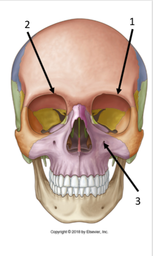
| Identify the structures | 1. Supraorbital margin 2. Supraorbital foramen 3. Infraorbital foramen |
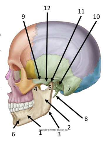
| Identify the structures 1-7 | 1. body of mandible 2. ramus of mandible 3. angle of mandible 4. coronoid process 5. condylar process 6. mental foramen 7. mastoid process |
| Identify the structures 8-12 | 8. styloid process 9. zygomatic process 10. external acoustic meatus 11. mandibular fossa 12. articular tubercle |
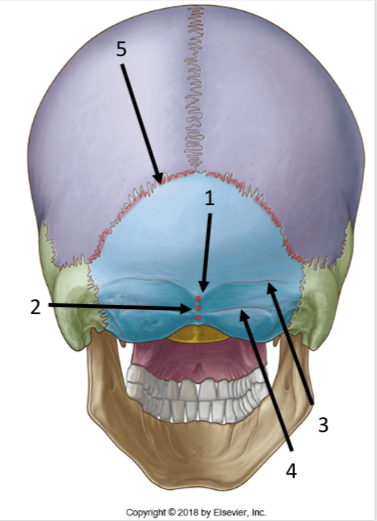
| Identify the structures | 1. External occipital protuberance 2. external occipital crest 3. superior nuchal line 4. inferior nuchal line 5. lambdoid suture |
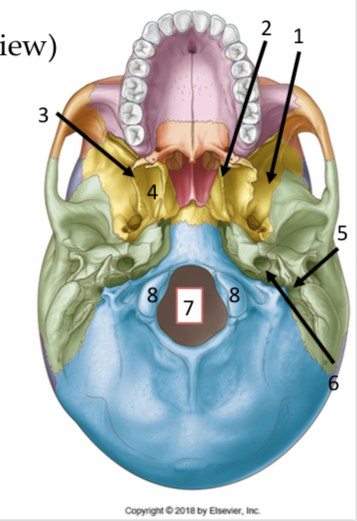
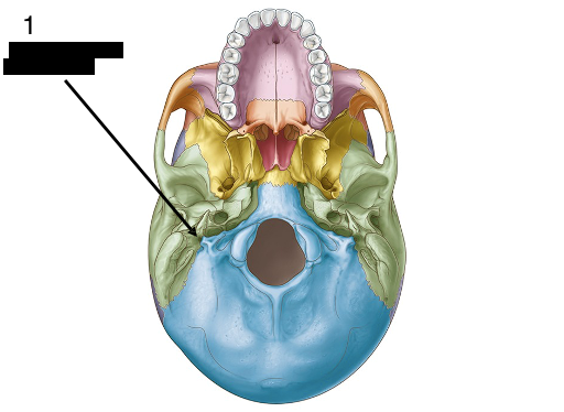
| Identify the structures | 1. Greater wing of the Sphenoid 2. medial plate of the pterygoid process 3. lateral plate of the pterygoid process 4. pterygoid fossa 5. Styloid process 6. carotid canal 7. foramen magnum 8. occipital condyles |
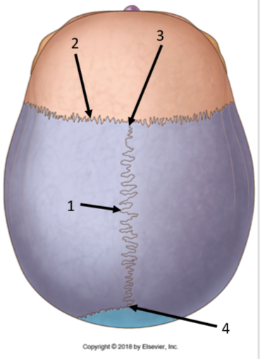
| Identify the structures | 1. Sagital Suture 2. Coronal Suture 3. anterior fontanel 4. posterior fontanel |
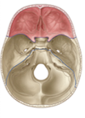
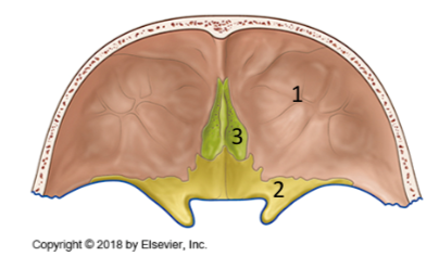
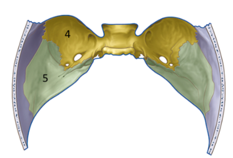
| What region is highlighted and what structures form it? | anterior cranial fossa. The frontal bones, Lesser wing of the sphenoid, and cribiform plate of the ethmoid |
| 1. Frontal bone 2. lesser wing of the sphenoid 3. Cribiform plate of the ethmoid | |
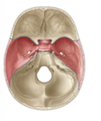
| What region is highlighted and what bones form it? | Middle Cranial fossa Greater wing of the sphenoid and temporal |
| 4. Greater wing of the Sphenoid 5. temporal bone | |
| Stylomastoid foramen | |
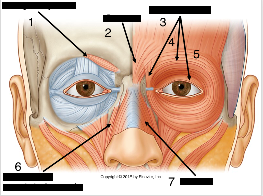
| 1. corrugator supercilii 2. Procerus 3. Lacrimal fibres of orbicularis occuli 4. Orbital fibres of orbicularis occuli 5. Palpebral fibres of orbicularis occuli | |
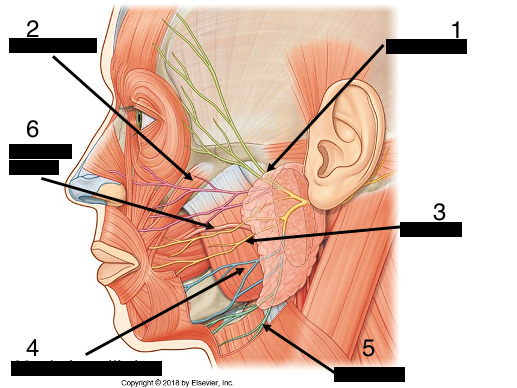
| 1. Temporal branch of the Facial N. 2. Zygomatic branch of the Facial N. 3. Buccal branch of the Facial N. 4. Marginal mandibular branch of Facial N. 5. Cervical branch of the Facial N. 6. Parotid duct | |
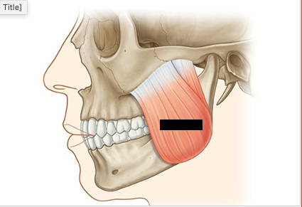
| OIIF & name | Masetter Origin: Zygomatic arch Insertion: Ramus of the Mandible, close to the body Innervation: Mandibular branch of the Trigeminal N. Function: Elevate the Jaw |
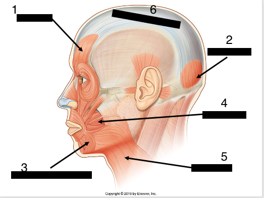
| 1. Frontalis 2. Occipitalis 3. depressor anguli oris 4. buccinator 5. Platysma 6. galea aponeurotica | |
| OIIF of 4 | Buccinator: O- Maxilla and Mandible I- Corner of mouth blending with orbicularis oris I- Facial N. F- push bolus away from cheeks and helps with whistling |
| OIIF of 3 | Depressor Anguli Oris: O- mandible I- corner of mouth I- Facial nerve F- lowers the angle of the mouth |
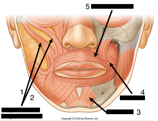
| 1. Zygomaticus Major 2. Zygomaticus Minor 3. Mentalis 4. Risorius 5. Orbicularis Oris | |
| OIIF for zygomaticus major and minor | O: zygomatic bone I: Upper lip and corner of the mouth I: Facial n. F: smile |
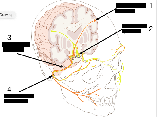
| 1.Upper motor neuron in motor cortex 2. Lower motor neuron in Brainstem 3. Internal Acoustic meatus 4. Stylomastoid foramen Significance is that if a stroke were to occur that damages the facial nerve, do to crossover and mixed control, only the contralateral lower section of the face will be without innervation. | |
| OIIF of Orbicularis Oris | O: maxilla I: skin of the lips, forming a circle around the mouth I: Facial nerve F: closes and protrudes lips |
| OIIF of Platysma | O: Fascia of the upper thoracic region I: fascia of the face and lower margin of the mandible I: Facial Nerve F: open the jaw and increase tension of the skin of the neck |
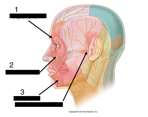
| 1. Supraorbital N. 2. Infraorbital N. 3. Mental N. 4. Auriculotemporal N. | |
| The Infraorbital N. is the terminal branch of the... | Maxillary division of the trigeminal |
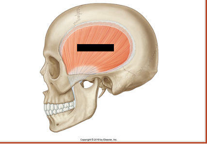
| Temporalis O: Temporal Fossa I: Coronoid process I: Mandibular branch of Trigeminal N. F: Elevate and retract the mandible | |
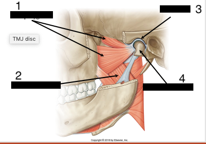
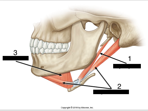
| 1. Lateral Pterygoid 2. Medial Pterygoid 3. TMJ disc 4. neck of the mandible | |
| OIIF for Lateral pterygoid | O: Greater wing of the sphenoid and some fibres on the pterygoid process (Superior head) Posterior surface of the maxilla with some fibres on the pterygoid process (inferior head) I: intra articular disc within the TMJ (superior head) neck of the mandible (inferior head) I: Mandibular branch of trigeminal F: Protusion of the mandible and intra-articular disc, as well as some lateral movement of the mandible |
| OIIF for Medial Pterygoid | O: Pterygoid process of the sphenoid bone (superior head) Posterior surface of the Maxilla (inferior head) I: medial surface of the ramus of the mandible, close to the angle I: Mandibular branch of trigeminal N. F: elevate and protrude the mandible. |
|
Image:
Tmj (binary/octet-stream)
|
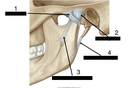
1. lateral temporomandibular ligament 2. joint capsule 3. Sphenomandibular ligament 4. stylomandibular ligament |
| What kind of a joint is the TMJ | a modified synovial-hinge joint |
| 1. stylo-hyoid 2. Digastric 3. Mylo-hyoid | |
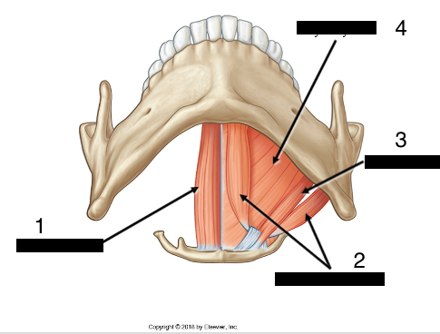
| OIIF for digastric | O and I: Posterior head is the medial surface of the mastoid process and the anterior head is the posterior surface of the body of the mandible I: Posterior head = Facial N. Anterior head = mandibular branch of Trigeminal N. F: elevate the hyoid and open the mouth |
| OIIF for mylohyoid | O: Mylo-hyoid on the inner surface of mandible I: body of the hyoid I: Mandibular branch of Trigeminal F: Elevate the hyoid or depress the mandible |
| OIIF for Stylo-hyoid | O: Styloid process I: Lesser horn of hyoid I: Facial N. F: Elevate the hyoid |
| OIIF for Genio-hyoid | O: 2 bony elevation on the inside surface of the mandible I: Body of the hyoid I: 1st cervical nerve F: Elevates the hyoid or depresses the mandible |
| 1. Genio-hyoid 2. Digastric 3. Stylo-hyoid 4. Mylo-hyoid | |
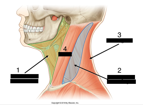
| 1. Anterior region (anterior triangle) 2. Lateral region (posterior triangle) 3. Posterior region 4. Sternocleidomastoid | |
| OIIF for SCM | O: Manubrium and medial 1/3rd of clavicle I: Mastoid process and lateral 1/3rd of superior nuchal line I: Spinal Accessory Nerve F: Unilateral contraction = ipsilateral side flexion of head and contralateral rotation. Bilateral contraction = Flexion of the head |
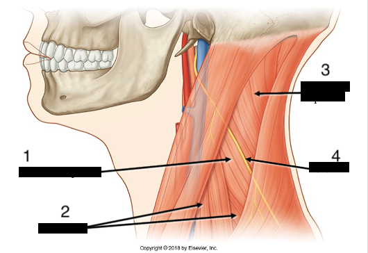
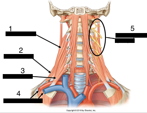
| 1. Levator Scapula 2. Anterior and Middle Scalenes 3. Splenius Capitis 4. Spinal accessory nerve | |
| OIIF for Levator Scapula | O: Transverse processes of C1-C4 I: Superior angle of the scapula and some of the medial border I: Direct branch from the brachial plexus known as the dorsal scapular nerve. F: Elevates the scapula |
| OIIF for anterior scalenes | O: Transverse processes of C3-C6 I: first rib I: Brachial plexus F: Lift the first rib and ipsilateral side bending and rotation of the neck |
| OIIF for middle scalenes | O: Transverse processes of C2-C7 I: first rib I: Brachial plexus F: Lift the first rib and ipsilateral side bending and rotation of the neck |
| OIIF for posterior scalenes | O: Transverse processes of C5-C7 I: 2nd rib I: brachial plexus F: Lift the second rib and ipsilateral side bending and rotation of the neck |
| What structures doe the scalenes act as a landmark for? | The roots of the cervical plexus and the subclavian artery and vein (which passes dorsal and ventral to the anterior scalene respectively) |
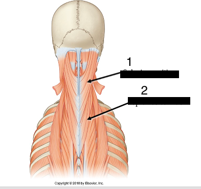
| 1. Splenius Capitis 2. Splenius Cervicis | |
| OIIF for Splenius Capitis | O: Lower part of the Nuchal ligament and spinous processes of the lower cervical and upper thoracic vertebrae I: lateral half of superior nuchal line and Mastoid process I: not mentioned in lecture F: Unilateral contraction = Ipsilateral side flexion and rotation of the head Bilateral = extension of head and neck |
| 1. Middle Scalene 2. Posterior scalene 3. Anterior scalene 4. Subclavian artery and vein 5. Cervical plexus | |
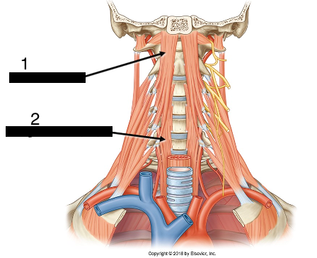
| 1. Longus Capitis 2. Longus Cervicus | |
| OIIF for Longus Capitis | O: transverse processes of C3-C6 I: inferior surface of basilar part of occipital I: Ventral rami of cervical spinal N. F: Flexion of the head |
| OIIF for Longus Cervicus | O: bodies of upper thoracic and lower cervical vertebrae I: Upper cervical vertebrae I: Branches from the ventral rami of cervical spinal nerves F: Flexion of the neck |
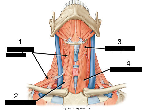
| 1. Omo-hyoid 2. Sterno-hyoid 3. Thyro-hyoid 4. Sterno-thyroid | |
| OIIF for Omo-hyoid | O and I: Body of the hyoid, intermediate tendon (which should be attached to the clavicle by a fibrous band), superior border of the scapula I: Ansa Cervicalis F: Depress the hyoid |
| OIIF for sterno-thyroid | O: Manubrium of the sternum I: Thyroid cartilage I: Ansa Cervicalis F: Depress the thyroid cartilage |
| OIIF of the sterno-hyoid | O: Manubrium of the sternum I: body of the hyoid I: ansa cervicalis F: depress the hyoid |
| OIIF for Thyro-hyoid | O: Thyroid Cartilage I: Greater horn of the hyoid I: direct branch from C1 F: Elevate the thyroid cartilage or depress the hyoid bone |
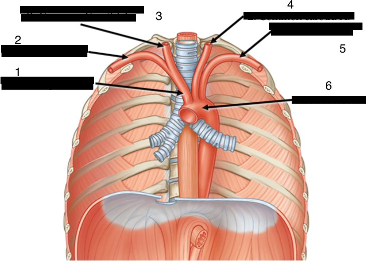
| 1. Brachiocephalic artery 2. Right Subclavian artery 3. Right Common Carotid 4. Left Common Carotid 5. Left Subclavian 6. arch of aorta | |
| 1. Subclavian artery (once it passes the first rib it becomes the axillary artery) 2. Suprascapular artery 3. Transverse cervical artery 4. Internal thoracic artery 5. thyrocervical artery/trunk 6. Costocervical artery 7. Vertebral artery | |
| What does the vertebral artery pass through? | the transverse formina of C1-C6, and then enter the cranium through the Foramen magnum |
| What are the branches of the thyrocervical trunk/artery and what do they supply? | Transverse cervical artery (which supplies the Trapezius) and the suprascapular artery (which supplies the infraspinatus and supraspinatus) |
| Provide the path of the internal thoracic artery. | 1. enters thorax 2. descends along either side of sternum 3. gives rise to anterior intercostal arteries 4. passes through diaphragm and becomes the superior epigastric artery |
| What does the costocervical artery supply blood to? | the first two posterior intercostal spaces and the deep muscles of the upper back (lev scap and splenius muscles) |
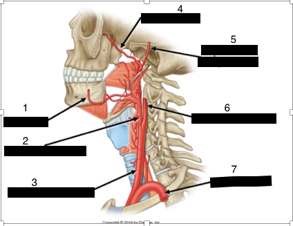
| 1. Facial A. 2. External Carotid A. 3. Common Carotid A. 4. Maxillary A. 5. Superficial Temporal A. 6. Internal Carotid A. 7. Subclavian A. | |
| At roughly what level does the common carotid branch into the internal and external? | C4 |
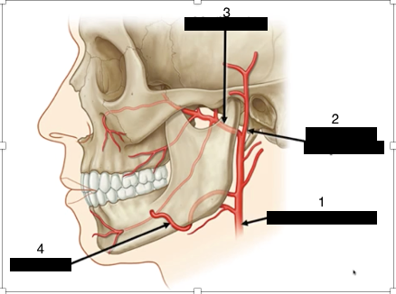
| 1. external carotid 2. Superficial temporal artery 3. Maxillary artery 4. Facial artery | |
| Where does the facial artery enter the face? | close to the anterior border of the insertion of the masetter muscle and the lower border of the mandible, it then travels upwards passing 1 inch away from the corner of the mouth, enters the groove between the nose and maxilla, then ends at the medial corner of the orbit. |
| What does the maxillary artery supply? | the cavities of the face (oral, orbital and nasal) |
| What is the largest branch of the external carotid A. | the maxillary artery |
| Describe the path of the superficial temporal artery | ascends directly in front of the external acoustic meatus and reaches to the lateral side of the scalp |
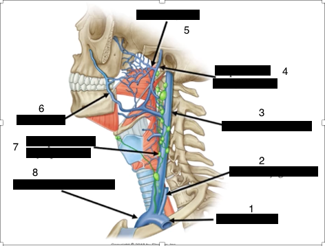
| 1. Subclavian V. 2. External Jugular V. 3. Internal Jugular V. 4. Superficial Temporal V. 5. maxillary v. 6. Facial v. 7. Lymph node 8. Brachiocephalic v. | |
| Where does the anterior jugular vein drain into? | either the external jugular or the subclavian |
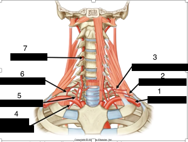
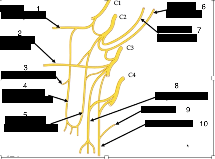
| 1. Branch to Genio-hyoid 2. Transverse cervical 3. Branch to Thyro-hyoid 4. Superior arch of Ansa Cervicalis 5. Inferior arch of Ansa Cervicalis 6. Lesser Occipital 7. Greater Auricular 8. supraclavicular 9. Contribution from C5 10. Phrenic nerve | |
| Which nerve branches of the cervical plexus are superficial? Deep? | superficial: Lesser Occipital, Greater Auricular, Transverse Cervical, Supraclavicular Deep: Branch to Geniohyoid, Branch to thryo-hyoid, phrenic, ansa cervicalis |
| The ansa cervicalis arises from which spinal nerves? | Superior root: C1 Inferior root: C2 and C3 |
| Where does the phrenic nerve run in relation to the scalenes | anterior to the anterior scalene muscle |
| What structure does the loop of ansa cervicalis rest on? | the internal jugular vein |
| What is the punctom nervosum? | the exit point of the superficial/sensory branches of the cervical plexus |
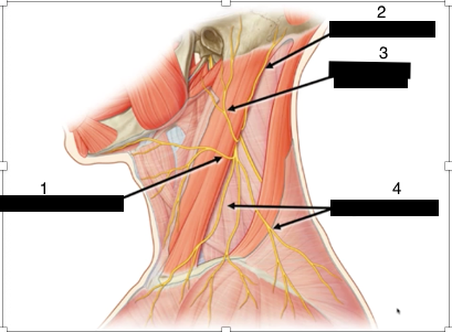
| 1. transverse cervical 2. lesser occipital 3. greater auricular 4. supraclavicular | |
| how would you landmark the spinal accessory nerve? | The spinal accessory nerve runs deep to the SCM and trapezius muscles (supplying these), and passes over the levator scapula. it can be landmarked as passing from the proximal 1/3rd of the posterior border of the SCM to the distal 1/3rd of the anterior border of the trapezius. |
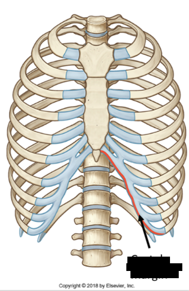
| Costal margin | |
| How many pairs of ribs are considered to be true ribs | 7 attach directly to the sternum, 3 pairs attach via the 7th rib cartilage, and 2 have no anterior attachments (floating) |
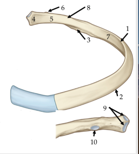
| 1. superior border 2. inferior border 3. costal groove 4. head 5. neck 6. tubercle 7. shaft 8. angle 9. demi facets 10. facet | |
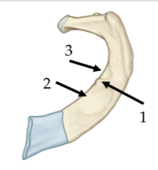
| 1. scalene tubercle 2. groove for the subclavian vein 3. groove for the subclavian artery | |
| What is atypical about the first rib? | this rib is flattened from above downward and neither has an angle nor a costal groove, with it's head only bearing an articular surface for the body of T1 |
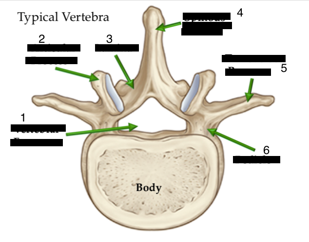
| 1. Vertebral canal 2. Articular process 3. lamina 4. Spinous process 5. Transverse process 6. Pedicle | |
| What is distinct about cervical vertebrae? | 1. They have a small, square shaped body 2. Triangular vertebral foramen 3. Transverse foramen 4. Bifid process 5. Lateral and anterior lips (uncinate processes) |
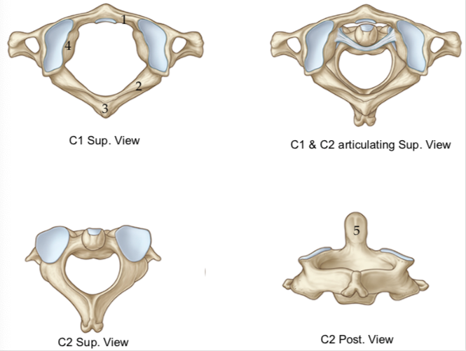
| 1. Anterior arch of the Atlas 2. Posterior arch of the Atlas 3. Tubercle of the Atlas 4. Lateral Masses of Atlas 5. Odontoid process | |
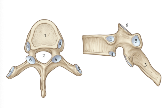
| 1. Body 2. Vertebral Foramen 3. Spinous process 4. Demi-facets (costal facets) 5. Full Facets (articulates with tubercle of ribs) 6. Superior articular processes 7. Inferior articular processes | |
| What is unique about T11 and T12? | They have no facets on their transverse processes |
| Which direction do the inferior articular processes of the twelfth thoracic vertebrae face? what is this akin to? | They face more laterally just like the inferior articular processes of the lumbar vertebrae. |
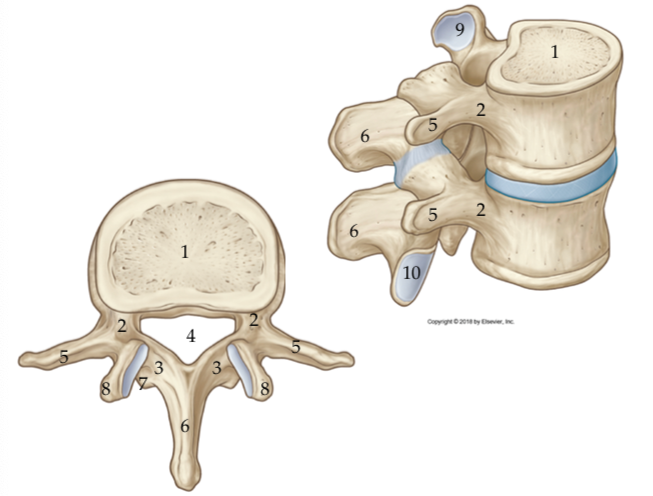
| 1. Body 2. Pedicles 3. Laminae 4. Vertebral canal 5. Transverse processes 6. spinous processes 7. Accessory process 8. Mammillary process 9 and 10. Articular surfaces | |
|
Image:
Sacrum (binary/octet-stream)
|
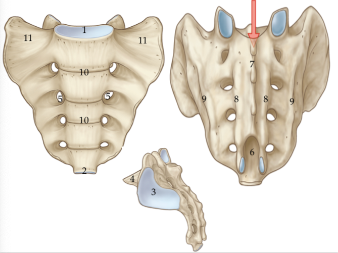
1. Base 2. Apex 3. Auricular surfaces 4. Sacral promontory 5. sacral foramina 6. sacral hiatus 7. Median sacral crest 8. Intermediate/medial sacral crest 9. Lateral sacral crests 10. transverse ridges 11. Sacral ala |
Want to create your own Flashcards for free with GoConqr? Learn more.