26147622
Description
Flashcards by Diamond Sapphire, updated more than 1 year ago
|
|
Created by Diamond Sapphire
over 3 years ago
|
|
| Question | Answer |
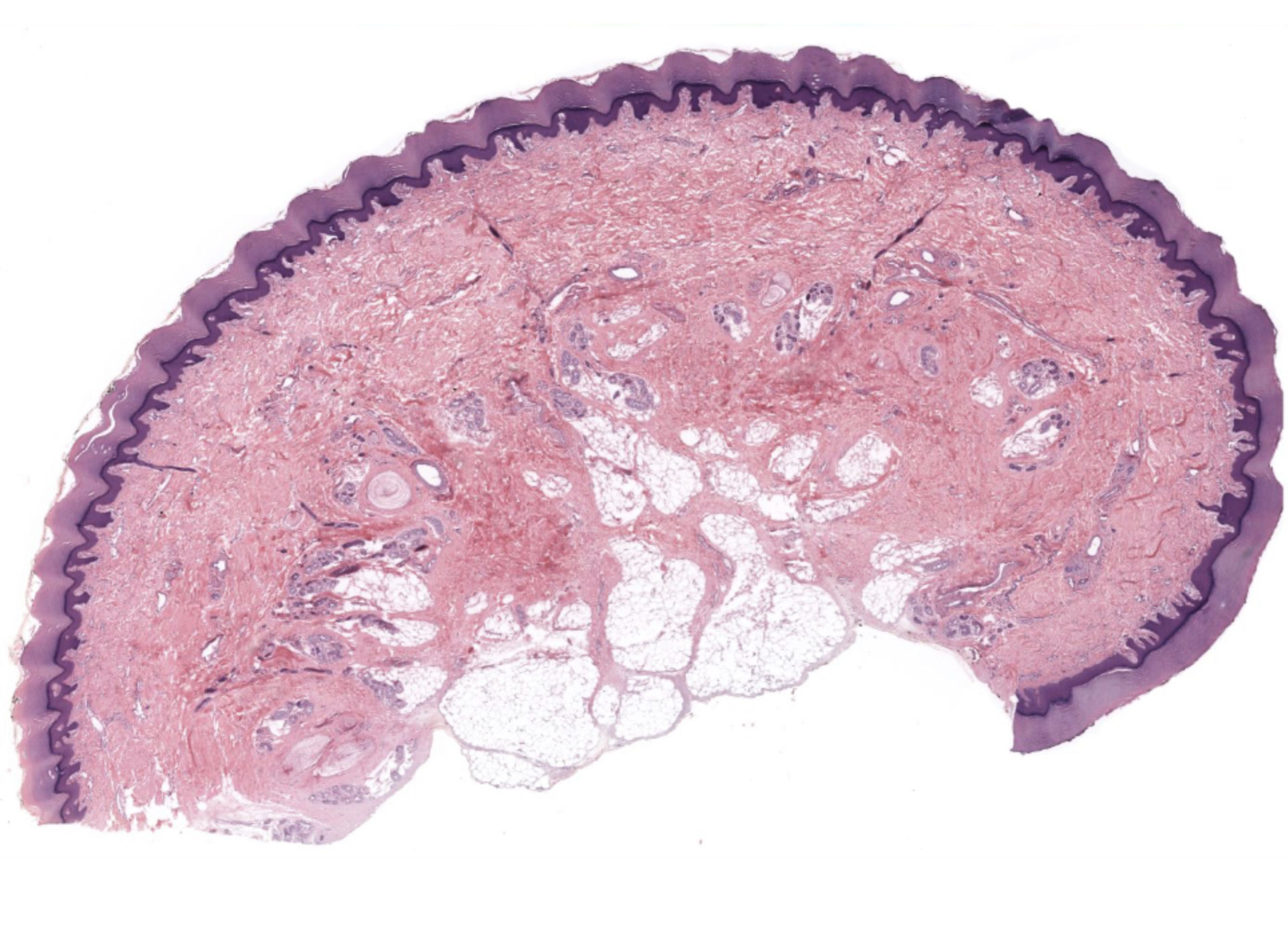
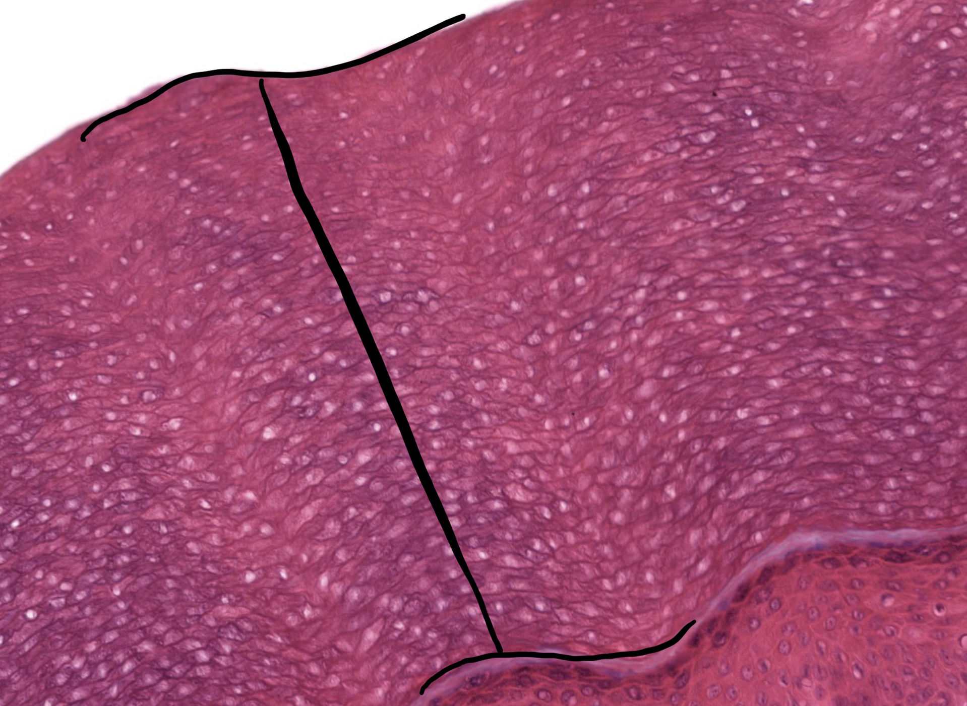
| what type of skin is represented by this slide | Thick skin |
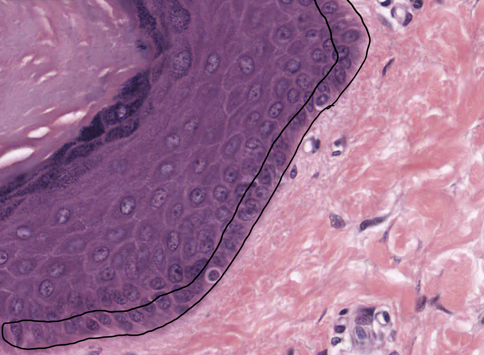
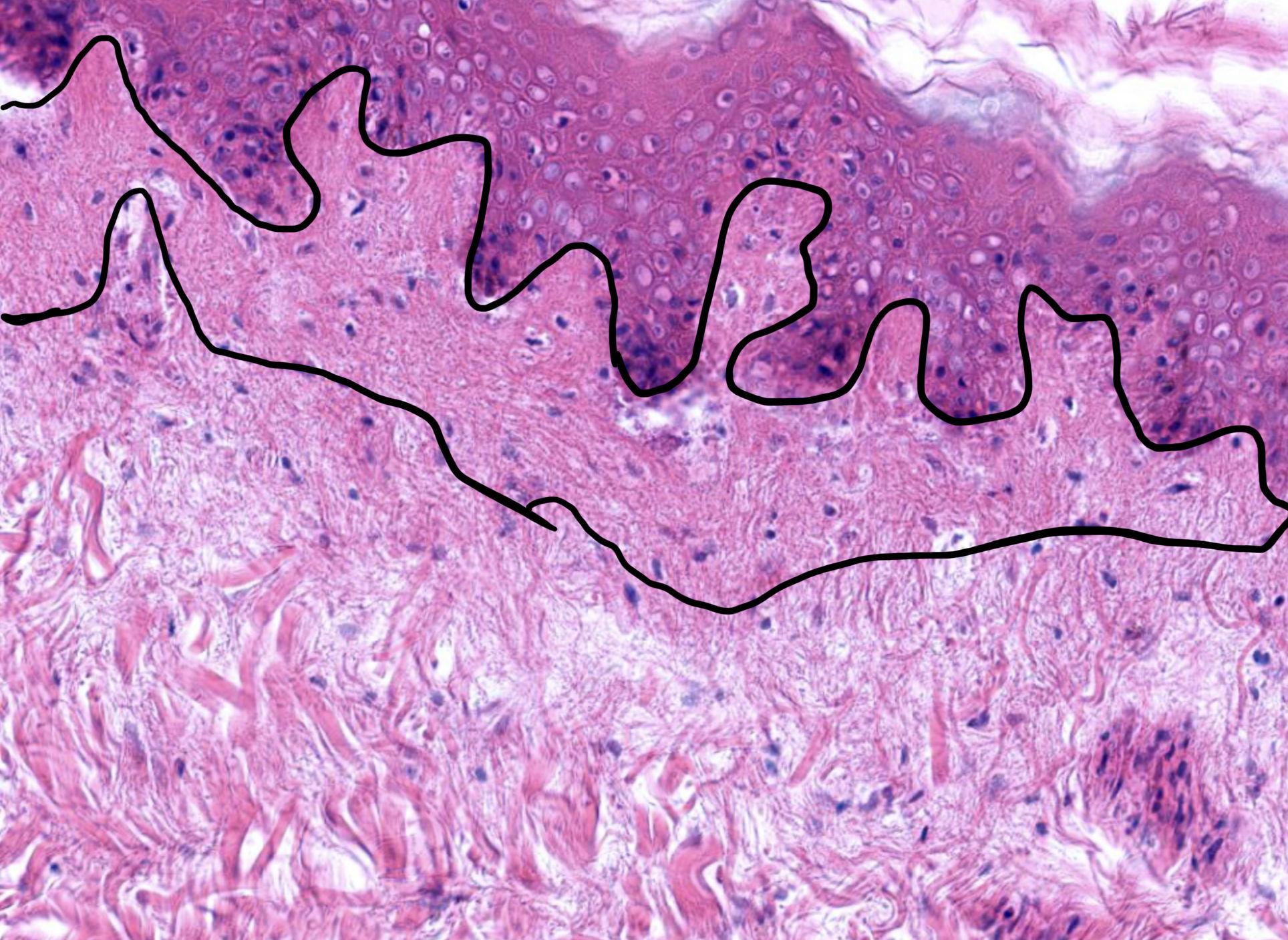
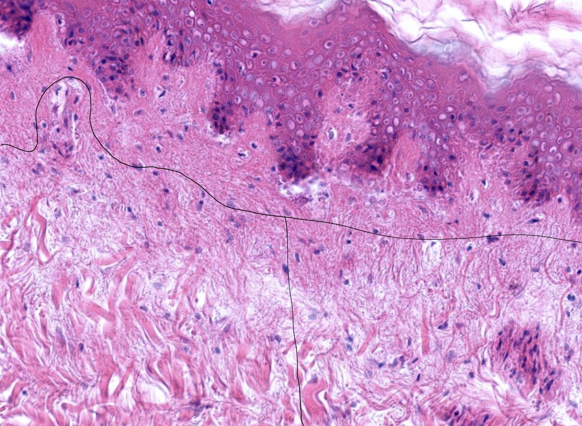
| single layer of germinal cells resting on the basement membrane which is attached to the dermis | Stratum basale |
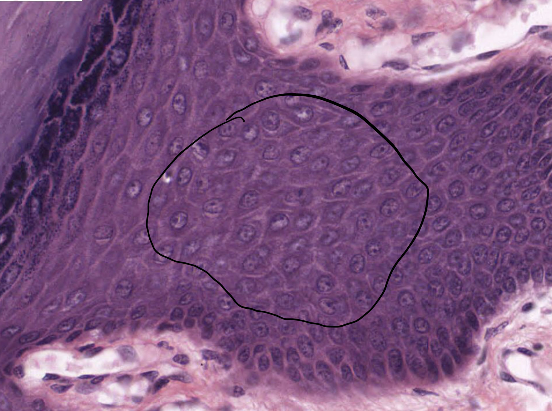
| keratinocytes attached to each other by desmosomes on spiny processes | Stratum spinosum |
| highly refractive zone only seen in very thick skin. | Stratum lucidum |
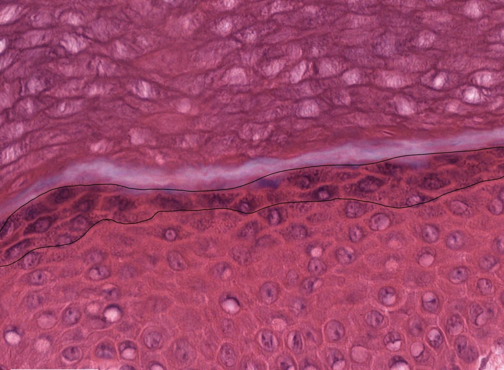
| keratinocytes with numerous basophilic granules in their cytoplasm | Stratum granulosum |
| thick layer of dead cells devoid of nuclei and organelle | Stratum corneum |
| What are the 3 types of nonkeratinocytes found in the epidermis? | Melanocytes, Merkel cells, and Langerhans cells |
| Melanocytes | In the basal layer of the epidermis; make melanin |
| Merkel cells | In the basal layer of the epidermis; sensory cells |
| These cells are located in the spinous layer of the epidermis and are known as antigen presenting cells | Langerhans cells |
| Melanocytes produce melanins which are packed into ___ and transported to the tips of dendritic processes | Melanosomes |
| Keratinocytes in the spinous layer are interconnected by extensive ___ | Desmosomes |
| Which layer of the epidermis is absent in the mucous membrane of the mouth? | Granular layer |
| connective tissue firmly attached to the epidermis by the basement membrane | Papillary layer of the dermis |
| less organized connective tissue that supports the papillary and the epidermis | Reticular layer of the dermis |
| This isn’t technically a layer of the skin, but helps anchor the dermis to muscle or bone; loose CT that contains small to large adipose depots called Panniculus adiposus | Hypodermis |
| Regarding hair, this part is the part above the skin surface | The hair shaft |
| Regarding hair, this part is the part below the skin surface | Hair root |
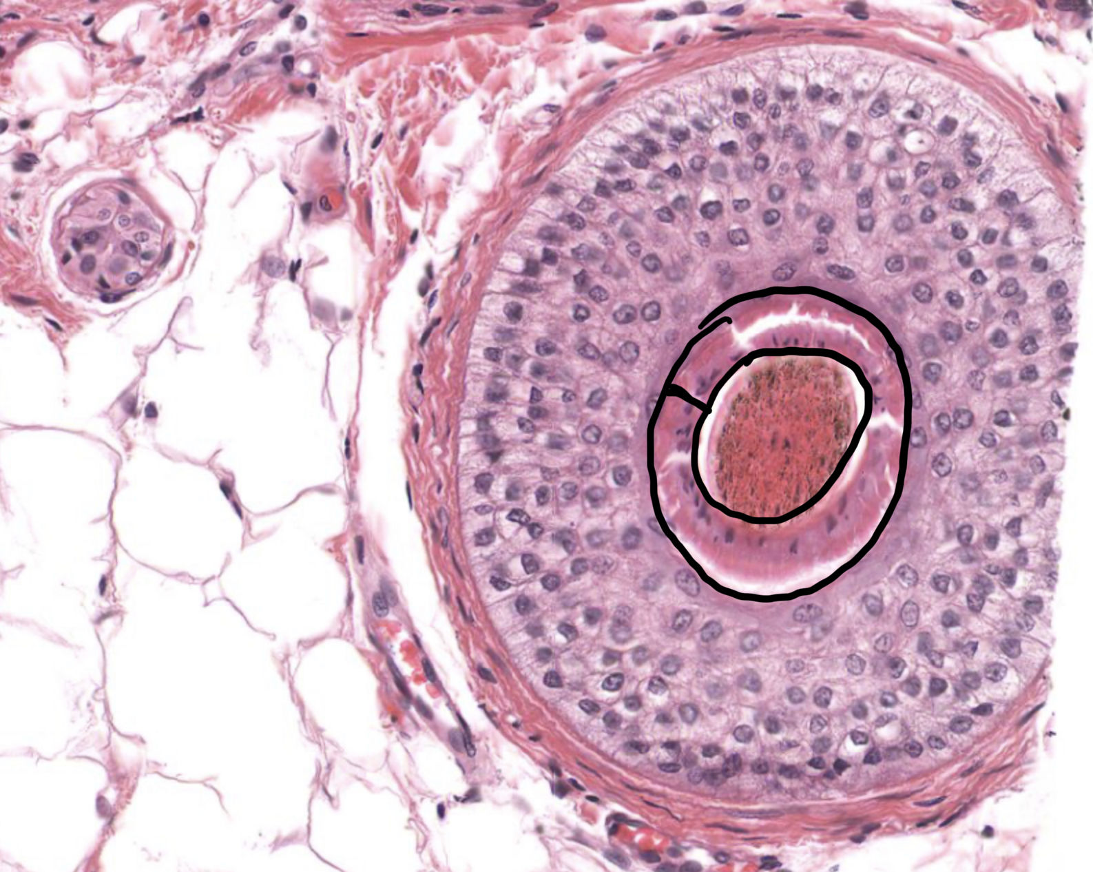
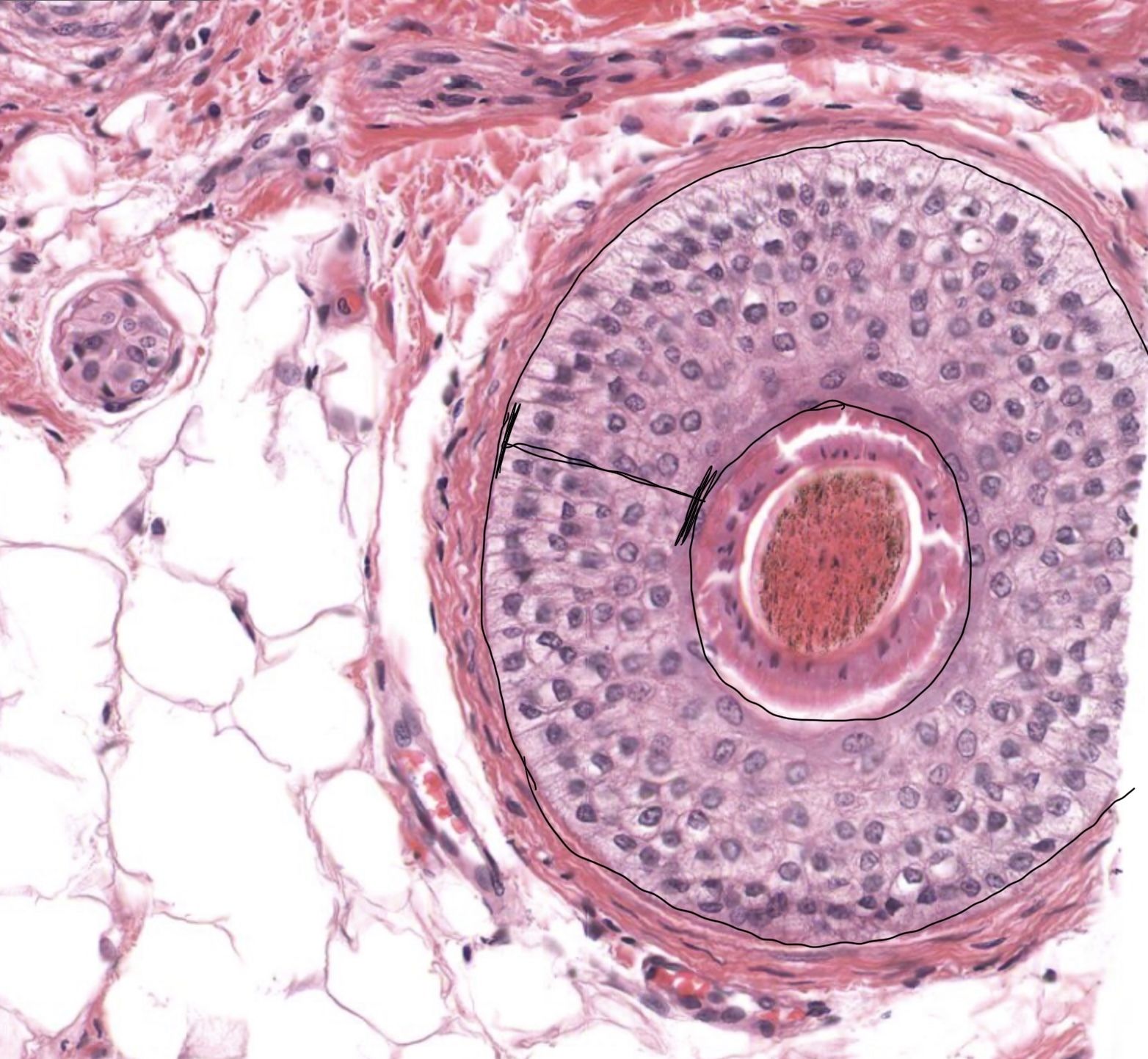
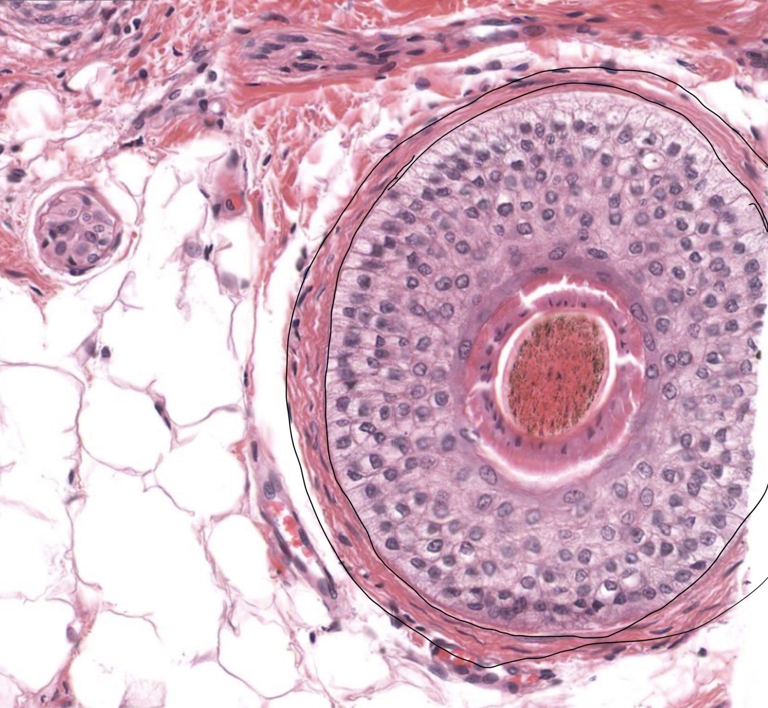
| The hair root is ensheathed in the ___ | Hair follicle |
| The hair follicle is the site of __ and is located in the __ | Hair growth; dermis (sometime extends into the hypodermis) |
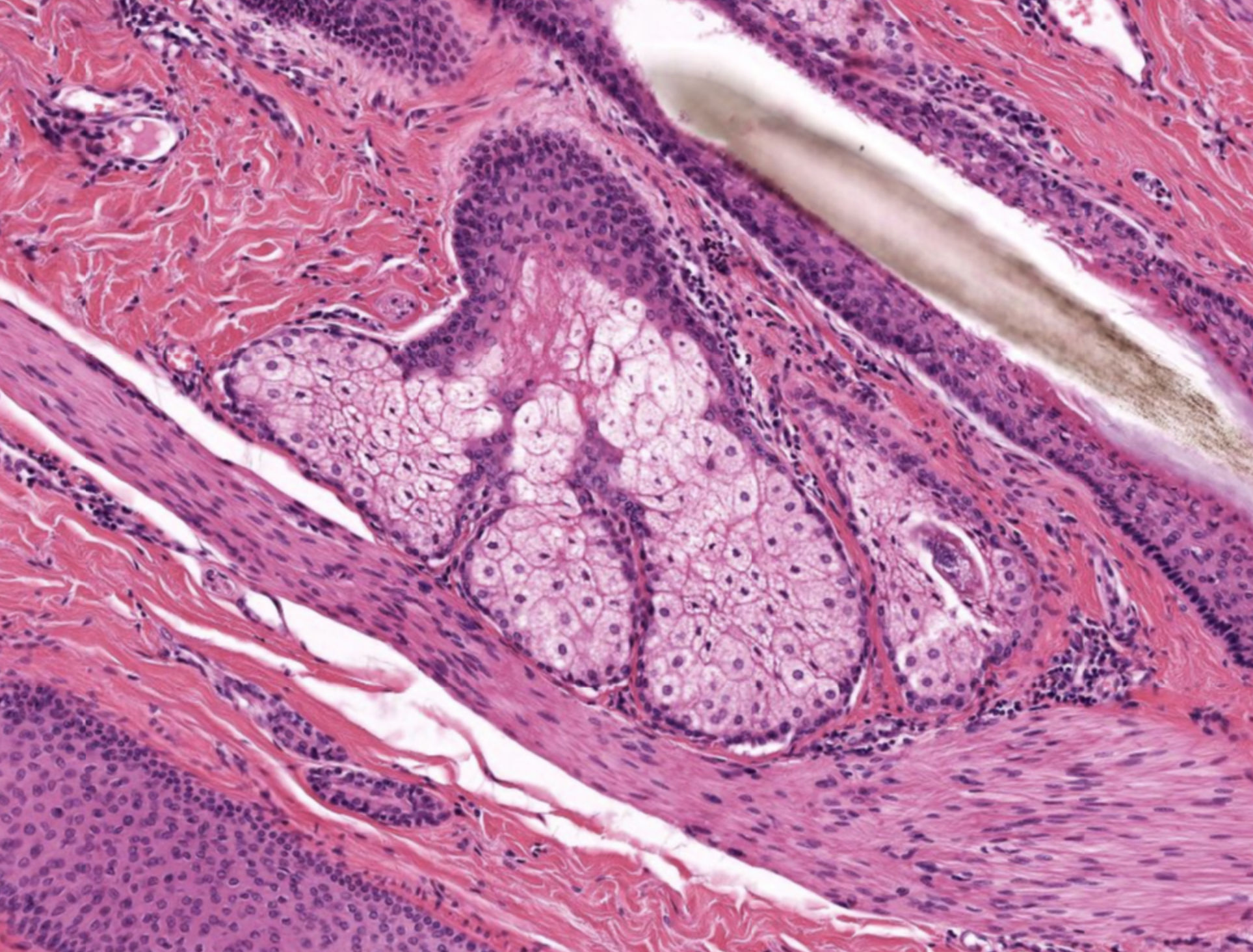
| produce a lipid rich secretion called sebum that is released into the hair follicle. Large cells with a central nucleus and a foamy appearing cytoplasm | Sebaceous gland |
| coiled, tubular sweat-producing glands and ducts with simple or stratified cuboidal epithelium. | Eccrine sweat gland |
| small muscles attached to hair follicles that cause hair to stand on end. | Arrector pili muscle. |
| extends from the hair bulb to the level of sebaceous glands. It is divided into Huxley's layer (inner layer of flattened cells) and Henle's layer (outer single layer of cuboidal cells). | Inner root sheath |
| layers of cells continuous with the epidermis In a hair follicle | Outer root sheath |
| thick basement membrane that separates the hair follicle from the dermis | glassy membrane |
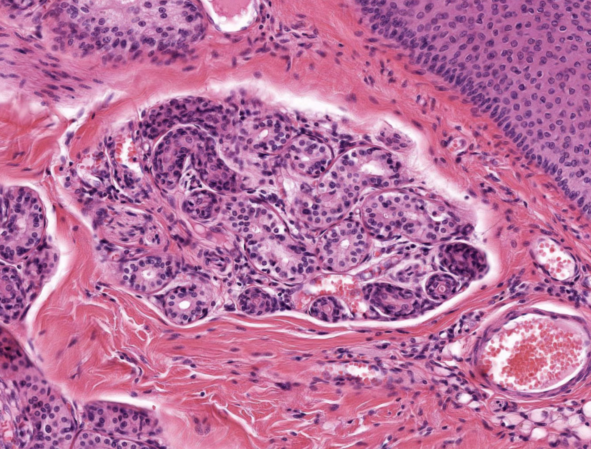
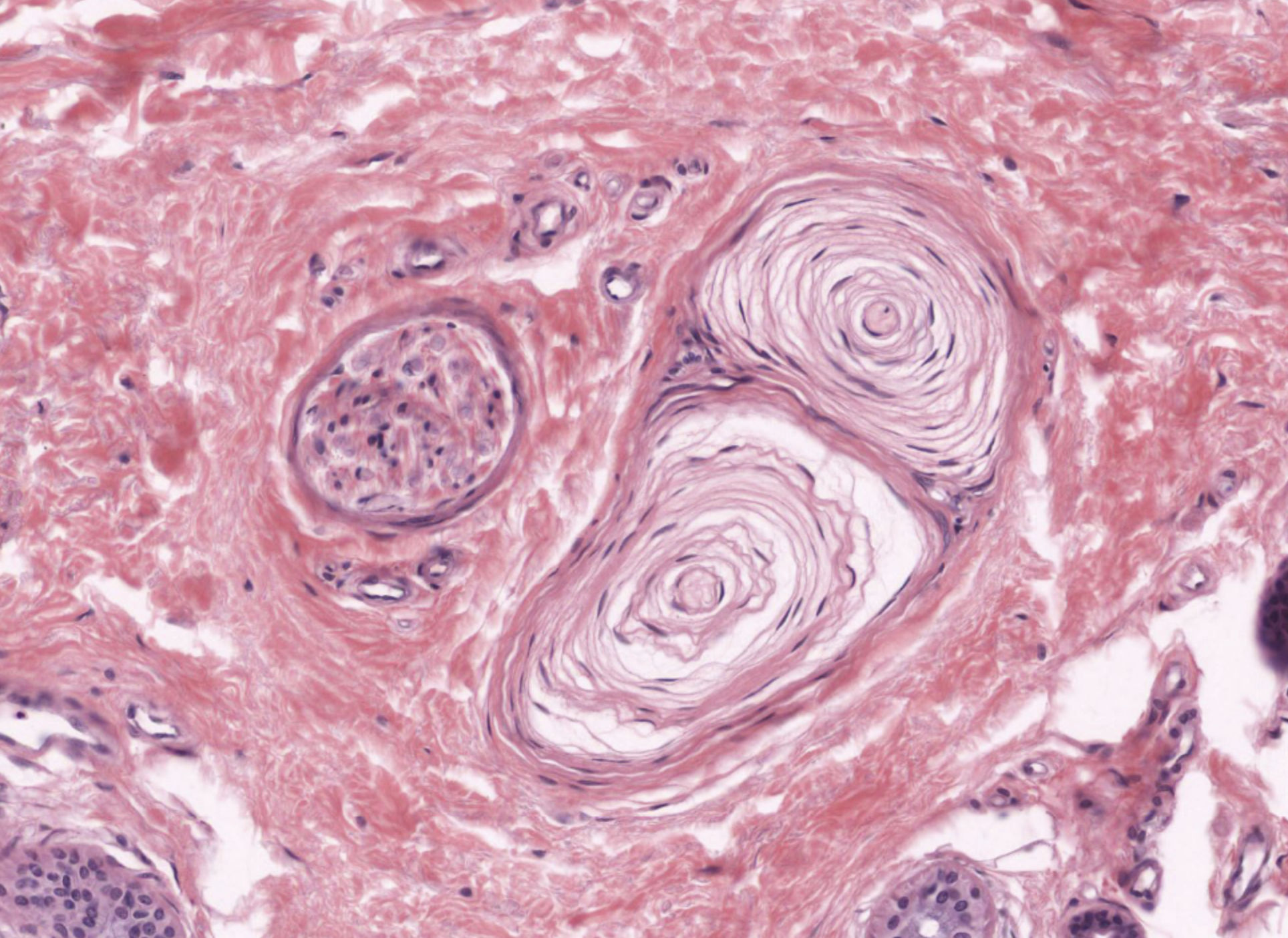
| nerve endings in skin responsible for sensitivity to vibration and pressure. | Pacinian Corpuscles |
| nerve endings in skin responsible for sensitivity to light touch. Located in dermal papillae. | Meissner's Corpuscles |
| Continuation of lactiferous sinus; lining epithelium is bi-layer stratified cuboidal to columnar | Teat sinus (teat cistern, cavity of teat) |
| Teat sinus empties into here; opens to teat surface; lining epithelium is stratified squamous | Teat canal (streak canal or papillary ducts) |
| in mammary glands, ___ are clustered to form lobules | Secretory alveoli |
| ___ cells surround alveolar epithelial cells contract for discharge of milk | Myoepithelial |
| Milk proteins released by ___ (think modes of secretion) | Merocrine/eccrine |
| Milk lipids released by ___ (think modes of secretion) | Apocrine |
| 3 layers of hoof wall | Stratum externum, stratum medium, stratum internum |
| Stratum externum | Perioplic epidermis; thin layer |
| Stratum medium | Coronary epidermis; tubular and intertubular horn; thick layer |
| Stratum internum | Lamellatum; primary and secondary epidermal laminae; interdigitates with laminar corium |
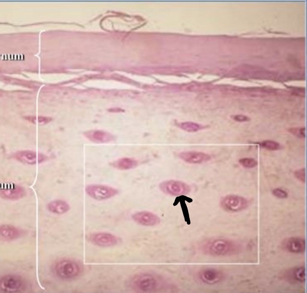
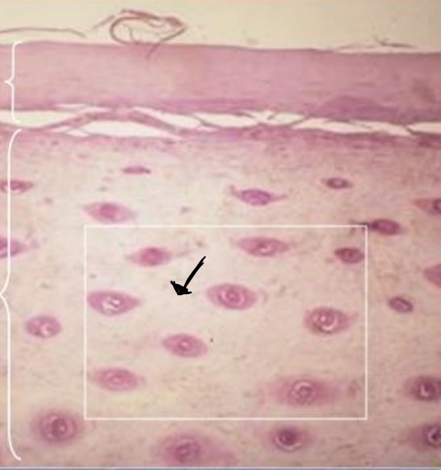
| What structure within the hoof wall is the black arrow pointing at? | Tubular horn |
| What structure within the hoof wall is the black arrow pointing at? | Intertubular horn |
| A crippling disease caused by inflammation in stratum internum | Laminitis |
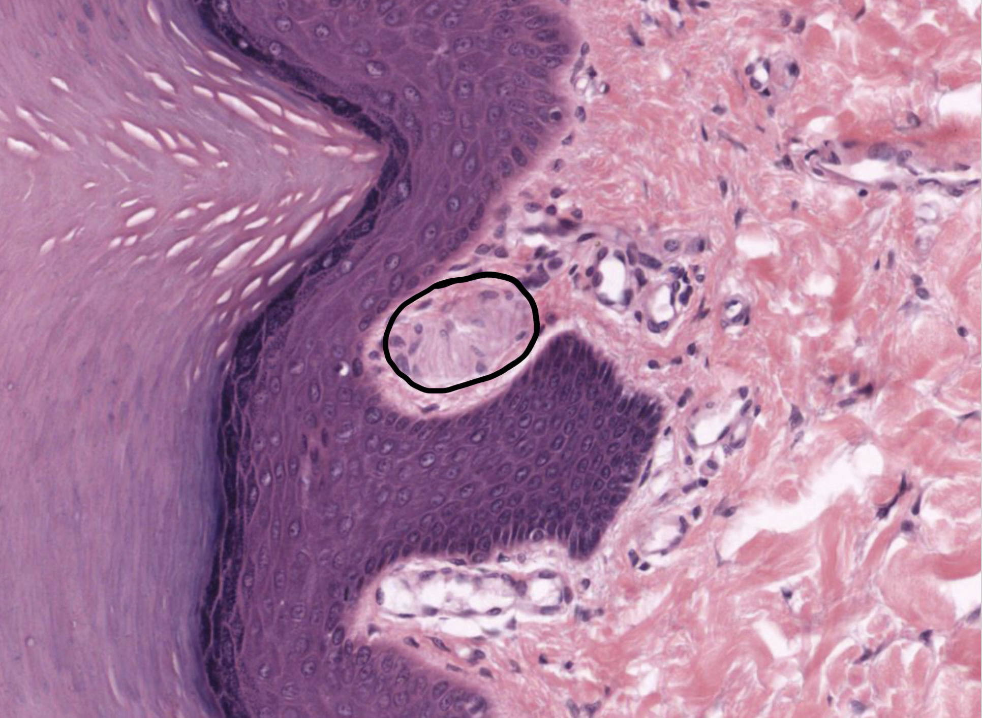
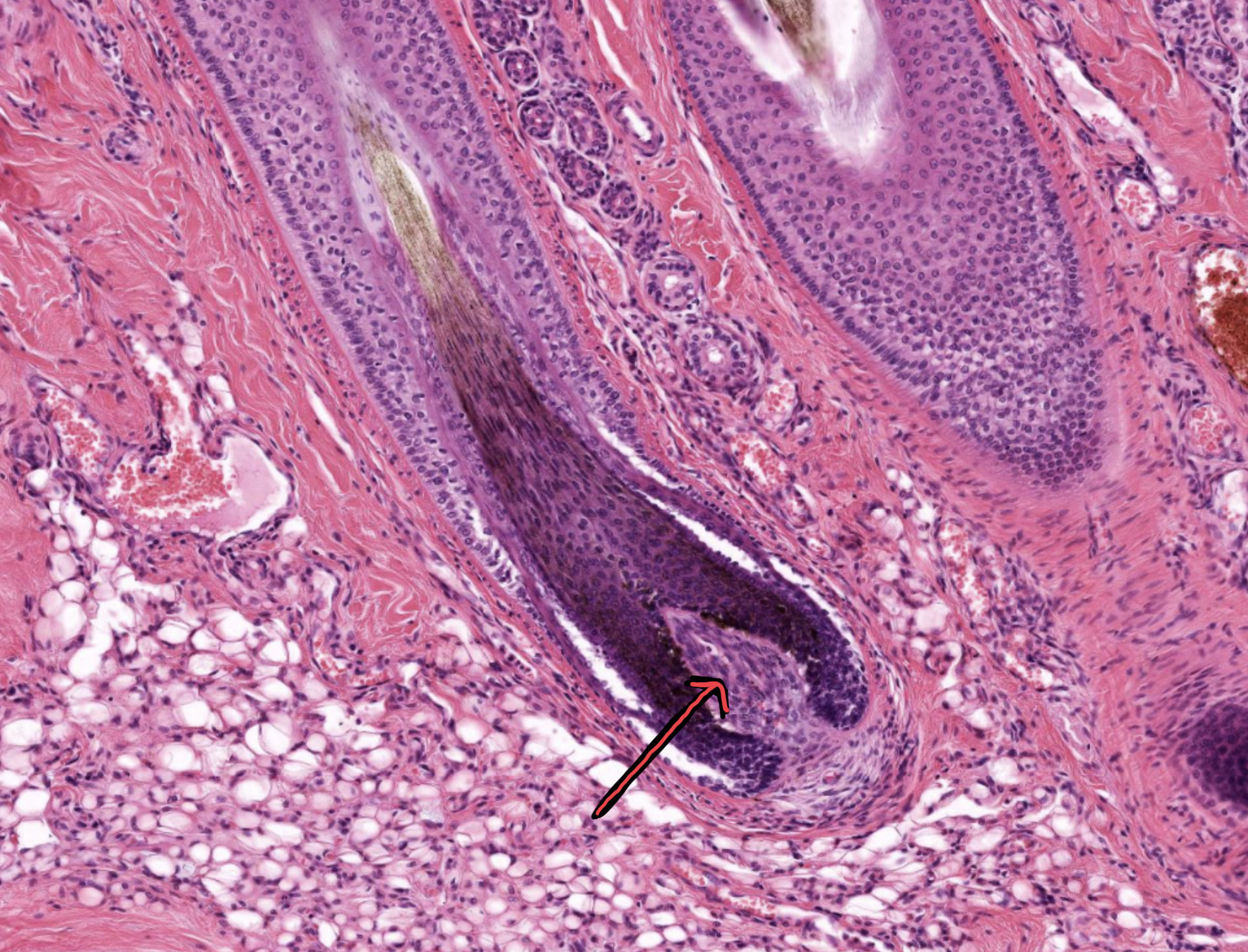
| What structure is the black/red arrow pointing at? | Dermal papilla |
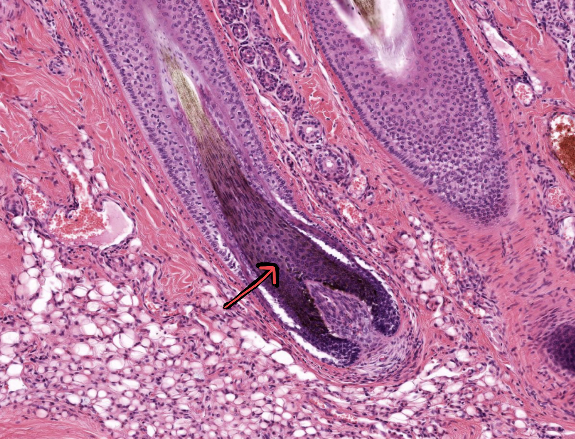
| What structure is the black/red arrow pointing at? | Hair matrix cells |
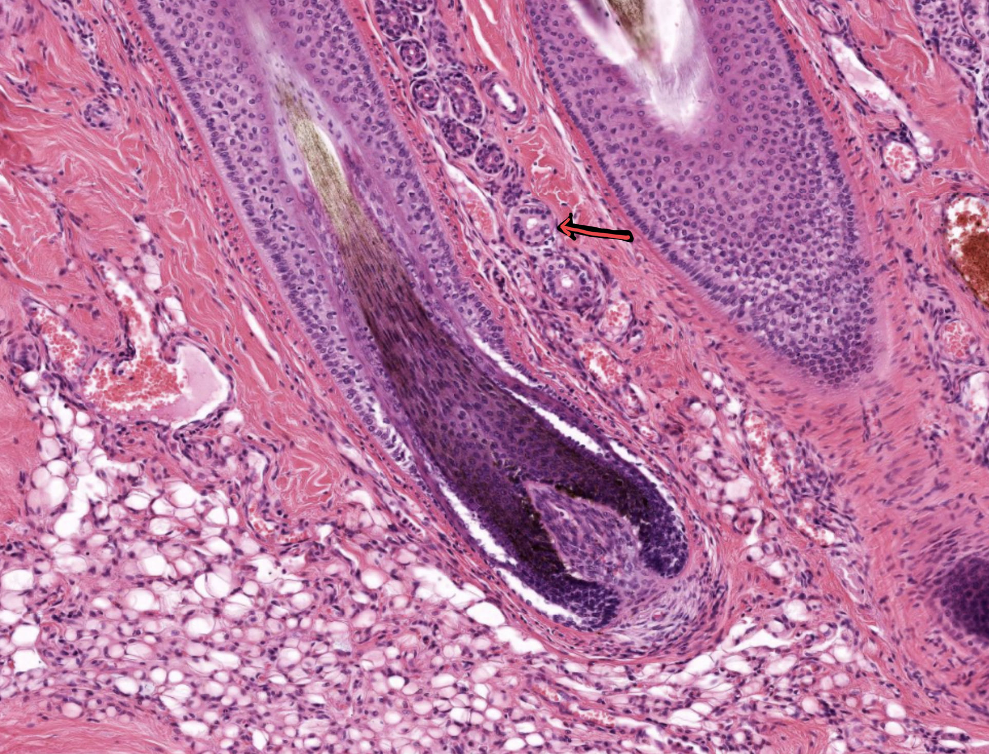
| What structure is the black/red arrow pointing at? | Sweat gland |
| Anagen | Growing stage |
| Catagen | Transition stage |
| Telogen | Resting or quiescence stage |
Want to create your own Flashcards for free with GoConqr? Learn more.