27141566
Description
Flashcards by luke werren, updated more than 1 year ago
|
|
Created by luke werren
over 5 years ago
|
|
| Question | Answer |
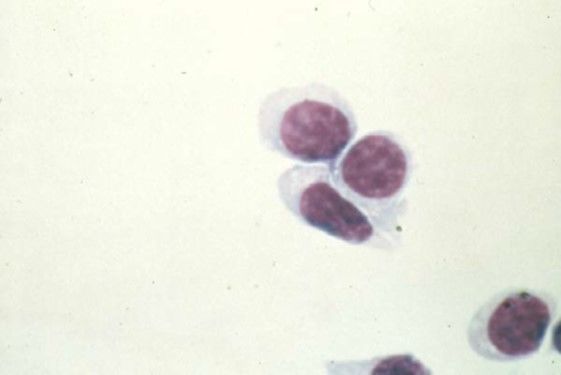
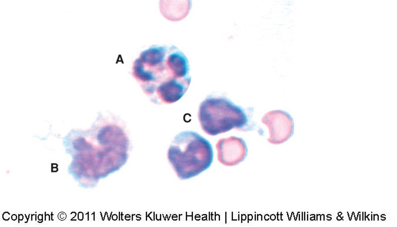
| Normal CSF | |
| A ==> Neutrophil B ==> Monocytes C ==> Lymphocyte | |
| Traumatic Tap OR Subarachnoid Bleed (all types of cells seen in peripheral will be seen with traumatic/bleeds) | |
| Erythrophagocytosis blue-black = hemosiderin granules yellow = hematoiden crystals evidence of a bleed | |
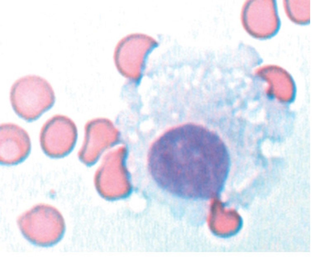
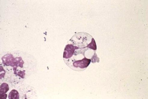
| Macrophage | |
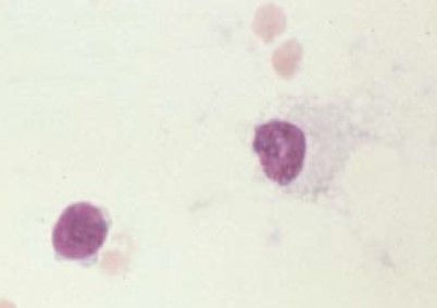
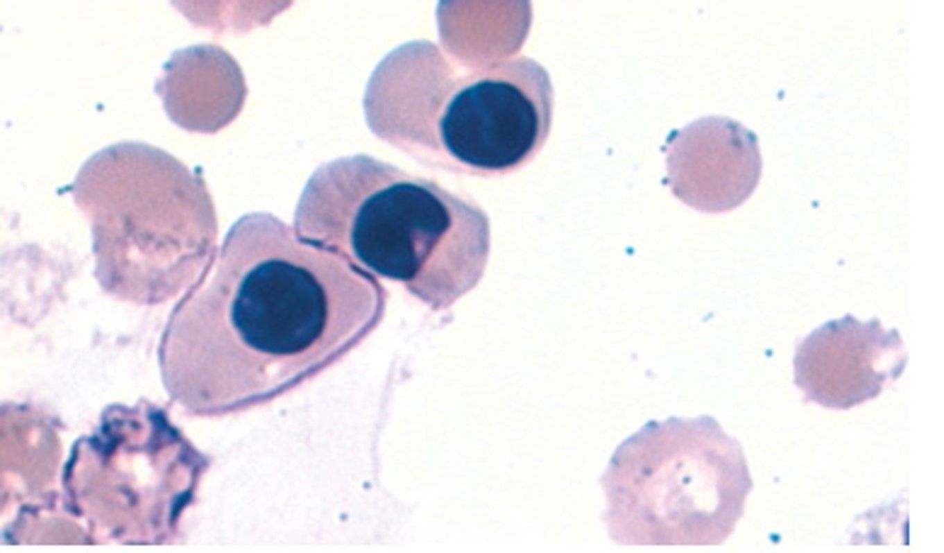
| Ventricular cells | |
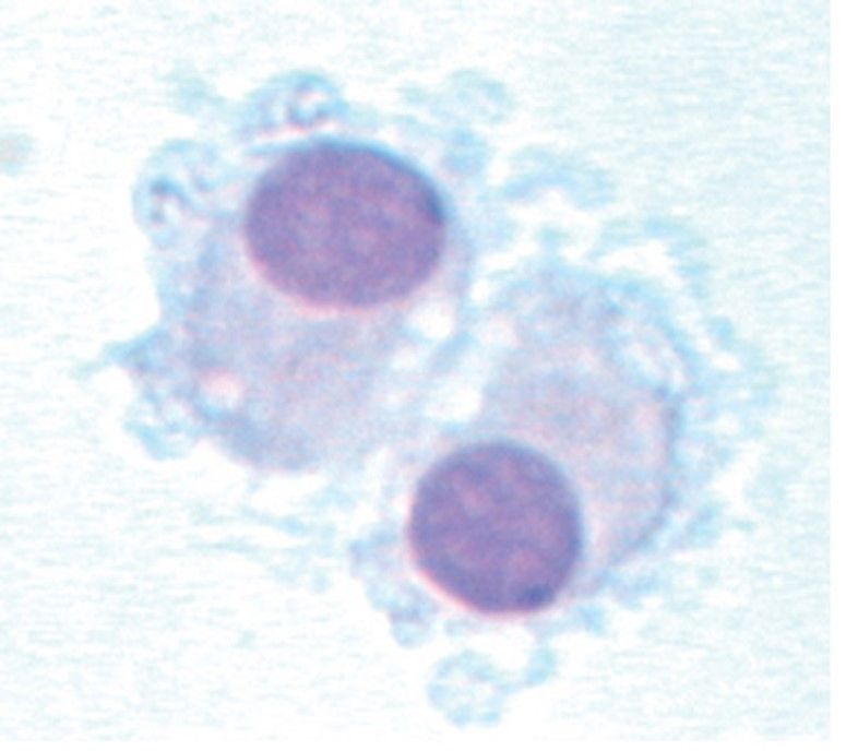
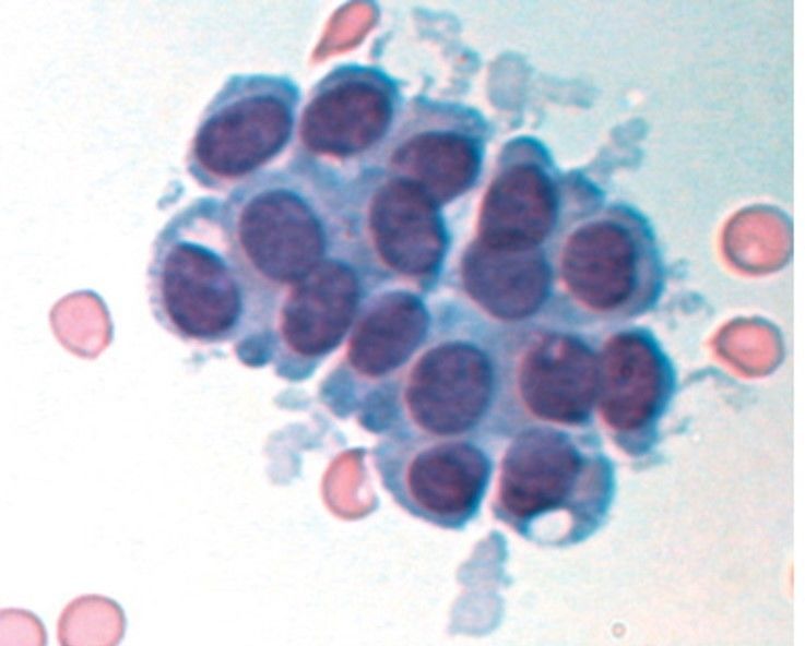
| Ventricular cell (may be mistaken for malignant cells due to their tendency to appear in clusters) | |
| Ventricular cell | |
| Ventricular cell (may be mistaken for malignant cells due to their tendency to appear in clusters) | |
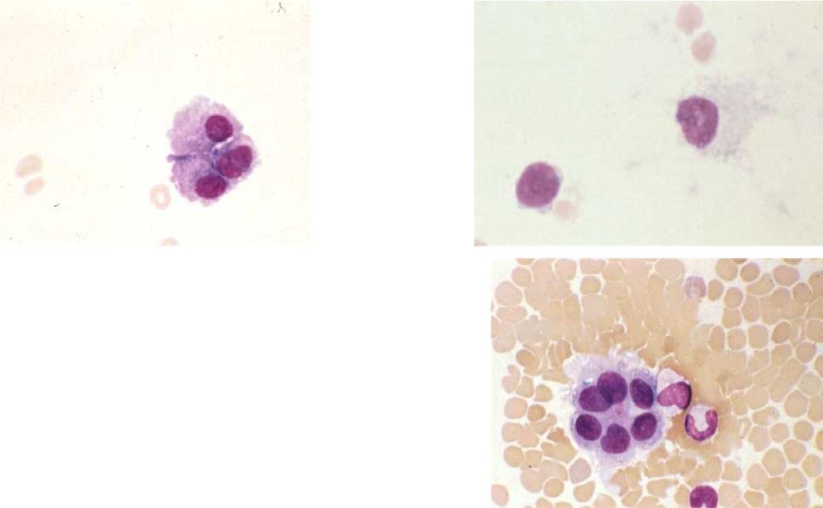
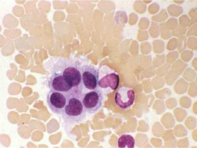
| Ventricular cell Ependymal Cells | |
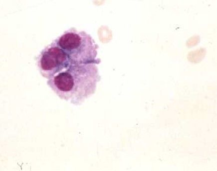
| Ventricular cell Choroid plexus "clumped" | |
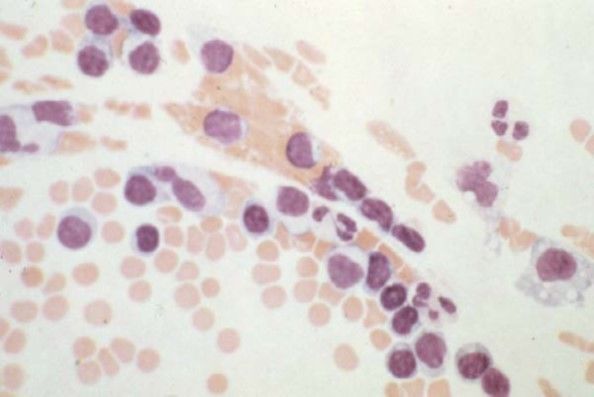
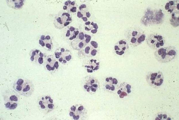
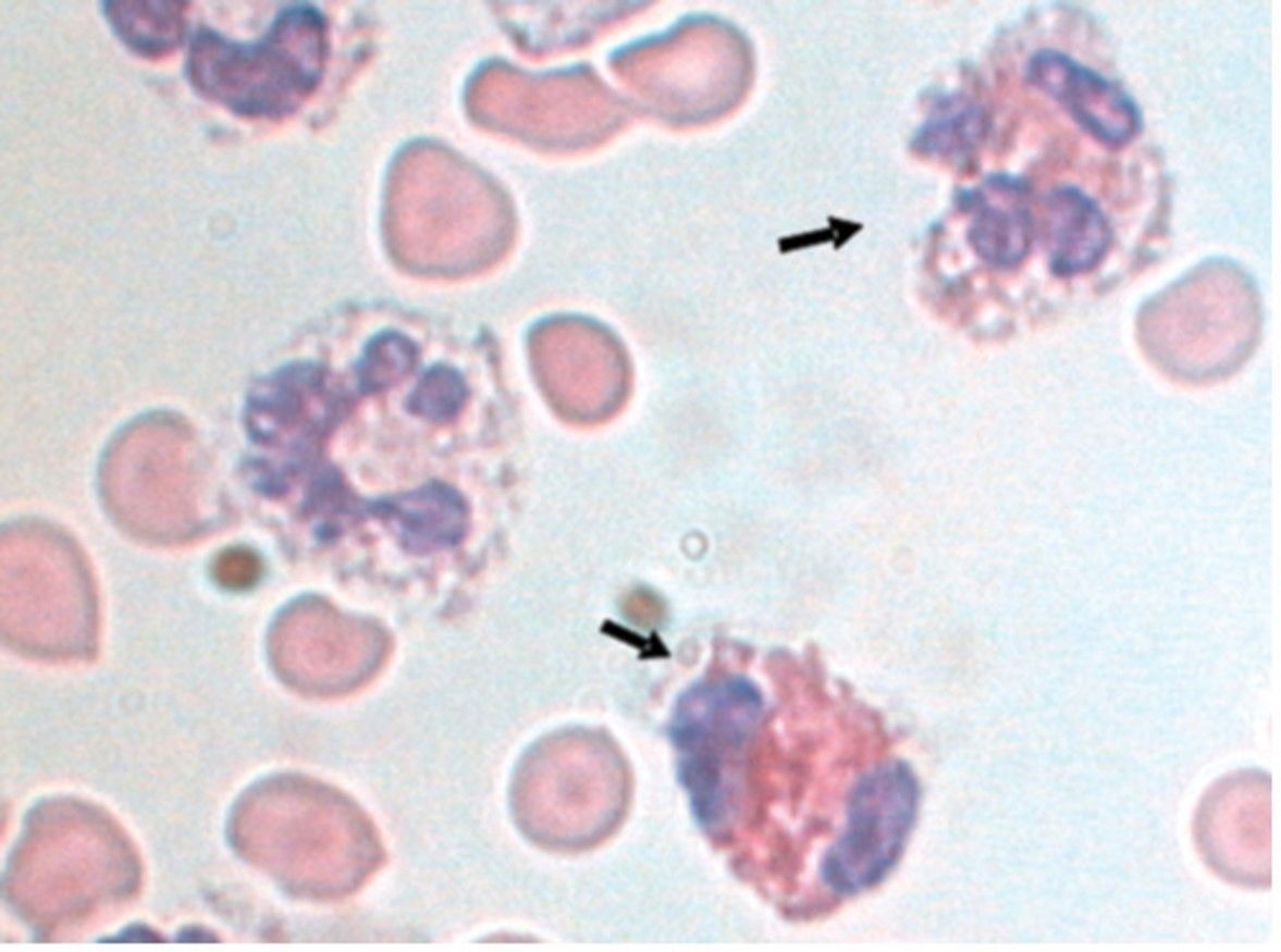
| Bacterial meningitis (note the intracellular bacteria in the neutrophils) | |
| Bacterial meningitis (note the intracellular bacteria in the neutrophils) (harder to see here) | |
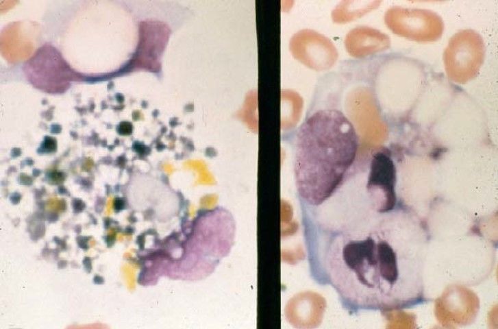
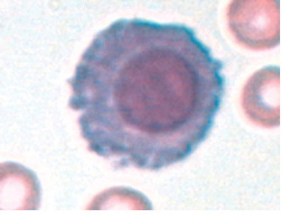
| Cryptococcus neoformans (fungal meningitis) Has characteristic "sunburst" appearance, which looks like a fuzzy halo with the Wright Stain | |
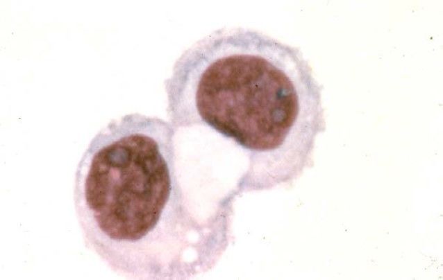
| Blast Cell | |
| Lymphoma Cells | |
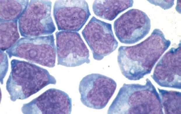
| Tumor cells | |
| Eosinophilia: Parasitic and Fungal Infections | |
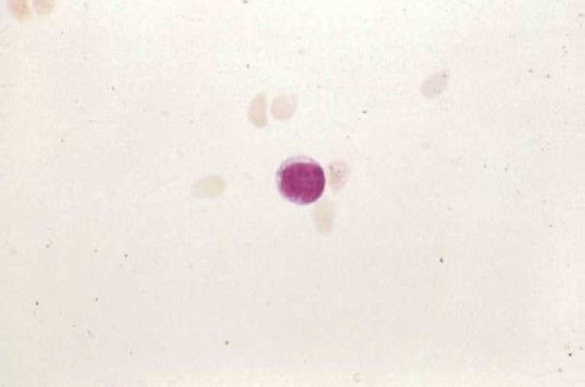
| Plasma cell Multiple Sclerosis | |
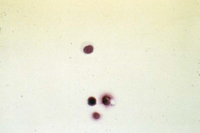
| nRBC's Traumatic tap caused by nicked vertebrae |
Want to create your own Flashcards for free with GoConqr? Learn more.