351971
| Question | Answer |
| List the 5 different types of bones. | 1. Long bones 2. Short bones 3. Flat bones 4. Irregular bones 5. Sesamoid bones |
| Describe long bones. | - It is longer than it is wide. - Consists of a shaft and two ends. - Largest bones in human body. - Usually more complex than other bones. - An example is the femur or humerus. |
| Describe short bones. | - Rough and cube like in structure. - Consists mainly of spongey bone with compact bone making up surface layer. - Found in the bones of the wrist (carpal bones) and ankle (tarsal bones). |
| Describe flat bones. | - Flat, thin & usually slightly curved. - Spongey inner structure with an outer parallel compact bony surface. - Examples include scapula, most bones of the skull, the breastbone & ribs. |
| Describe irregular bones. | - They don't match any of the descriptions for other bones. - Examples include the spinal vertebrae and the bones of the pelvis. |
| Describe sesamoid bones. | - Specialised short bones. - Often embedded in a tendon. - Alter the direction and pull of a tendon. - An example of this is the patella. |
| Bones are not living connective tissue. True or False? | False. Bones are living connective tissue, just very hard and dense living tissue. |
| How many bones in an average child and how many in an average adult? | Child - 270 Adult - 206 (As the body grows, many bones fuse together). |
| 'Form follows function'. Is this statement True or False? | True. The 'form' (shape) of a bone develops due to the 'functions' it carries out. |
| The stress applied to a bone, develops only the internal shape, not the external. True or False. | False. The stress applied to a bone develops both the internal & external shape, structure and strength. |
| Bones need stress placed upon them to reach their full potential in both strength and size. True or False? | True. There are 5 or 6 different type of loading forces placed on bones. |
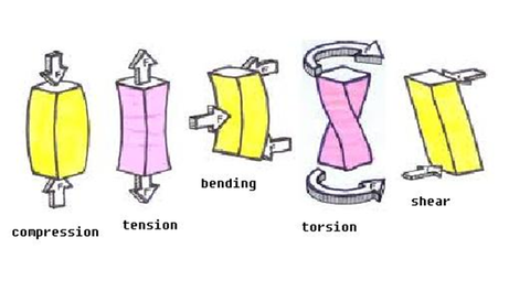
| List the forces placed on bones. | 1. Compression. 2. Tension. 3. Torsion. 4. Rotation. 5. Shear. |
| Describe compression. | - Weight bearing down through the bone due to gravity ↓↓↓ |
| Describe tension. | - Pulling forces applied to the bone from muscles ↑↑↑ |
| Describe torsion. | - Where one end of the bone is turned in the opposite way to the other end. |
| Describe rotation. | - A rotary force. The whole bone rotates along it's axis. - Sometimes interchanged with torsion but in rotation both ends of the bone are twisting the same way. |
| Describe shear. | - A direct opposing force → ← |
|
Image:
Bones (image/png)
|
Bending is different from shear because in bending, both ends of bone are fixed and force is centrally placed. Rotation is not included in the picture. |
| What happens when bones aren't stressed enough? | - Bone tissue can demineralise. - The bone will get softer and lose strength. However if stress is placed on the bone again, overtime, the bone density can increase with progressive loading of everyday life and physical exercise. |
| Describe compact bone. | - Very dense, hard structure on outside. - Inside comprised of less dense material. - The inside can be spongey bone (looks like little bubbles of air surrounded by arches of bone). - The inside absorbs shock, lighter than compact but still strong and acts like internal framework inside and under compact bone. |
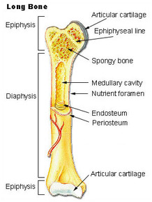
| What is the diaphysis? | - Shaft of the bone that makes up tubular long or sagittal axis of bone. - Very strong (due to collagen in compact bone) - Surrounds the medullary cavity. |
| What is the medullary cavity? | - Hollow section of bone. - Warehouse for yellow bone marrow for long bone fat storage. |
| What are the epiphysis? | - Rounded ends - Move with end of another bone to form a bony joint. - Located at proximal and distal ends of each long bone. - Contains spongey bone. - Contains epiphyseal line (epiphyseal plate in kids). - Surface covered by articular hyaline cartilage. |
| What is articular cartilage? | - Smooth shiny ends of long bone. - Smooth to reduce friction as it moves against articular cartilage of another bone. - Provides support, flexibility & resistance. - Cushions each epiphysis. - Absorbs compressive forces placed on bone (including gravity). |
| What is the periosteum? | - Double layered membrane, covers entire long bone, except epiphysis. - Attached to the bone by strong collagen fibres called Sharpey's fibres. - Outer layer is dense connective tissue that allows for the attachment of tendons. - Inner layer consists of osteoblasts and osteoclasts. - Supplied with nerve fibres & blood vessels. This allows for diffusion of minerals, in & out long bone. - Glistening white colour, visible to human eye. |
|
Image:
Long_Bone (image/jpg)
|
Picture of long bone. |
| What is the epiphyseal plate (a.k.a. the growth plate)? | - Hyaline cartilage situated in epiphysis of children. - Where most long bone growth occurs. - Complex if fractures as can interfere with bone growth. - The epiphyseal line is the 'grown up' version of this. |
| What are the two types of bone marrow? | 1. Red - Produces red and white blood cells. Found in spongey bone in long bones and in diploe of flat bones. Hematopoiesis (blood cell formation) in adults occurs at the head of the femur and humerus. 2. Yellow - Fat storage in long bones (for nutrition of bones). Does not produce blood cells. |
| There are 11 functions of bones, grouped into three areas. List these areas. | 1. Mechanical functions 2. Synthetic functions. 3. Metabolic. |
| List the four functions under mechanical. | 1. Protection - The rib cage protects the internal organs. 2. Shape - Bones give the structural scaffold that gives the body its form. 3. Movement - By articulating with another bone and being pulled by muscles. 4. Transfer sound - Bones conduct sound waves as vibrations in the inner ear. |
| List the function under synthetic. | 5. Blood production - the red marrow produces blood cells through heamatopoiesis. |
| List the functions under metabolic. | 6. Mineral storage - bones store minerals, mainly calcium and phosphorus. 7. Growth factor storage - bones can store some human growth factors that can assist with cell growth. 8. Fat storage - Yellow marrow stores fatty acids. 9. Acid-based balance - bones can assist maintaining blood pH via release of minerals. 10. Extra physiological regulation - Bones contain components that can assist in physiological regulation, via mineral release, when bones release too much calcium in the absence of certain hormones, bones can become osteoporotic and fracture more easily. 11. Detoxification - Can remove some heavy metals and store them. However this can have adverse affects as lead can be stored but critical organs such as brain, kidney and bone marrow can get lead poisoning if there is prolonged exposure. |
| Bone is a dynamic tissue and is constantly being remodelled by the actions of osteoblast and osteoclasts. True or False? | True. |
| Define ossification. | The developmental process of bone formation. |
| Define osteoblast. | - Specialised bone cells that lay down new layers of bone. - If a bone is fractured, more osteoblasts will be sent to that area. - If gap between two bones is very close & kept immobile, osteoblasts lay down new layer of bone. (This can take 6-8 weeks depending on size of fracture) |
| Define osteoclast. | - Specialized cell that absorbs and removes bone. - Allows for the development of new bone & maintenance of bone strength. |
| What is intramambranous ossification? | The bone develops from embryonic "mesenchymal cells" into a collagen membrane, in which specialised cells become osteoblasts which form spongy bone inside and compact bone outside at ossification centres. |
| What is endochondral ossification? | The same embryonic cells as in intramambranous become chondroblasts (produce cartilage).The chondroblasts produce a hyaline cartilage model in the rough shape of the final bone. Primary and secondary ossification centres exist. In long bones the secondary centres appear in the epiphyses. (When bones stop growing the growth plate or epiphyseal plate is known as the epiphyseal line.) |
| Broadly the three types of joints found in human bodies are what? | 1. Fibrous 2. Cartilaginous 3. Synovial |
| What are two common synovial joints? | Hinge joint - Two bones that are held together by ligaments to form the hinge. Usually allows movement in one plane only from the anatomical position (i.e. total of 2 movements) (Like a door hinge opening and closing). E.g. Elbow flexion/extension. Ball & socket joint - The ball of one bone fits into the socket made by another bone. Allows movement in all planes of movement (7 movements). E.g. The shoulder and hip joints They are both capable of: • Flexion/extension • Adduction/abduction • Internal & external rotation • Circumduction (a combination of all of the above movements at the same time, e.g. arm circles) |
| Describe a fibrous joint. | - Held together by only a ligament. Examples are where the teeth are held to their bony sockets and at both the radioulnar and tibiofibular joints. |
| Describe a cartilaginous joint. | - Occur where the connection between the articulating bones is made up of cartilage. For example between vertebrae in the spine. Synchondroses are temporary joints which are only present in children, up until the end of puberty. For example the epiphyseal plates in long bones. Symphesis joints are permanant cartilagenous joints, for example the pubic symphesis. |
| What are two common synovial joints? | Hinge joint - Two bones that are held together by ligaments to form the hinge. Usually allows movement in one plane only from the anatomical position (i.e. total of 2 movements) (Like a door hinge opening and closing). E.g. Elbow flexion/extension. Ball & socket joint - The ball of one bone fits into the socket made by another bone. Allows movement in all planes of movement (7 movements). E.g. The shoulder and hip joints They are both capable of: • Flexion/extension • Adduction/abduction • Internal & external rotation • Circumduction (a combination of all of the above movements at the same time, e.g. arm circles) |
| Describe a synovial joint. | Most common. - Highly moveable. - All have a synovial capsule surrounding the entire joint, a synovial membrane (the inner layer of the capsule) which secretes synovial fluid and cartilage known as hyaline cartilage which pads the ends of the articulating bones. |
| What are the joints that allow the most movement? | Synovial joints. They have a capsule that encases the joint and contains synovial fluid. This fluid acts as a lubricant for the joint to reduce friction between the two bones moving. Physical pressure from the bones assists our synovial membrane to produce this fluid. |
|
Image:
Joint (image/jpg)
|
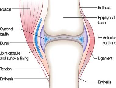
In this image, you can also see the ligaments that joint bone to bone. You can also see the tendon that joins muscle to bones through the enthesis |
| What is a bursa? | - Sac of fluid. - Runs between muscle & bone or tendon & bone to reduce friction. - If inflamed it is called bursitis. 'itis' means inflammation. |
| Define articulation. | The region where adjacent bones contact each other - another word for joint. E.g. The distal end of the femur articulates with the proximal end of the tibia. |
| Define condyle. | A large, rounded articular process. |
| Define crest. | A prominent ridge. E.g. The iliac crest on the hip bone. |
| Define diaphysis. | The long, relatively straight main body of a long bone; region of primary ossification. Also known as the shaft. |
| Define epicondyle. | A projection near to a condyle but not part of the joint. E.g. Epi means ‘upon’ so it is ‘upon’ the condyle. |
| Define facet. | A small, flattened articular surface. E.g. The facet joints in the spine. |
| Define foramen. | An opening through a bone. E.g. The neural foramen that the spinal cord runs down in the spinal column. |
| Define fossa. | A broad, shallow depressed area. E.g. The glenoid fossa on the scapula. |
| Define head. | The proximal articular end of the bone. |
| Define malleolus. | One of two specific protuberances of bones in the ankle. E.g. The medial malleolus on the inside of the ankle joint. |
| Define sinus. | Define sinus. A cavity within a cranial bone. E.g. The sinus cavity behind the nose. |
| Define spine. | A relatively long, thin projection or bump. E.g. The spine of the scapula bone. |
| Define suture. | Articulation between cranial bones.The sutures joining the head bones together. |
| Define tubercle. | A projection or bump with a roughened surface, generally smaller than a tuberosity. E.g. The greater and lesser tubercle on the humerus. |
| Define tuberosity. | A projection or bump with a roughened surface. E.g. The tibal tuberosity that the quadriceps tendon attaches to on the anterior, proximal part of the tibia. |
| Define trochanter. | One of two specific tuberosities located on the femur. E.g. The greater and lesser trochanter of the femur. |
|
Image:
upper_bones (image/jpg)
|
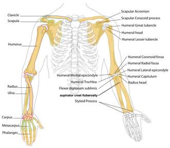
Upper bones. |
|
Image:
Skull (image/jpeg)
|
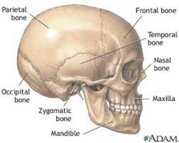
What does 'Full Pot Of Tea' stand for? The frontal, parietal, occipital and temporal bones. |
| There are four primary tissue types in the human body. List them. | 1. Epithelial Tissue 2. Connective Tissue 3. Muscle tissue 4. Nervous Tissue |
| What are the connective tissue of the skeletal system? | 1. Bone. 2. Ligaments - Join bone-to-bone, acting like a ‘hinge’ for the joint. Their role is to provide joint stability. they have a reduced blood supply. 3. Tendons - Join muscle to bone. Tendons allow muscles to act upon a bone when the muscle action contracts to shorten (concentric) or lengthen (eccentric). Tendons are more ‘elastic’ than ligaments to absorb and store energy. 4. Cartilage - protects joint surfaces, some types produce lubricating fluid etc. |
| Summarise embryonic connective tissue. | Mesenchyme and mucous. |
| Summarise adult connective tissue. | Connective tissue proper: Ligaments and tendons fit in here as they are dense, regular collagenous CT. Whereas fat or adipose tissue is Loose CT. Supporting CT: Bone and cartilage. Fluid CT: Blood and bone marrow. |
| Describe muscle tissue. | - Muscles also help stabilise joints but are not classified as connective tissue. - Has a very good blood supply. - Ability to shorten and lengthen under load. - When they ‘ pull’ they move two bones together that generates movement in the body’s limbs. - Muscles pull on tendons, which are attached to bones and move the bones. Most times one bone is stable, so it can move the other bone towards it |
| Describe nervous tissue. | - A specialised tissue capable of conducting electrical impulses. |
| Describe epithelial tissue. | - Covers and protects all outside and inside surfaces. - Varies in number of layers and cell structure. - A protective barrier to foreign antigens and abrasions. - Allow diffusion of nutrients. - Absorb and secrete substances in our bodies. |
| What differentiates connective tissue from the rest? | - The main difference is at a microscopic level. Connective tissue - Mostly made up of "matrix" with individual cells dispersed within this matrix. Eg. bone cells (osteocytes) are dispersed in a calcified matrix. Specialised Tissue - Mostly made up of individual cells (eg muscle cells or nerve cells) tightly packed together against each other with relatively less matrix . |
Want to create your own Flashcards for free with GoConqr? Learn more.