36273932
Description
Flashcards by Leanne van den Berg, updated more than 1 year ago
|
|
Created by Leanne van den Berg
over 3 years ago
|
|
| Question | Answer |
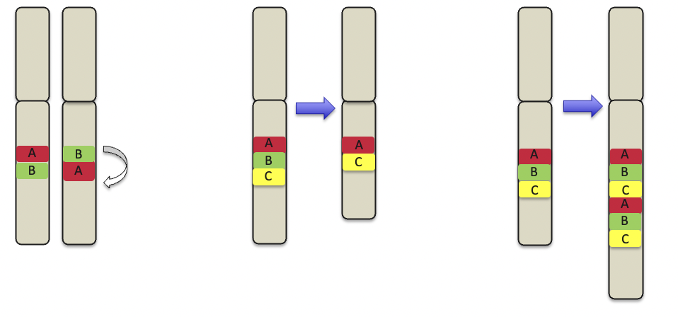
| What are these intrachromosomal abberations called? | Left= Inversion Middle= Deletion Right= duplication |
| what is the difference between intrachromosomal and inter chromosomal abberations? | Intrachrom. ab are changes within the same chromosome, in interchrom abberations, changes occur among multiple chromosomes Interchromosomal > translocation or insertion |
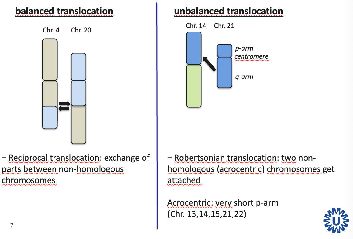
| What is the difference between a balanced and an unbalanced translocation? | Balanced = exchange of DNA between non-homologous chromosomes Unbalanced= 2 non-homologous chromosomes get attached, no exchange |
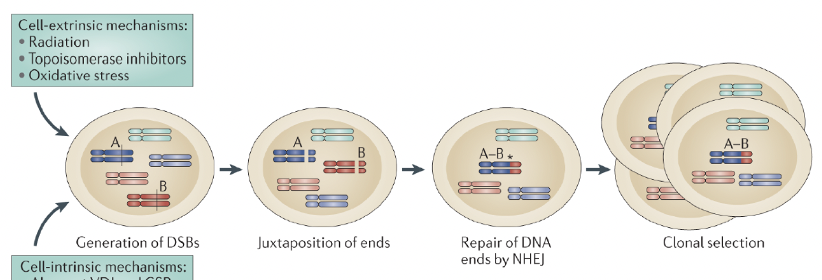
| Which 3 steps are required for gene fusions? | 1. ds DNA break 2. Illegal (wrong) recombination 3. Clonal expansion |
| Name 3 reasons for gene fusions | Radiation (causes ds breaks) Recombination events that have gone wrong (B and T cells) Vulnerable places lie close to each other in the nucleus |
| What are 3 consequences of oncogenic chromosomal translocations | 1. fusion protein as tyrosine kinase forms 2. Fusion protein as transcription factor forms 3. Oncogenes are translocated to a place where they are under the control of a stronger promoter (like the promotor of the BCR) |
| Which 3 methods can be used to detect translocations and fusions? | Karyotyping > only on high quality material FISH RNA based > RT-PCR and Archer RNA seq |
| Can FISH be done on FFPE or only of FF material | FISH can be done on both FFPE and FF |
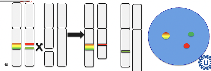
| What is the difference between a break apart FISH and a fusion FISH | Break apart: When gene is broken into pieces (translocated), the yellow dots form separate green and red dots. Fusion: Red and green dots lie close to each other. Each color marks a different genomic region |
| What chromosomal abberations can be seen with FISH? | Chromosomal breaks > translocation Fusions CNVs (amplification) |
| Why would you used RNA seq instead of DNA seq (NGS/ Sanger)? | Generally, RNA sequencing provides information about expression of genes. In addition, it gives information about the level of expression and at what moment genes are switched on or off. In tumor cells, RNA sequencing provides information about the (functionality of) fusion of genes, alternative splicing and/or exon skipping. |
| For translocation detection: When do you use RNA seq, and when FISH | FISH: When you need results quick (1 day), cheaper, but only applicable for 1/2 targets RNA seq: more info of multiple genes in 1 assay Both possible with FFPE |
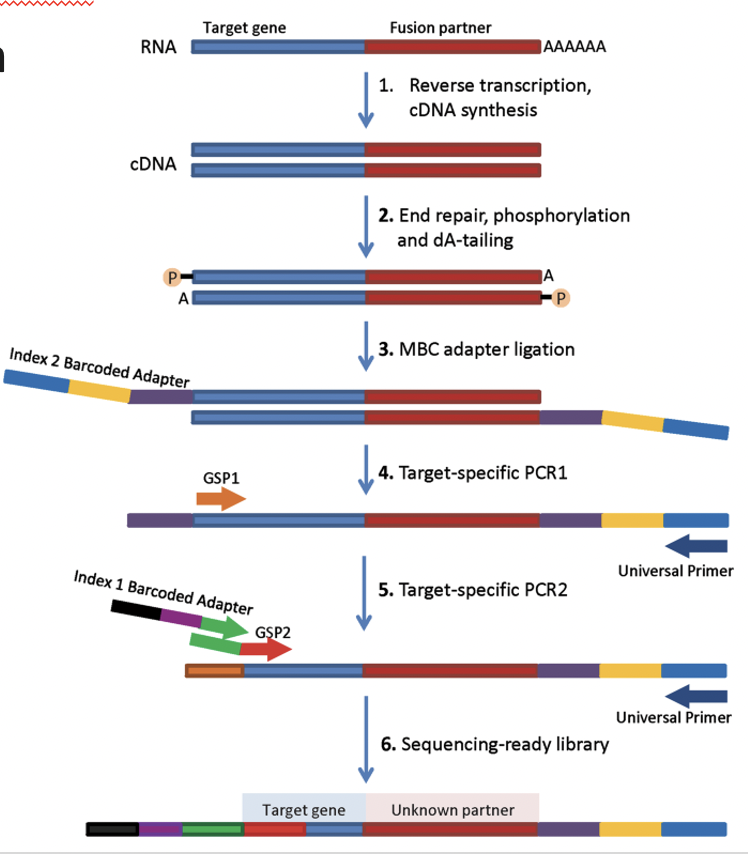
| How does Archer RNA seq work? | Archer can determine the unknown fusion partner of your target gene |
| Name 2 pros and 2 cons of Archer | Pros: 1. Unknown fusion partners can be detected 2. Fusion gene detection of multiple genes in 1 assay Cons: 1. Takes 1 week 2. Expensive per sample |
| What is Nanostring used for? | Gene expression miRNA expression Fusion gene detection CNV detection SNV detection Protein expression |
| Name 2 pros and 2 cons of Nanostring | Pros: 1. Relatively cheap 2. No amplification step needed Fast, can be done on FFPE etc. Cons: 1. Expensive machines needed 2. No detection of unknown fusion genes |
| Name 2 pros and 2 cons of RNA sequencing | Pros: 1. Transcriptome wide 2. Detects both over translocation, expression and SNVs Cons: 1. Only works well on FF 2. Slow (2 weeks) extra: Complex bio-informatics needed |
| What markers are used for the detection of B and T cells in IHC? | B cells: CD19, CD20 T cells: CD3 |
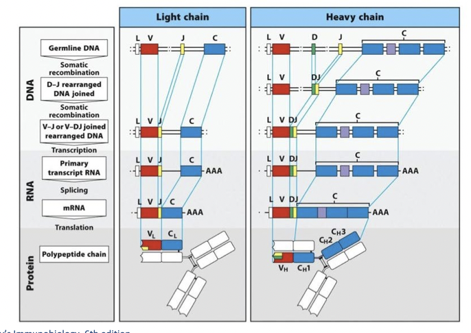
| BCR rearrangement involves combinations of different fragments. Which fragments are combined in the light and which in the heavy chain | Light: V and J Heavy: V, D and J |
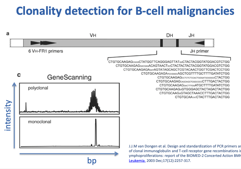
| On which region of the BCR gene do primers bind to detect clonality for B cell malignancies? | On the V region of heavy chain Consensus primer binds to the J region of the heavy chain |
| What are the pitfalls of gene rearrangement analysis using TCR/BCR? | 1. Low amount of T or B cells might look like monoclonality 2. difficult to distinguish between reactive B/T cells or malignant T/B cells 3. In case of a high mutation load, primers will not bind and no peak is shown for the malignant cells |
| What is the difference between lymphomas and leukemia? | Leukemia: lymphoid malignancy with involvement of bone marrow and usually peripheral blood Lymhomas: lymphoid malignancy arising as a discrete tissue mass |
| What 3 molecular things are testen in lymphomas | TCR/BCR gene rearrangement > reactive or malignant Translocations > classification and treatment Mutations > classification and treatment |
Want to create your own Flashcards for free with GoConqr? Learn more.