36376571
Description
Flashcards by Jessica Do, updated more than 1 year ago
|
|
Created by Jessica Do
over 3 years ago
|
|
| Question | Answer |
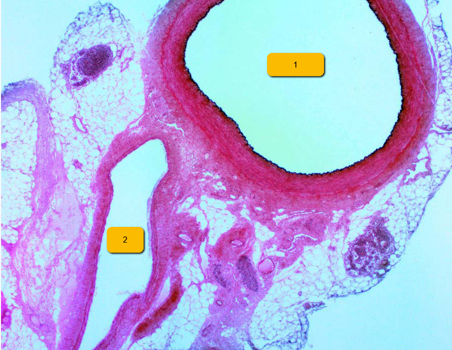
| 1. Artery 2. Vein | |
|
Image:
Tunica (binary/octet-stream)
|
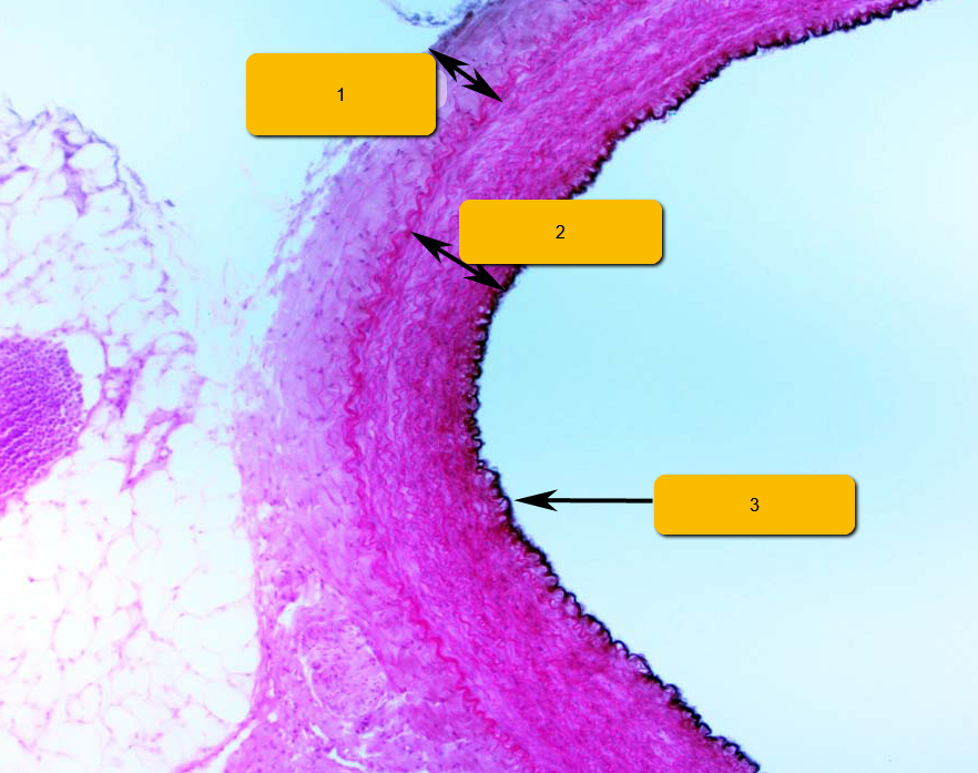
1. Tunica Externa 2. Tunica Media 3. Tunica Interna |
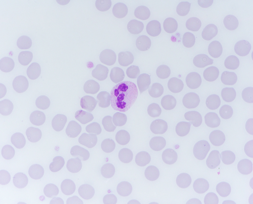
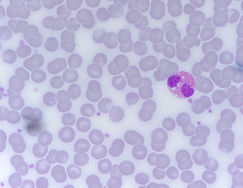
| Neutrophil | |
| Neutrophil | |
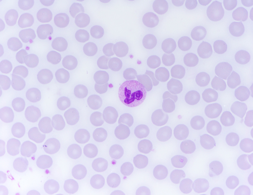
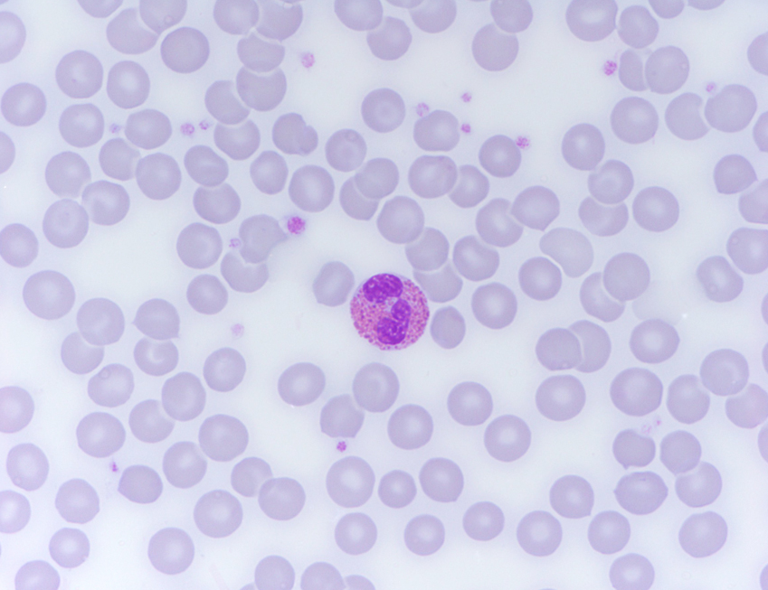
| Eosinophil | |
| Eosinophil | |
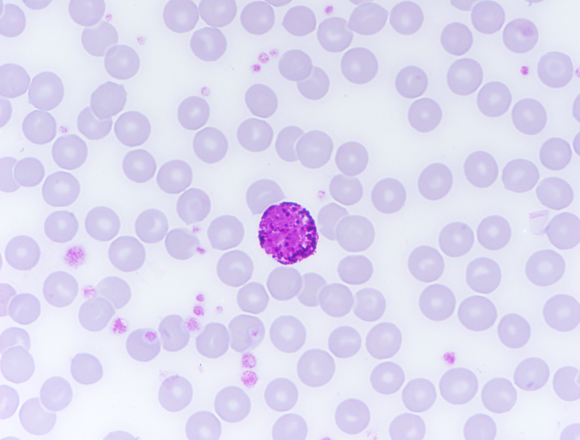
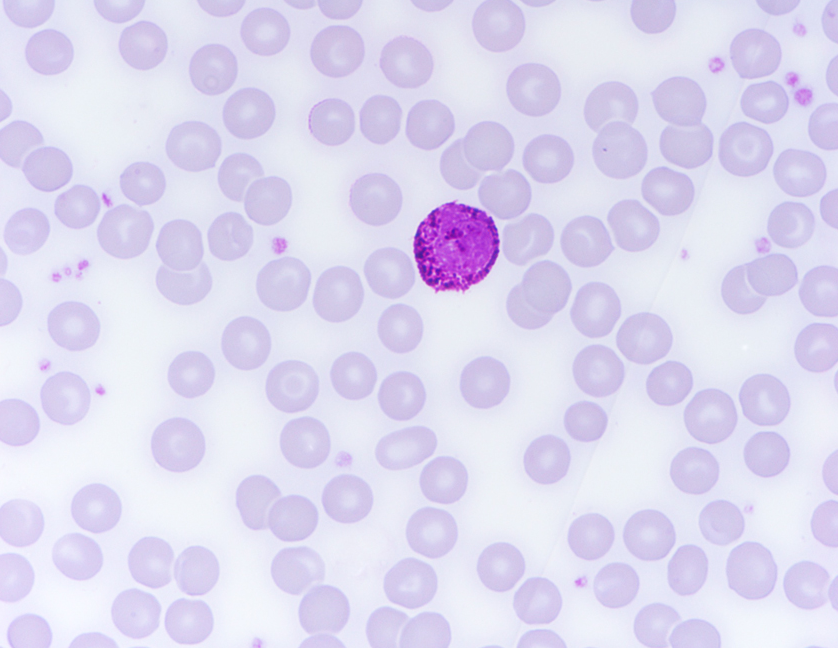
| Basophil | |
| Basophil | |
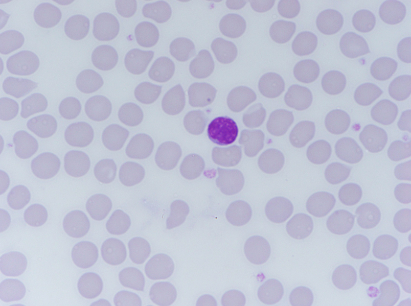
| Lymphocyte | |
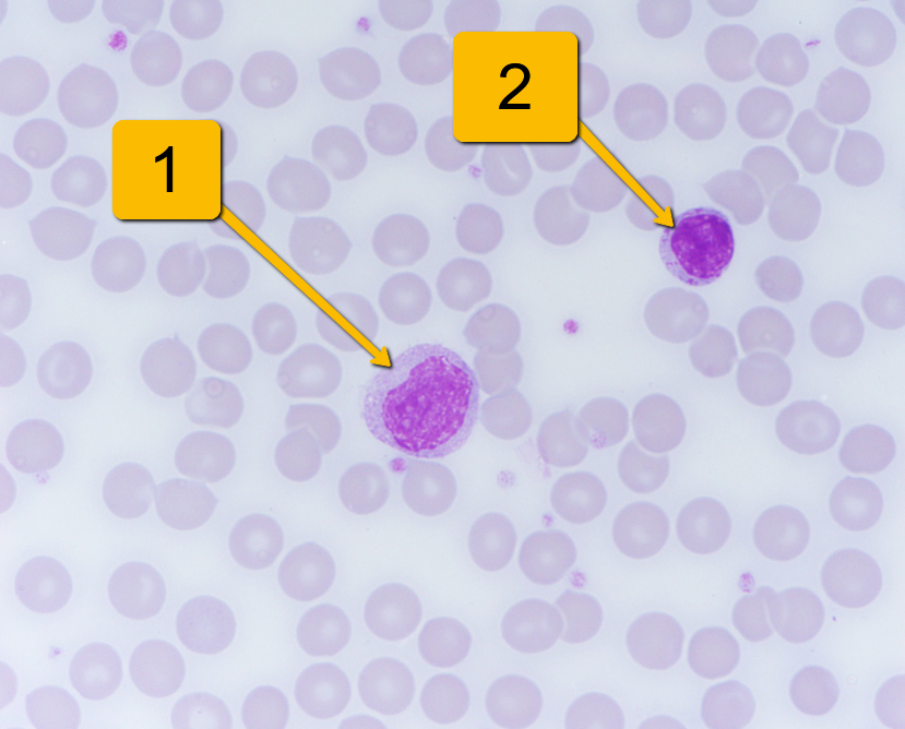
| 1. Monocyte 2. Lymphocyte | |
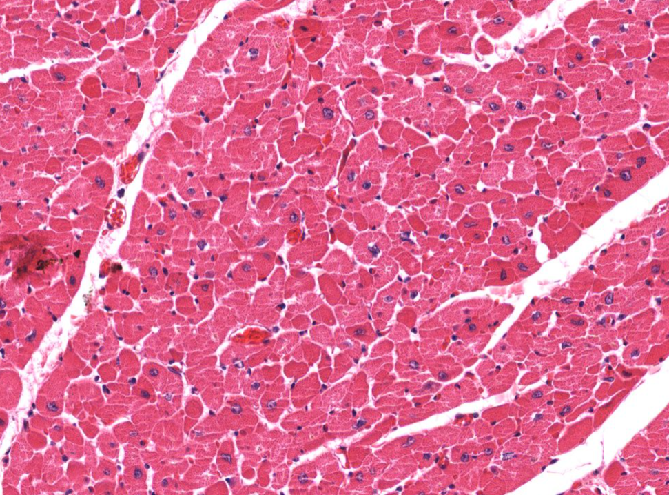
| Cross section view of cardiac muscle cells | |
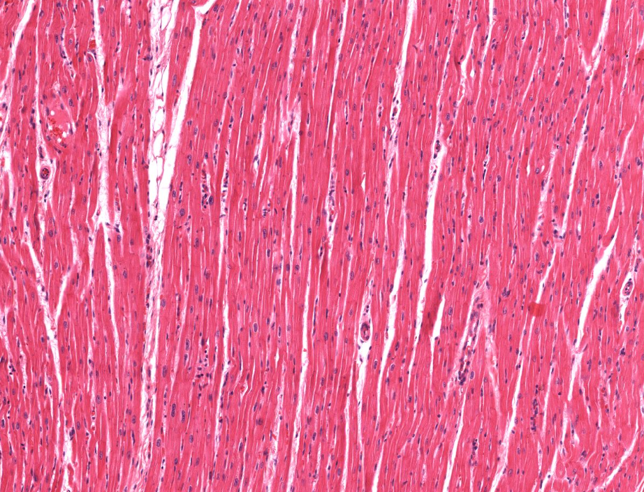
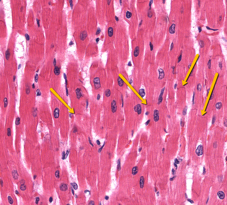
| Longitudinal View of Cardiac Muscles | |
| Intercalated discs of a cardiac muscle | |
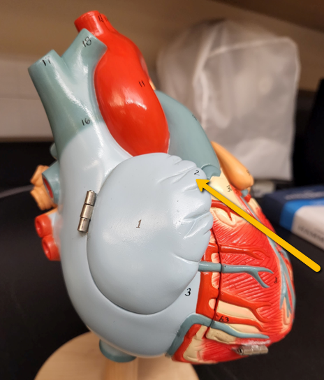
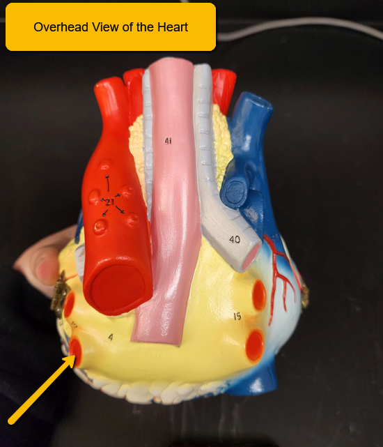
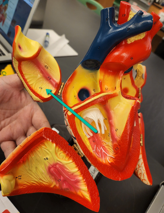
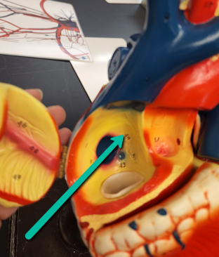
| Auricle of the Right Atrium - Flap that is located along the anterior margin of the atrium; gives us extra space for the atrium to pouch outward when it fills up with blood | |
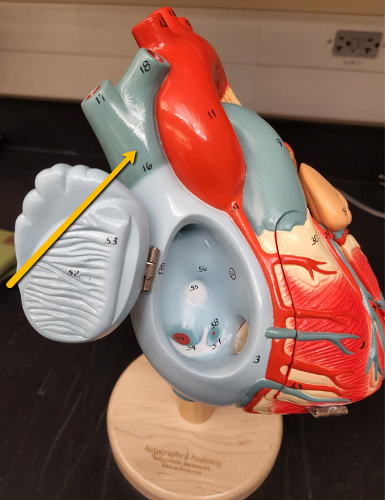
| Auricle of the Left Atrium - a muscular appendage of the atrium | |
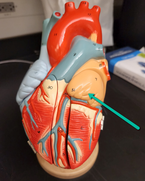
| Left and Right Ventricles | |
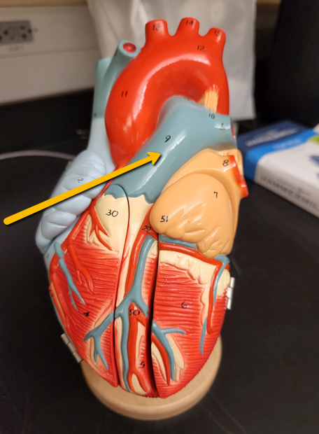
| Pulmonary Trunk | |
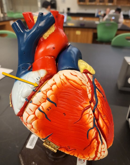
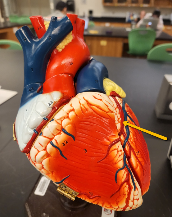
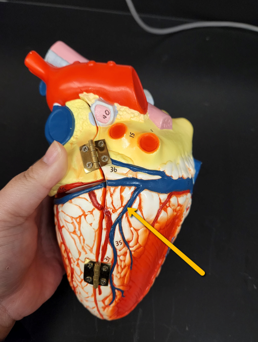
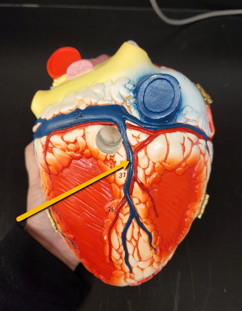
| Right Coronary Artery | |
| Left Coronary Artery | |
| Left Pulmonary Artery | |
| Right Pulmonary Artery | |
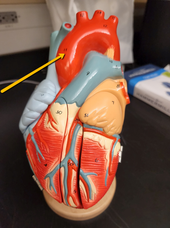
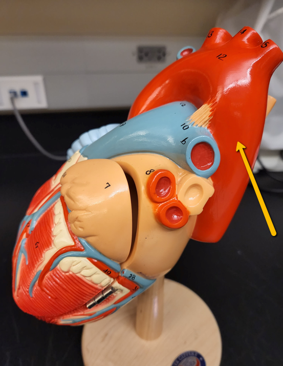
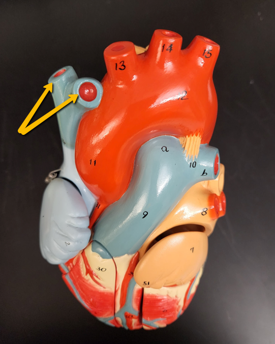
| Ascending Aorta | |
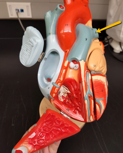
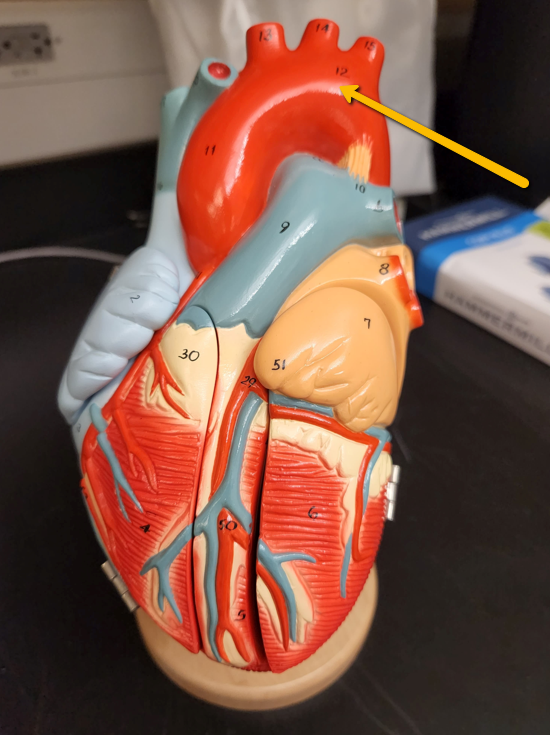
| Aortic Arch | |
| Descending aorta | |
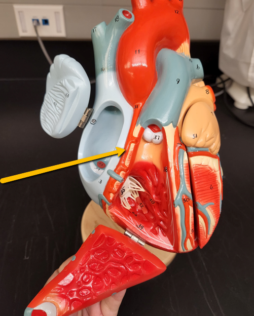
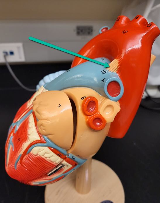
| Ligamentum Arteriosum | |
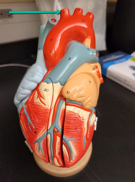
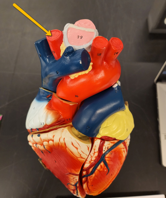
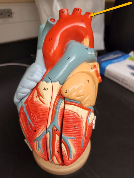
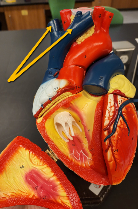
| Brachiocephalic Trunk | |
| Brachiocephalic Trunk | |
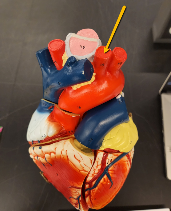
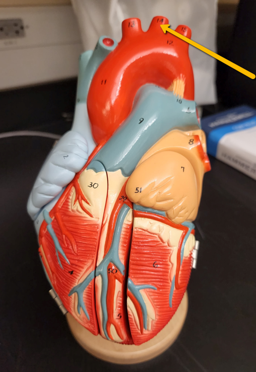
| Left Common Carotid Artery | |
| Left Common Carotid Artery | |
| Left Subclavian Artery | |
|
Image:
Sulcus (binary/octet-stream)
|
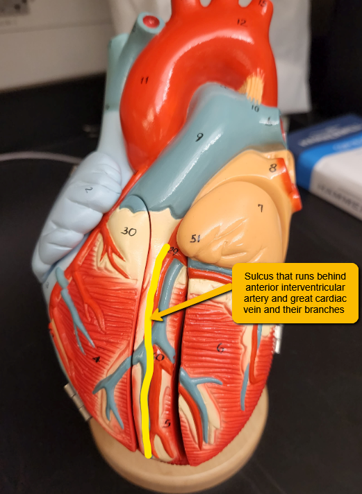
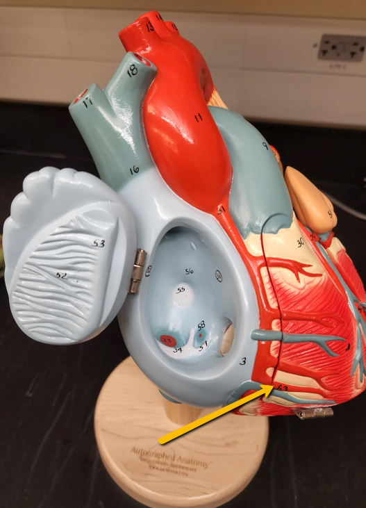
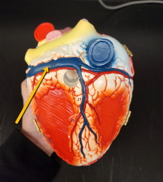
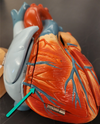
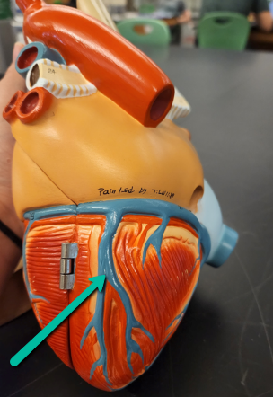
Anterior Interventricular Sulcus - Contains interventricular branch of left coronary artery/great cardiac vein |
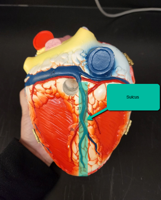
| Posterior Interventricular Sulcus - Contains coronary vessels including posterior interventricular branch of right coronary artery/middle cardiac vein | |
| Right Coronary Artery | |
| Left Coronary Artery | |
| Circumflex Branch of Left Coronary Artery | |
| Left Anterior Descending Artery (Anterior Interventricular Branch of Left Coronary Artery) | |
| Marginal Branch of Right Coronary Artery | |
| Right Coronary Artery | |
| Left Coronary Artery | |
| Posterior interventricular branch of right coronary artery | |
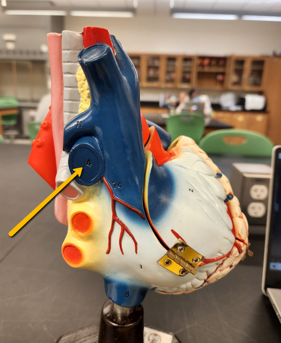
| Superior Vena Cava - Brings blood in from the head, neck, and upper extremities into the right atrium | |
| Inferior Vena Cava - Brings blood up from levels below the heart; From lower thoracic, abdominal and lower extremities into the right atrium | |
| Coronary Sinus - Vein that wraps around the posterior side of the heart in the coronary sulcus; brings blood into right atrium; underneath is the coronary sulcus | |
| Brachiocephalic Veins | |
| Brachiocephalic Veins | |
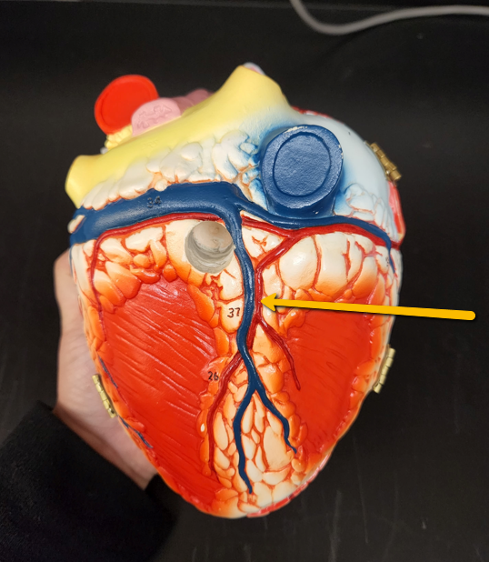
| Great Cardiac Vein | |
| Posterior Cardiac Vein | |
|
Image:
Middle (binary/octet-stream)
|
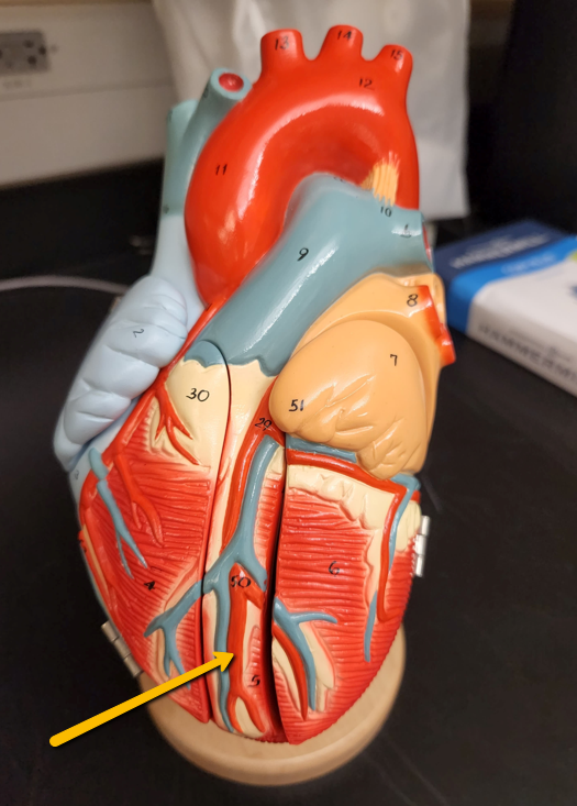
Middle Cardiac Vein |
| Small Cardiac Vein | |
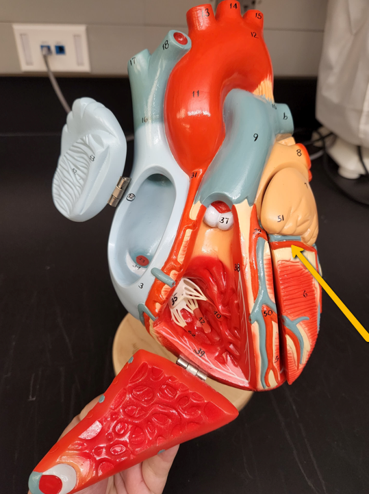
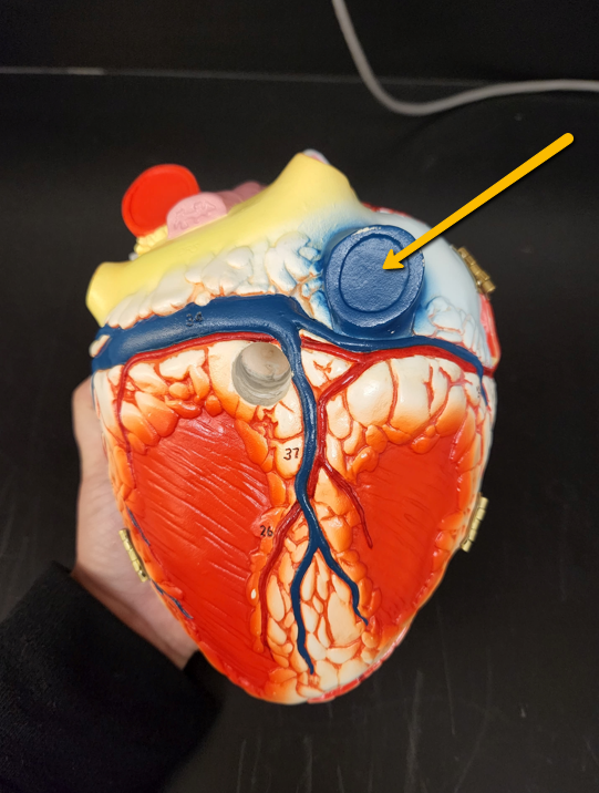
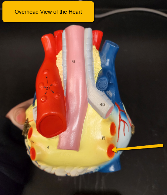
| Right Pulmonary Vein - bring in blood from the right lung into the left atrium | |
| Left Pulmonary Vein - bring in blood from left lung into the left atrium | |
| ***need an image | Myocardium |
| ***need an image | Endocardium |
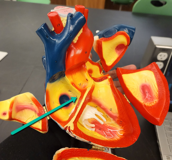
| Interatrial Septum - Important for keeping left and right atrium separate since deoxygenated blood enters the right atrium while oxygenated blood enters the left | |
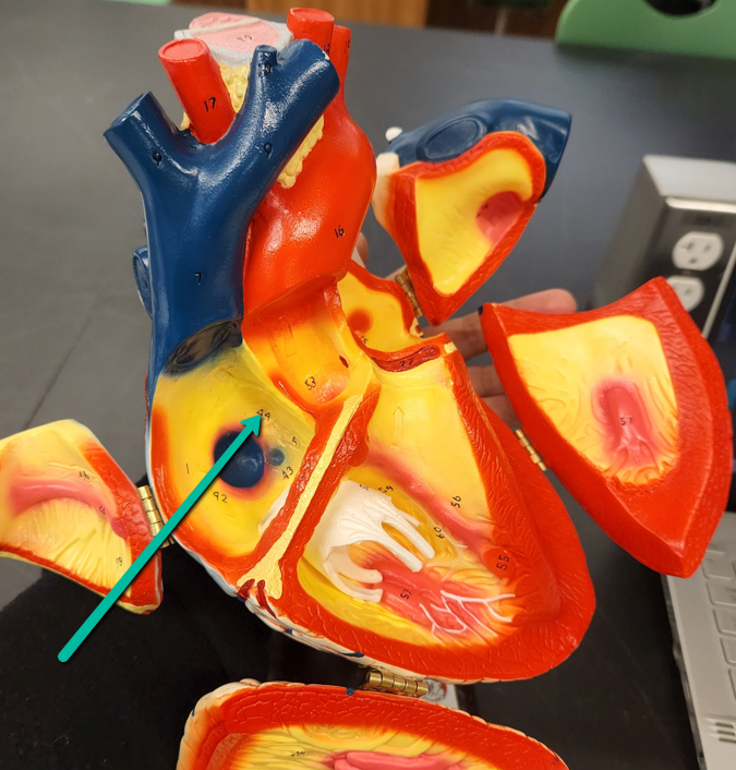
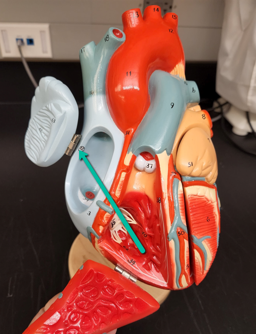
| Fossa Ovalis - An indentation in the right atrium; is a remnant from our fetal circulation (called foramen ovale when open) | |
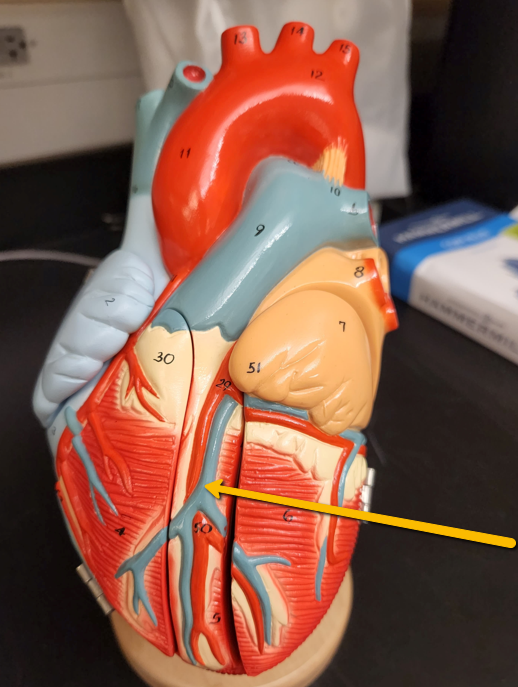
| Interventricular Septum | |
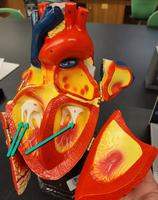
| Tricuspid Valve - Between the right atrium and right ventricle; prevents backflow | |
| Pulmonary Semilunar Valve - valve located between right ventricle and pulmonary artery/aorta and prevents backflow into ventricles. | |
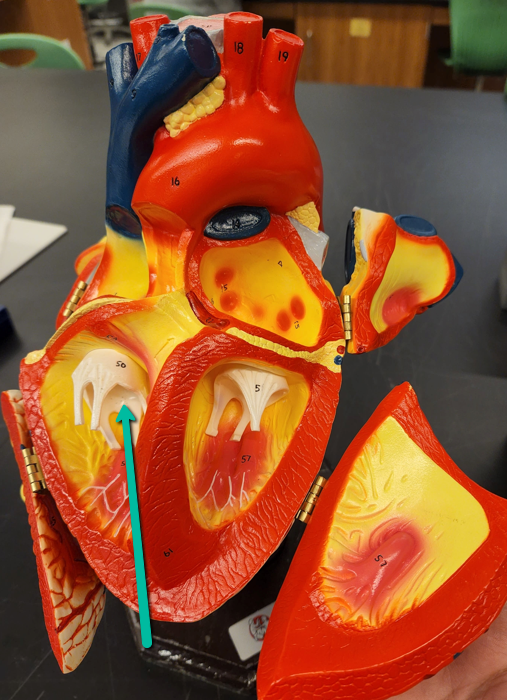
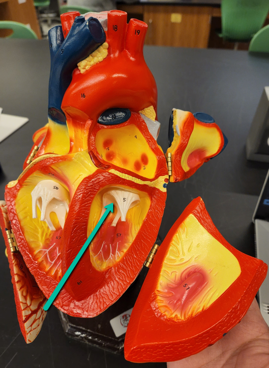
| Bicuspid Mitral Valve | |
| Aortic semilunar valve | |
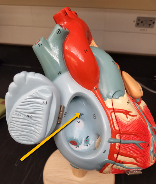
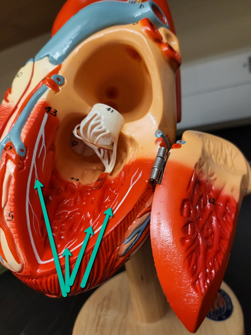
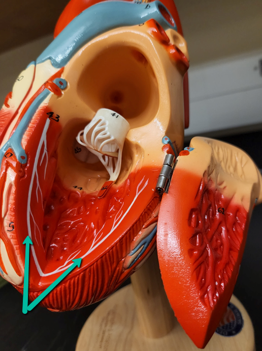
| Pectinate Muscles - lines the wall of the right atrium of the heart only | |
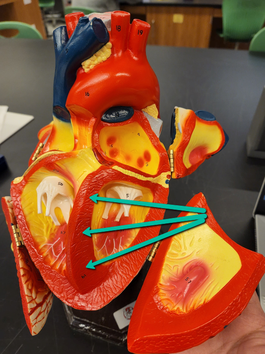
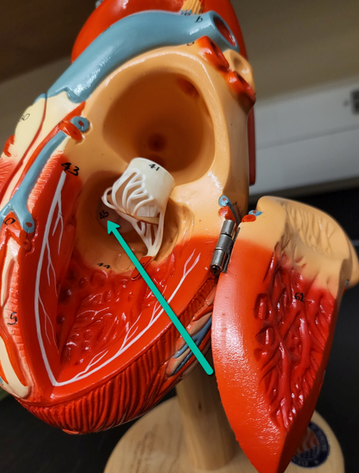
| Papillary Muscles | |
| Chordae Tendineae - Threads on the valves that extend down to papillary muscles on the wall of the ventricles; prevents valves from snapping back into atria | |
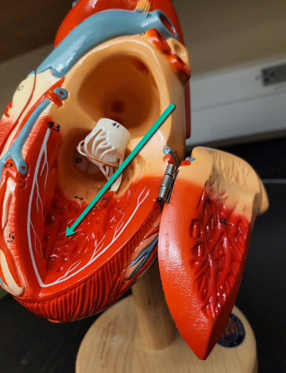
| Trabeculae Carneae - found in both ventricles; corkscrew motion to push blood upward | |
| Sinoatrial Node - contains pacemaker cells of the heart | |
| Atrioventricular node - receives conduction from internodal pathways and transmits to the Bundle of His | |
| Perkinje Fibers - Extend from right and left bundle branches; convey impulse to ventricular myocardial cells | |
| Bundle Branches | |
| Posterior Cardiac Vein | |
| Fossa Ovalis | |
| AV Node |
Want to create your own Flashcards for free with GoConqr? Learn more.