3708261
Description
Flashcards by Jennifer Lay, updated more than 1 year ago
|
|
Created by Jennifer Lay
about 9 years ago
|
|
| Question | Answer |
| Cardiovascular System | The heart, which pumps blood. Blood Vessels, through which blood flows |
| In humans, blood is always contained... | Within the Blood Vessels |
| Functions of Blood | *Remove Waste *Provides interstitial fluid with O2 and Nutrients cells require to live. |
| Interstitial Fluid | Or Tissue fluid is a solution that bathes and surrounds the tissue cells of multicellular animals. It is the main component of the extracellular fluid, which also includes plasma and transcellular fluid. |
| Extracellular Fluid | Extracellular fluid (ECF) or extracellular fluid volume (ECFV) usually denotes all body fluid outside of the cells. The remainder is called intracellular fluid. In some animals, including mammals, the extracellular fluid can be divided into two major sub compartments, interstitial fluid and blood plasma. |
| At the lungs, blood drops off___1__ Then picks up___2___ | 1.Carbon dioxide, C02 2.Oxygen |
| Blood is purified of its waste at | The kidneys...(Water and salts are retained as needed.) |
| Cardiovascular system:Regulation | Participates in the homeostasis of the body's conditions. (Temp, pH balance, water/electrolyte levels.) |
| Lymphatic System assist in... | Cardiovascular system. This is because lymphatic vessels collect excess interstitial fluid and return it to the cardiovascular system. |
| Lymphatic system | The network of vessels through which lymph drains from the tissues into the blood |
| Vessels | a tube or canal (as an artery, vein, or lymphatic) in which a body fluid (as blood or lymph) is contained and conveyed or circulated. |
| Tissues | Group of like cells that together carry out a specific function |
| As soon as fluid enters lymphatic vessels it is called... | Lymph, a fluid connective tissue |
| Connective Tissue | tissue that connects, supports, binds, or separates other tissues or organs |
| Types of Connective tissue | aerolar, blood, adipose, fibrous, bone, cartilage, hemopoietic (never talked about in class) |
| Three types of Blood Vessels | Arteries, veins, capillaries |
| Artery | blood vessel that transports blood away from the heart |
| Arterioles | Small arteries barley visible to the naked eye. The middle layer has some elastic tissue but it is composed mostly of smooth muscle. These muscle fibers encircle the arteriole. |
| When the fibers from the arteriole contract | the vessel constricts The constriction or dilation of arterioles control blood pressure. When they constrict blood pressure rises. |
| When arteriole muscle fibers relax... | the vessel dilates. Dilation of arterioles causes blood pressure to fall. |
| Arterioles Branch into | Capillaries, the smallest of the blood vessels |
| Veins | Blood vessels that return to the heart |
| Venules | Small veins that drain blood from capillaries and then join to form a vein |
| Veins often have | Valves which allow blood to only flow towards the heart when open and prevent backward flow when closed. |
| The heart | Cone-shaped muscular organ located between the lungs, directly behind the sternum(breastbone). the heart is tilted, so that the apex(the pointed end) is oriented to the left. |
| The major portion of the heart is the | Myocardium, the interior wall of tissue. Consisting largely of cardiac muscle tissue. |
| Myocardium | (The middle, muscular layer of the three layers of the walls of the heart) Interior wall of tissue. Consisting largely of cardiac muscle tissue. Receives O2 and nutrients from coronary arteries |
| The heart is surrounded by the | Pericardium, a thick and membranous sac that supports and protects the heart |
| Pericardium | A thick and membranous sac that supports and protects the heart |
| Four Chambers of the heart | The two upper thin walled atria(atrium) - the right and left. The two lower chambers are the thick walled ventricles - the right and left |
| Valves between the atria and ventricles(the chambers of the heart) are called the | Antrioventricular valves(AV valves) Tricuspid (right) Bicuspid/Mitral (left) |
| Atrioventricular Valves(AV valves) | Supported by strong fibrous strings, the chordae tendinaea |
| Chordae Tendinaea | Strong fibrous strings that anchors valves and prevents them from inverting when heart contracts. |
| Semilunar valves | Has flaps shaped like half moons. Lie between ventricles and attached vessels. Two types: Pulmonary and Aortic |
| Pulmonary Semilunar valve | lie between the right ventricle and pulmonary trunk |
| Aortic semilunar valve | lies between the left ventricle and aorta. |
| Coronary arteries | serve the heart muscle itself. The first branches off the aorta. Originate just above the aortic semilunar valve. Lie exterior to the surface of the heart. |
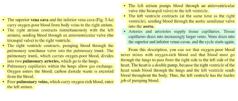
| Passage of Blood through the heart |
Want to create your own Flashcards for free with GoConqr? Learn more.