4118841

| Question | Answer |
| :D بسم الله نبدأ | xD ربنا يستر |
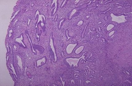
| where am I from the universe o.O?! TISSUE TYPE: see glands and stroma... UTERUS PATHOLOGY: hyperplastic glands with different sizes and shapes DIAGNOSIS: endometrial hyperplasia KEY: FIND THE SWISS CHEESE XD.. | |
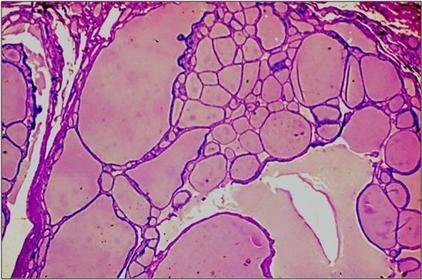
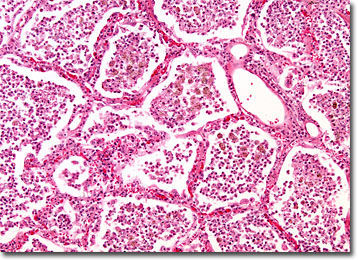
| TISSUE TYPE: glands filled with colloid PATHOLOGY: glands increased in size , stretched with colloid (flat epith.) DIAGNOSIS: Nodular Goiter KEY: homogeneous big pinky patches everywhere everywhere ^^ (so unique) | |
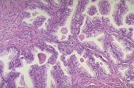
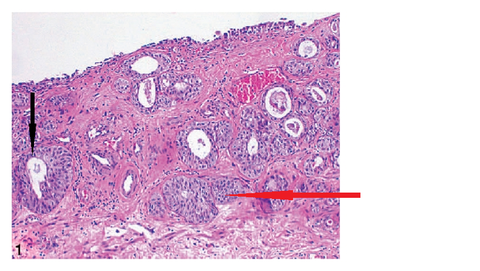
| TISSUE TYPE: gland and stroma... specific glands of prostate PATHOLOGY: hyperplasia of gland and stroma DIAGNOSIS: benign prostatic hyperplasia KEY: U CAN SEE CORPORA AMYLECIA | |
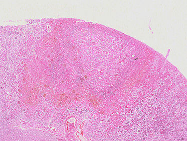
| TISSUE TYPE: see glomeruli PATHOLOGY: see area with increased eosinophilia and preserved outlines .. NECROSIS DIAGNOSIS: kidney infarction KEY: EOSINOPHILIA.. NO CASEATION .. NO PUS.. COAGULATIVE NECROSIS | |
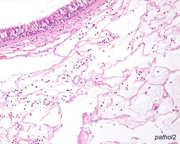
| o.O o.O !! eyh el hezar da .. xD keep caaalm .. TISSUE TYPE: respiratory epith. 4ayfo ya Doc. ?? :D .. NOSE PATHOLOGY: Oedematous C.T can find ulceration of epith. or squamous metaplasia DIAGNOSIS: allergic nasal polyp KEY: RESPIRATORY EPITH. | |
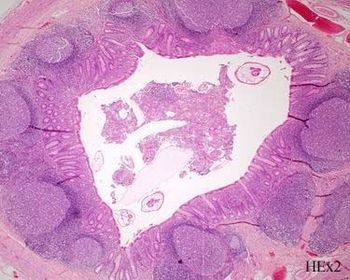
| TISSUE TYPE: circular organ .. with lymph follicles .. u see the complete circle... APPENDIX PATHOLOGY: necrotizing tissue in the lumen ... hyperplastic lymph tissue DIAGNOSIS: acute suppurative appendicitis KEY: COMPLETE CIRCLE | |
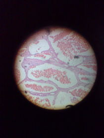
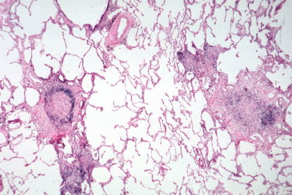
| TISSUE TYPE: dots on white page :D... white page= alveolar spaces.. u can see carbon particles (anthracosis)..LUNG PATHOLOGY: inflamatory cells in alveolar spaces .. DIAGNOSIS: acute lobar pneumonia KEY: WHITE BACKGROUND .. | |
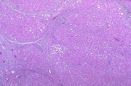
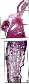
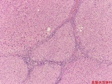
| TISSUE TYPE: O.o (loss of architecture) PATHOLOGY: thick fibrous speta with inflamatory cells .. separate nodules of undefined cells. (steatosis) Diagnosis: liver cirrhosis xD KEY: CAN'T SEE ANY SPECIAL THING ABOUT LIVER CAUSE ACTULLAY IT'S DEAD :D | |
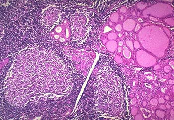
| TISSUE TYPE: colloid of..... THYROID PATHOLOGY: inflammatory cells, hyperplastic lymphoid follicles, cells lining acini called oncocytes ,Hurthle or askanazy cells DIAGNOSIS: Hashimoto's thyroditis KEY: ACINI + LYMPH FOLLICLES N.B: differentiate it from Goiter (no lymph follicles) | |
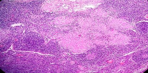
| TISSUE TYPE: lymphocyets every where... where am I ??? xD PATHOLOGY: inflamatory granuloma with caseation in the center .. eosinophilic Langhan's cells (an orphan cell in the center) ^^ DIAGNOSIS: tuberculous lymphadenitis KEY: LYMPHOCYTES + CASEATION | |
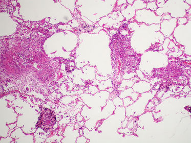
| TISSUE TYPE: alveolar spaces ... LUNG PATHOLOGY: abnormal tubercle between alveoli ... consist of epithelioid cells and langhan's gaint cells .. and caseation DIAGNOSIS: TB of lung N.B: differentiate it from pneumonia .. (solid pink masses between alveoli NOT dots IN alveoli..) | |
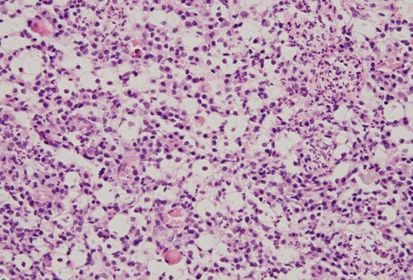
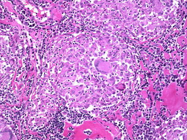
| TISSUE TYPE: lymphocytes every where ... lumph node PATHOLOGY: granuloma with NO caseation .. langhan's cells (with horse shoe arrangement of nuclei) are seen and epithelioid cells.. DIAGNOSIS: Sarcoidosis N.B: NO caseation in contrary to TB. special charecters : Schaumann and asteroid bodies (Don't even think about seeing them xD).. may be isolated in a question . | |
| TISSUE TYPE: by epith. which doesn't exist here xD ... respiratory .. or can be squamous due to metaplasia... NOSE PATHOLOGY: inflamatory cells of Mickulicz and Russel bodies in plasma cells (more eosinophilia) in lower part of section DIAGNOSIS: Rhinoscleroma KEY: RUSSEL BODIES | |
| TISSUE TYPE: u can see transitional epith. (not clear here) ... urinary bladder PATHOLOGY: black arrow: cystitis cystica red arrow: Brunn's nest of course u will see ova (not clear here) DIAGNOSIS: blihariziasis KEY: OVA N.B: differentaite between bladder and ureter Bilhariziasis .. just ureter will be complete circle ..(with same epith. changes) .. in intestine u can see ova or even the worm itself and no epith. changes .. squamous metaplasia can also be seen | |
| DEMO | ^^ |
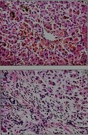
| TISSUE TYPE: unique architecture of live is preserved PATHOLOGY: brownish pigment is deposited in the sinuses DIAGNOSIS: obstructive jaundice KEY: BROWN.. | |
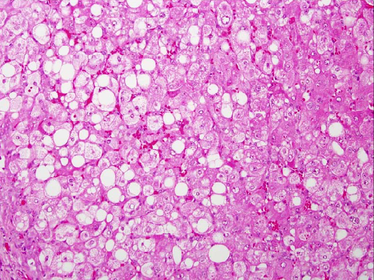
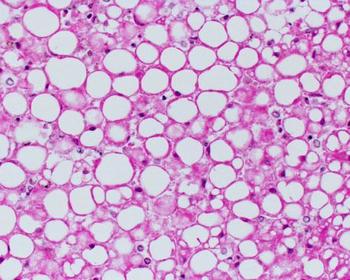
| TISSUE TYPE: preserved liver PATHOLOGY: vacuoles of fatty subestance DIAGNOSIS: liver steatosis KEY: VACUOLATION | |
| TISSUE TYPE: unique shape of atrium and ventricle .. heart PATHOLOGY: dystrophic calcification of valve DIAGNOSIS: choronic rheumatic vavulitis | |
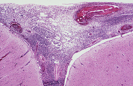
| TISSUE TYPE: Gyri and subarachinoid space... brain PATHOLOGY: inflamatory cells in subarachinoid space DIAGNOSIS: acute suppurative meningitis | |
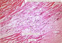
| TISSUE TYPE: periphrally we can see muscle fibrs .. HEART PATHOLOGY: centrally .. fibrosed area with some inflamatory cells DIAGNOSIS: myocardial infarction (healed by fibrosis) KEY: VIABLE PART OF MUSCLE AND FIBEROSED PART | |
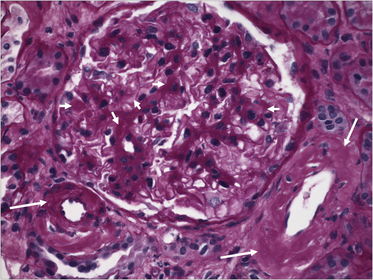
| TISSUE TYPE: glomeruli... kidney PATHOLOGY: depsition of foreign material in wall of capillaries .. DIAGNOSIS: Amyliodosis ( can be found in tubules ... afferent arterioles or in interlobular a.) N.B: Amyloidosis in kidney... sarcoidosis in lymph nodes.. | |
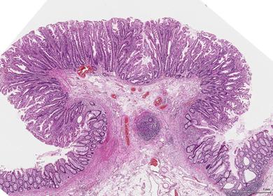
| TISSUE TYPE: villi of intestine PATHOLOGY: protrusion of mucosa and submucosa upwards.. bilharizial ova in sub. DIAGNOSIS : bilharizial intestinal polyp KEY: TREE LIKE PROTRUSION.. | |
| NOW TRY BY YOUR SELF ;) | .. |
| liver cirrhosis | |
| Fatty liver | |
| Acute lobar pneumonia | |
| Sarcoidosis | |
| TB in lung | |
| DONE ^_^ | " ما رميت اذ رميت و لكن الله رمى" ^^ |
Want to create your own Flashcards for free with GoConqr? Learn more.