4158583
Description
Flashcards by Vando Sousa, updated more than 1 year ago
|
|
Created by Vando Sousa
about 10 years ago
|
|
| Question | Answer |
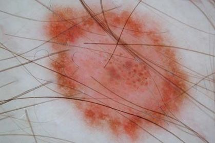
| Clinicamente - atipico Dermatoscopicamente - nevo comum Clinically this nevus reveals border irregularity and an asymmetric shape (A), while dermoscopy (B) shows a striking symmetry of colors (brown) and structures (reticular pattern). The red globule-like structure (arrow) is a small angioma in close vicinity to the nevus. | |
| Nevo comum. Dermoscopy of a nevus showing a multifocal pigment distribution characterized by small areas revealing dark and light brown color | |
| Nevo comum. Nevus showing dermoscopically eccentric hypopigmentation. This pigmentation type is quite infrequent, but it is important to differentiate it from an eccentric focus of regression. This can be facilitated by examining the surrounding normal skin. The structureless hypopigmented area will have the same or slightly darker hue as compared with the normal uninvolved skin. | |
| Nevo comum. Dermoscopy of a nevus showing central hypopigmentation. | |
| Nevo comum. Dermoscopy of a nevus showing central hyperpigmentation | |
| Nevo comum. Dermoscopy of a nevus revealing eccentric hyperpigmentation. Note that the pattern (in this case a reticular pattern) corresponding to the area of hyperpigmentation is the same as in the rest of the nevus | |
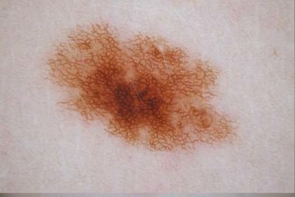
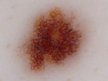
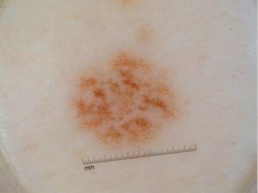
| Nevo comum. Dermoscopy of a reticular nevus reveals a regular network with regular network meshes and holes. The pigmented lines typically fade out at the periphery. | |
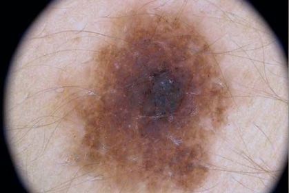
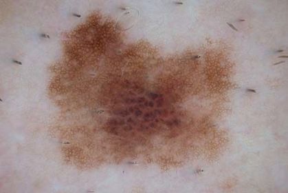
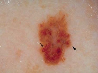
| Nevo comum. Classic appearance of a nevus of an individual with dark skin type (so-called black nevus) exhibiting dark brown and black color, reticular network, and central hyperpigmentation. The central hyperpigmentation is caused by a black lamella (arrow) corresponding histopathologically to pigmented parakeratosis. | |
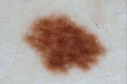
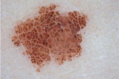
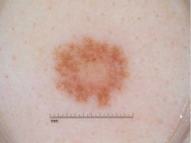
| Nevo comum. Dermoscopy of a nevus composed of brown to gray globular and cobblestone pattern. | |
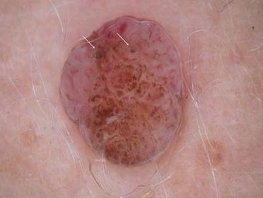
| Nevo comum intradermico. a striking asymmetry of colors and structures. The nonpigmented area reveals comma-like vessels of different size and diameter, and the pigmented area reveals a mixture of structureless homogenous pattern and residual globules and cobblestones. Terminal hairs or few comedo-like openings (arrows) are often present in such nevi | |
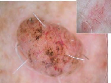
| Nevo comum intradermico. Only residual pigmentation appearing as irregularly distributed brown-to-gray blotches are visible. The short but large caliber vascular pattern (comma-like vessels) appears blurred, which is an important criterion to differentiate dermal nevi on the face from nodular basal cell carcinoma. In the latter, the vessels are typically elongated and sharply in focus | |
| Nevo comum. Globular–reticular nevi are composed of a central dermal component with globules surrounded by a regular reticular network | |
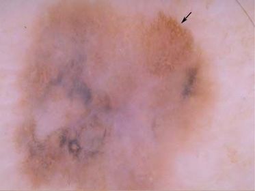
| Nevo comum. Example of globular-reticular nevus exhibiting in the center predominantly structureless hypopigmentation with a few globules | |
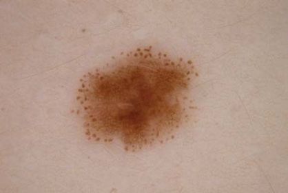
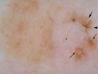
| Nevo comum. The peripheral rim of small brown globules is indicative for a growing nevus. | |
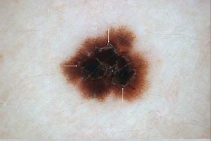
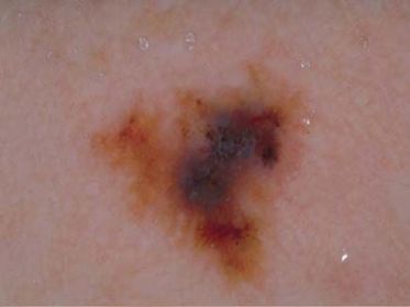
| Nevo atípico. features concerning for melanoma (asymmetric growth and blue-white veil). The lesion was excised and the pathology report was that of a dysplastic nevus | |
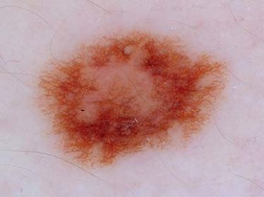
| Nevo atípico. Dermoscopy reveals a diffuse reticular pattern nevus. Dots overlying the network lines can be ignored. They represent small junctional nests located at the tips of rete ridges—a feature that speaks in favor of a benign nevus. | |
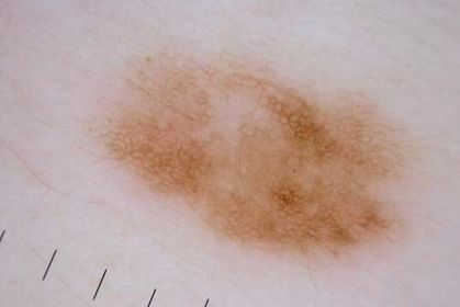
| Nevo atípico. Dermoscopy reveals a patchy network pattern nevus. | |
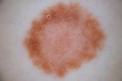
| Nevo atípico. This nevus has a pattern with peripheral network with central hypopigmentation. This pattern is commonly seen in fair skin individuals | |
| Nevo atípico. Dermoscopy reveals a nevus with peripheral reticulation (network) and central hypopigmentation. | |
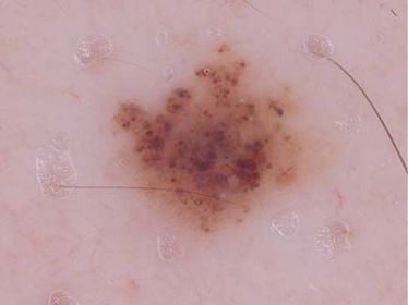
| Nevo atípico. Dermoscopy of this biopsy-proven dysplastic nevus reveals a globular pattern | |
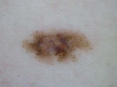
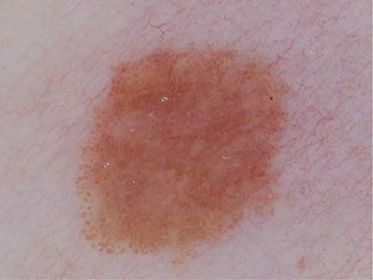
| Nevo atípico. This nevus has a homogenous pattern. | |
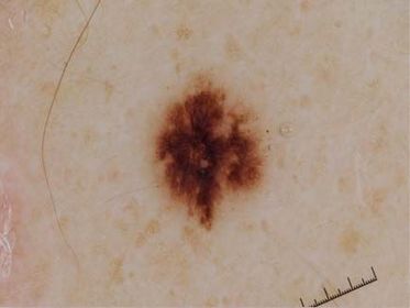
| Nevo atípico. Dermoscopy reveals a starburst pattern consisting of peripheral globules that are tiered, one on top of the other. Notice how this differs from the peripheral globular pattern shown in Figure 7b.19 where only one rim of globules are present. This tiered globular patter can be seen in Spitz nevi and in DN with spitzoid features. This lesion was biopsied and the pathology was consistent with a dysplastic nevus with spitzoid features. | |
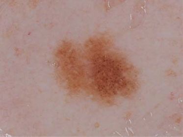
| Nevo atípico. Dermoscopy reveals a two- component pattern nevus | |
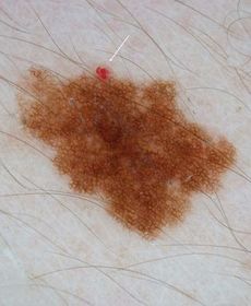
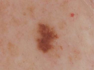
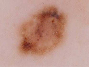
| Nevo atípico. This nevus has a relatively symmetric multicomponent pattern. It has network, blotches, and a few globules. It is clinically often diffi cult to rule out melanoma in lesions with this pattern. If this is a clinical or dermoscopic outlier lesion then a biopsy would be the prudent management decision. However, if the pattern in this nevus is similar to the patient’s other nevi then monitoring would be an acceptable alternative to biopsy | |
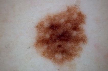
| Nevo atípico. Dermoscopy reveals a symmetric multicomponent pattern. This acquired lesion has network, globules, homogenous areas, and blue-white veil. The differential diagnosis for this lesion is between dysplastic nevus and melanoma. Excisional biopsy and pathology proved that this was a dysplastic nevus. | |
| This is an asymmetrically patterned multicolored lesion. It lacks symmetry of pattern over every axis through the center of the lesion (see chap. 7b) and has more than one color; scored from tan, dark brown, gray, black, blue, and red. It has the positive melanoma features of a broadened atypical network (thick arrow), and the pseudopod (thin arrow), and radial streaming (red arrow) are very subtle. Note the peripheral light brown structureless area occupying more than 10% of the lesion surface from 3 to 7 o’clock in position. Diagnosis: in situ superfi cial spreading melanoma. | |
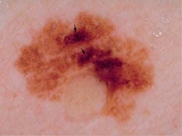
| This asymmetrically patterned multicolored lesion has the positive melanoma feature of broadened atypical network (arrows). Broadened network is usually seen focally rather than in a uniform distribution. It is probably the most important (sensitive) feature of early nonregressing superfi cial spreading melanoma (SSM). Note the blue pigmentation formed by confl uent melanophages in the dermis. Diagnosis: in situ SSM. | |
| This asymmetrically patterned multicolored lesion has the one positive melanoma feature of focal broadened network (arrows). Note the amelanotic contiguous nevus. Diagnosis: superfi cial spreading melanoma, Breslow thickness 0.35 mm with a contiguous compound nevus | |
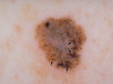
| This asymmetrically patterned multicolored lesion (dark brown, tan, and gray) has the positive features of focal broadened network (thin arrow) and subtle radial streaming (large arrow). Diagnosis: superfi cial spreading melanoma, Breslow thickness 0.35 mm. | |
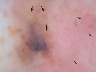
| This asymmetrically patterned multicolored lesion has the positive features of scar-like depigmentation, blue-white veil, and focal broadened network (arrow). In addition, note the widespread peripheral light brown structureless areas occupying more than 10% of the lesion surface. Diagnosis: regressing superfi cial spreading melanoma, Breslow thickness 0.4 mm. | |
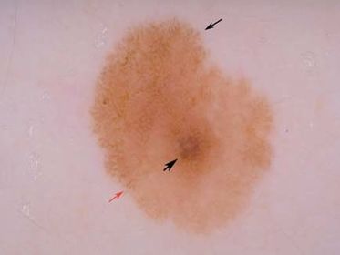
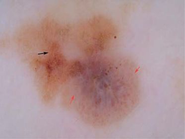
| This asymmetrically patterned multicolored lesion has the positive melanoma features of multiple brown dots, found in the classical focal distribution (thin arrows), radial streaming (thick arrows), and multiple blue-gray dots (asterisk). Note the contiguous banal nevus at the left half of the lesion. Diagnosis: regressing superfi cial spreading melanoma, Breslow thickness 0.45 mm with a contiguous compound nevus | |
| This melanoma has the positive feature of multiple (5–6) colors. Diagnosis: superfi cial spreading melanoma, Breslow thickness 0.55 mm. | |
| This hypomelanotic asymmetrically patterned multicolored lesion has the positive features of extensive scar-like depigmentation and blue-white veil. Note the pin-point (dotted) vessels within the amelanotic component (small arrow). Also note the negative pigment network (large arrow), which is a signifi cant fi nding in both Spitz nevi and invasive melanoma (Menzies et al., 1996b). Diagnosis: superfi - cial spreading melanoma, Breslow thickness 0.7 mm. | |
| This amelanotic lesion has some linear irregular and arborising vessels. Because of overlap between amelanotic melanoma and other malignant amelanotic lesions, it is easier to distinguish all malignant amelanotic/hypomelanotic lesions from all benign lesions rather than to attempt to discriminate melanoma from other malignant lesions, such as basal cell carcinoma. This illustrates the importance of biopsy prior to any nonsurgical treatment of amelanotic lesions. Diagnosis: amelanotic superfi cial spreading Figure 8a.16a Clinical image. melanoma, Breslow thickness 0.8 mm. | |
| This asymmetrically patterned multicolored SSM has the positive melanoma features of focal multiple brown dots (arrows) and small areas of blue-white veil. Note also the extensive negative pigment network (red arrow). Diagnosis: SSM, Breslow thickness 1.2 mm with associated compound nevus. Abbreviation: SSM, superfi cial spreading melanoma. |
Want to create your own Flashcards for free with GoConqr? Learn more.