4379938
Description
Flashcards by Rachael Jones, updated more than 1 year ago
|
|
Created by Rachael Jones
almost 10 years ago
|
|
| Question | Answer |
| Name the 4 locational terms for the brain (above/below/in front/behind) | Dorsal, Ventral, Anterior/Rostral, Posterior/Caudal. |
| Define Medial and Lateral. | Medial - closer to the centre. Lateral - further from centre. |
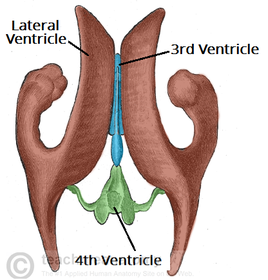
| How many ventricles are part of the Ventricular System. (describe anatomy) | 4: 2 lateral ventricles, the 3rd and the 4th. *hollow cavities that contain cerebrospinal fluid |
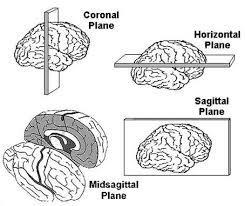
| Name the three techniques of slicing the brain. | Horizontal, Sagittal and Frontal (creating a horizontal, Sagittal and Transverse slice). |
| What is a Myelin? (for example the corpus callosum). | A fatty white substance that surrounds the axon of some nerve cells forming an electrically insulating layer. (axons are nerve fibres- carry Impulses between nerve cells/neurons). |
| What is grey matter? | cell bodies and dendrites e.g. cortex, basal, ganglia, thalamus. |
| What is white matter? | Myelinated axons e.g. the corpus callosum. |
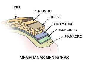
| What are meninges? | 3 layers of tissue that protect the brain and spinal cord. |
| What is the function of Cerebrospinal Fluid (CSF). | clear liquid that fills the subarachnoid space that functions as a shock absorber/buoyancy. |
| How do the capillaries in the brain differ from the capillaries of the rest of the body? (blood-brain barrier) | capillaries in the brain do not have any 'gaps', so only soluble substances like glucose can travel through. |
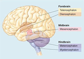
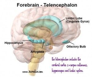
| Name the main structures of the Fore-Brain (Prosencephalon) (2). | The Telencephalon and the Diencephalon. |
| Name the main structures of the Mid-Brain(2). | The Tectum and the Tegmentum. |
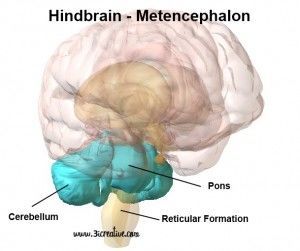
| Name the main structures of the Hind-Brain (Rhombencephalon) (2). | The Metencephalon and the Myencephalon. |
| Define Gyri and Sulci. | (*knuckle trick)- gyri=bump, sulci=groove. |
| Name the major nuclei of the Telencephalon(3) | Limbic system, Basal Ganglia (and obs the Cerebral Cortex). |
| Name the major nuclei of the Diencephalon (2). | The Thalamus and the Hypothalamus. |
| Name the 2 major structures of the Metencephalon. | The Cerebellum and the Pons. |
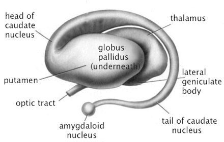
| Name the 3 major structures of the Basal Ganglia. | 1.Caudate nucleus (nucleus with a tail) 2.Putamen (pit/stone like fruit) 3. Globus Pallidus (a plate for the putamen) |
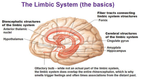
| Name the 5 major structures of the limbic system. | 1. Limbic Cortex 2. The Hippocampus 3. The Amygdala 4. The Fornix 5. Mammillary Bodies |
| Function of the Basal Ganglia? | Important for movement/rewards system. |
| Function of Limbic System? | Important for emotion and learning/memory. |
| Function of the Thalamus and the Hypothalamus? | Thalamus- receives information and relays back to the cortex. Hypothalamus- controls autonomic nervous system& fight or flight functions. *also responsible for digestion and other unnoticed functions. Important for generating emotional response & has been associated with aggressive behaviour. |
| Functions of the Tectum? | The 'Superior colli-culi' relates to vision, and the 'Inferior Colli-culi' relates to hearing. |
| Functions of the Tegmentum? | net-like structure that protects other surrounding structures such as the cerebral aquaduct. |
| Functions of the Pons and Cerebellum (2 parts of the Metencephalon). | Pons- sleep and arousal, relays info to and from cortex. Cerebellum- co-ordination of movement. |
| What are neurons? - and 3 different types of neuron? | Specialised nerve cells that transmit impulses within nervous system. They do all information processing/transmitting. |
| Between structures- define Afferent and Efferent. | Afferent(ARRIVE) - brings info into a structure. Efferent(EXIT) - takes info away from a structure. |
| Describe Glia (including Astrocytes, Ogliodendrocytes, and Microglia). | non-neuronal cells that: maintain homeostasis, form myelin, & provide support/protection for neurons in the CNS/PNS. They also supply nutrients/oxygen to neurons, destroy pathogens, and remove dead neurons. |
| Define membrane potential. | the difference in electrical potential inside and outside the cell. MP is balanced by: 'diffusion', and 'electrostatic pressure'. |
| What is an ion channel? | A membrane spanning protein that gates the flow of ions across the cell membrane. |
| Define ions, and 2 types of ion. | Ions are electrically charged molecules. - Cations = positively charged. - Anions = negatively charged. |
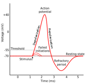
| Describe resting potential. | When the inside of a neuron is 70Mv less than the outside. The K+ inside wants to move outside, and the Na+ and Cl- want to move inside (electrostatic level-). |
| Describe action potential. | neurons undergo quick rapid changes in polarization, this is how neurons send electrical signals. |
| Define depolarisation and hyperpolarisation. | D- increase from normal resting potential (brings membrane closer to 0). H- decrease relative to resting potential (more negative voltage). |
| The stages of Action Potential (changes in polarisation). | 1.Threshold excitation reached, Na+ channels open, Na+ begins enter cell (MP rises towards 0). 2.K+ channels open, K+ begins to leave cell(increase slows). 3.Na+ channels become refractory (difficult), no more Na+ enters cell. (peak of action potential) 4.K+ continues leave cell, causes membrane potential return to resting. 5.K+ channels close, Na+ channels reset. 6.Extra K+ outside diffuses away. |
| Define Saltatory Conduction. | the propogation of action potentials along myelinated axons from inbetween nodes(gaps) increasing the conduction velocity of action potentials. *more energy efficient *fast conduction |
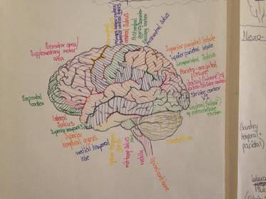
| The 'groove' that separates the temporal and frontal lobes? | The lateral fissure. |
| The 'groove' that separates the Parietal and Occipatal lobes? | Parieto-occipital sulcus. |
| The 'groove' that separates the Frontal and the Parietal lobes? | The Central Sulcus. |
| Tripartite Synapse? | Glia are not merely passive neuronal support cells, but instead play an active role by removing NT's leftover in synaptic gap/cleft. |
| What are Ionotropic receptors & Metabotropic receptors. | Both receptors contain a binding site. Ionotropic receptors open the ion channel when a molecule attaches to the binding site. Metabotropic receptors initiate a chain reaction that eventually opens ion channels. |
| Define 'Excitatory Neural Integration' and 'Inhibitory Neural Integration'. | Excitatory increases the likelihood of a neuron firing. |
| What are amino acids? | Simple organic compounds that contain both a Carboxyl (-COOH) and an Amino (-NH^2) group. Are also able to transmit a nerve message across a synapse. |
| What do the poisons Curare and Muscarine do? | Curare prevents muscle contraction by blocking the action of Acetycholine (ACh), an agonist. muscarine imitates the action of ACh (agonist) |
| What is Acetycholine (ACh) | functions as a NT at neuromuscular junctions (muscular contraction). |
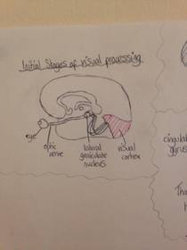
| V1 visual processing is mediated by 2 streams: | - Dorsal (visuo-spatial= 'where'/ visuo motor= 'how') - Ventral (object analysis= 'what') *lesions to posterior parietal impairs object location (where) but not discrimination (what) *lesions to inferior temporal lobe lesions in macaques impair object discrimination ('what') but NOT location. |
| Milner & Goodale * patients with occipito-temporal brain damage | - patients with occipito-temporal brain damage show severe forms of visual agnosia (deficits in aspects of visual perception without blindness) BUT intact visually guided actions. - patients with posterior-parietal lobe lesions show optic ataxia (deficits in visually guided reaching) with otherwise intact visual function. |
| Describe Face Cells | Some neurons in inferior temporal lobe show highly selective responses to individual faces. This suggests that neurons may act as 'gnostic units' that recognise individual entities. |
| Describe the Medial Temporal Lobe (MTL) | at the end of visual processing hierarchy combines inputs from ventral and dorsal stream, and requires additional inputs from other sensory modalities. It elaborates visual representations further and generates multi-modal representations. |
| Describe the function of the Amygdala. | important for fear & aggression. |
| Describe Kluver-Bucy syndrome | monkeys with temporal lobes removal= absence of fear/aggression, hypersexuality, visual recognition problems, oral tendencies shows that Amygdala is critical for changes in emotional states. |
| Describe the Ventromedial Prefrontal Cortex (orbitofrontal cortex) | important for emotional feeling, social interactions and decision making (Phineas Gage, 1848) |
| The Parietal Lobes split into 3 main parts: | Somatosensory cortex and the somatosensory association cortex (superior parietal lobule & inferior parietal lobule). |
| Describe the main role of the somatosensory cortex | processing information about the body (somatic sensations) i.e. pressure, pain, temperature, sensation of muscle movement/position. Sensations of different areas are localised in specific areas of SC. -INPUT: spinal cord> Thalamus> SC -OUTPUT: motor cortex(frontal L)> posterior parietal areas |
| Describe plasticity and phantom limbs | patients with arm amputations feel sensation of the missing arm. Cells in arm region do not receive any input neighbouring cells expand into territory so when touching the face activation spreads into the 'arm' region of the SC (brain interprets this as sensations coming from the arm. |
| Describe the role of the superior parietal lobule | elaboration and integration of sensory information from the somatosensory cortex (maintains representation of the body state that is used during voluntary/intentional limb movement). *also has a role in controlling attention. |
| Describe the role of the inferior parietal lobule | multi-modal sensory integration (1. somatic sensation, 2. visual info, 3. auditory information & CALCULATION of language. *sense of self (self/other distinction) |
| Damage to the left posterior inferior parietal lobule? | causes language impairment including expressive aphasia (production deficit and non-fluent) and receptive aphasia (comprehension deficit and 'fluent but wrong words') |
| Differences between Unilateral and Bilateral damage to the posterior parietal lobules. | Unilateral- Hemispatial neglect Bilateral- Balint's Syndrome involves: *Optic Ataxia(difficulties looking/reaching) *Ocular Apraxia (visual motor disturbance/gaze) *Simultanagnosia (difficulties seeing more than one object at once) |
| What 3 landmark areas can the Frontal lobe be split into? | - Prefrontal cortex - Pre-motor area/supplementary motor area - Primary motor cortex |
| Describe the function of the primary motor cortex | -controls voluntary movement (similar organisation into map -*Input from somatosensory cortex, premotor and supplementary areas -*output to the somatosensory cortex and spinal cord |
| Describe function of Premotor area/ Supplementary motor area | planning of complex movement sequences, selection of appropriate actions (intentions) |
| Describe function of Prefrontal Cortex | - is connected to all sensory & motor areas and exerts a 'top-down' influence into these areas. - cognitive control (WM, problem solving etc.), Emotion, Language, and Decision-Making, |
| Properties of Schizophrenia | - affects approx. 1% of world's population - persecution, poverty of speech, social withdrawal, difficulty sustaining attention, anhedonia |
| Describe the Dopamine Hypothesis (Pharmacology of Schizophrenia) | Dopamine antagonists diminish positive symptoms (and agonists induce +symptoms)- shows that symptoms are caused by over-activity of neurons. |
| What is Anhedonia? | an inability to feel pleasure in normally pleasurable activities |
| Describe Bipolar Disorder and Unipolar Disorder | - (B) alternating periods of mania and depression, 1% of population are afflicted at some point in their life, equally as frequent in men and women - (U) depression without mania |
| Rosenthal (1971) genetic inheritance | 10 times more likely to suffer from an affective disorder if a close relative does. |
| Gershan et al. (1976) (Affective Disorders) | - Mz concordance = 69% - Dz concordance = 13% |
| Monoamine Hypothesis (pharmacology of Affective disorders) | - depression is caused by low activity of MonoAmine neurons. Lithium is often used as treatment. *- MOA enzyme destroys MA like Serotonin, Dopamine, Norepinephrine - Iproniazid inhibits MOA thus higher level of MA's |
| Broca's area & Broca's Aphasia | -Broca's area = anterior speech region -Broca's Aphasia = syndrome that results from damage to Broca's area, can't articulate the words but CAN write them down, so non-fluent spontaneous speech and good comprehension (so is down to the production of speech) |
| Wernicke's area and Wernicke's Aphasia | area of the cortex that receives information from the ear behind Broca's area, patients spoke fluently but with no sense, they could hear but could NOT understand WHAT was said to them. (fluent spontaneous speech and poor comprehension). |
| A model of language processing? | 1. Auditory information sent to Wernicke's area (sounds to sound images) 2. Sound images are transmitted along the 'Arcuate fibres' to BA 3. Information is sent from Broca's area (representative of speech movements) to control the mouth muscles. |
| Conduction Aphasia | when Arcuate fibres are damaged, comprehension is maintained (and speech sounds) but speech is impaired (difficulty repeating what is said to them). |
| Define types of memory: episodic, semantic, working, procedural, & perceptual. | -(E) specific times/events -(S) facts -(W) short term rehearsal -(Pr) motor (skills + habits) -(Pe) familiarity with stimuli |
| Memory described as Declarative and Non-Declarative | -(D) episodic and semantic memory -(ND) perceptual and procedural |
| Describe Korsakoff's syndrome | Thiamine deficiency due to alcoholism/poor diet/& impaired absorption of thiamine from intestine. This all produces bilateral degeneration of mammillary bodies. |
| What is a Temporal Lobectomy | a bilateral removal of temporal lobes for patients with uncontrollable seizures. |
| The role of the hippocampus | enables consolidation of new memories which are stored elsewhere (does not store) |
| The hypothalamus' role in sleep? | -studied the brains of those who had died from the virus 'Encephalitis Lethargica' (headaches + drowsiness leading to a coma). -those who had difficulty sleeping often had damage to the anterior region of H -those who had difficulty staying awake had damage to the posterior region of H |
| The role of the Reticular system in sleep - Mouzzi & Morgan (1949) | The reticular formation (raphe-nuclei, magnocellular nuclei, parvocellular nuclei) connects the spinal cord/ cerebrum/ cerebellum is part of the Reticular Activating System (RAS) that is responsible for regulating wakefulness & sleep-wake transitions. M&M found that stimulating the reticular formation in sleeping cats' brains caused them to wake up. |
| Hetherington & Ranson (1940) (ventral medial H) | lesion to the ventral medial hypothalamus results in hyperphagia (over-eating & obesity) *a month or 2 after destruction a rat recovers spontaneous eating. |
| Anand & Brobeck (1951) (lateral H) | *bilateral lesions to the lateral hypothalamus leads to Aphagia (cessation of eating) *Lateral Hypothalamus syndrome: aphagia and adipsia (cessation of drinking) *lesions to the lateral hypothalamus also produces a variety of motor disturbances, and a lack of responsiveness. |
| What is a sodium-potassium pump? | the process of moving sodium and potassium ions across the cell membrane as an active transport process. |
| What is repolarisation? | the change in membrane potential that returns it to a negative value just after the de-polarisation phase of an action potential has changed the membrane potential to a positive value. |
| How will people with frontal lobe damage perform in the stroop task? | *responsible for planning future actions- they cannot ignore the word and read this instead of the ink colour. |
| Describe EEG patterns through stages 1-4. | increases in amplitude and decreases in frequency |
| What are simple cells? | cells in the primary visual cortex that responds primarily to orientated edges and gratings. *has an elongated receptive field and responds best to a stationary line in the correct orientation. |
| Describe the fluctuation of voltage during action potentials | |
| Describe what happens as an action potential reaches the pre-synaptic terminal. | 1. depolarisation of pre-synaptic terminal causes opening of voltage-gated Ca^2+ channels 2. influx of Ca^2+ through channels 3. Ca^2+ causes vesicles to fuse with pre-synaptic membrane 4. NT released into cleft via exocytosis 5. NT binds to receptor molecules in postsynaptic membrane 6. opening or closing of post-synaptic channels 7. postsynaptic current causes excitatory or inhibitory postsynaptic potential that changes the excitability of postsynaptic cell 8. removal of NT by glial uptake of enzymatic degradation 9. retrieval of vesicular membrane from plasma membrane |
| what is the most abundant excitatory NT in the CNS. | Glutamate, it can bind to a number of receptors (involved in learning and memory) *always excites the postsynaptic cell |
| what is the most abundant inhibitory NT in the CNS | GABA |
| Bechara et al. (2000) Iowa gambling task | found that patients with lesions to the ventromedial prefrontal cortex performed poorly on the Iowa gambling task. |
| What is the function of the ventricular system? | It allows the exchange of materials between blood vessels and brain tissue . |
| Define: *a commissure *retino-topic map *V1 modules | *a pathway that connects the left and right sides of the hemisphere. *an orderly mapping of retina/ visual field onto the visual cortex. *V1 is divided into small columnar modules that combine neurons sensitive to different aspects of stimuli presented in a small part of the visual field. |
| *What parts of the brain constitute the visual association cortex? *auditory association cortex? | *the inferior temporal lobe and occipital lobe. *the superior temporal lobe. |
| What components are part of the Papez circuit? | * Neocortex * Fornix * Cingulate Gyrus * Thalamus * Hypothalamus * Hippocampus |
| Brodmann's areas? | a map of psychological functions across the brain. |
| What is the Insula (part of the brain) responsible for? | Is important for the experience of pain and several basic emotions (especially disgust), also has an important role in translating visceral states into subjective feelings. |
| Joseph LeDoux states that...(emotional processing) | there are 2 routes of emotion responses: *emotional stimulus> thalamus> cortex> amygdala> emotional response. (INDIRECT) *stimulus> thalamus> amygdala> emotional response. (DIRECT) |
Want to create your own Flashcards for free with GoConqr? Learn more.