5031023
Description

Flashcards by Hayley Pfeffer, updated more than 1 year ago
|
|
Created by Hayley Pfeffer
over 9 years ago
|
|
| Question | Answer |
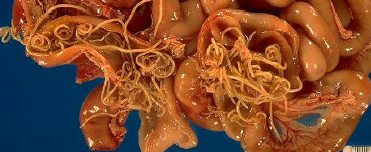
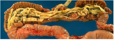
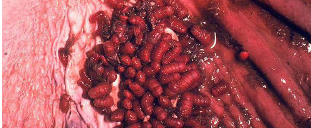
| Toxocara canis | |
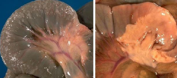
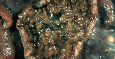
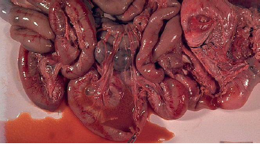
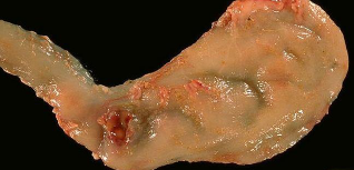
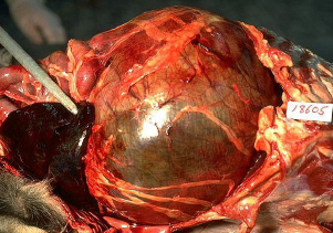
| small intestinal carcinoma | |
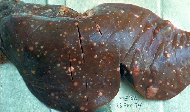
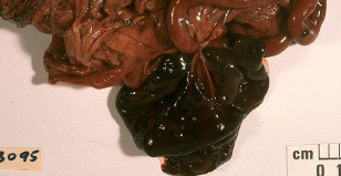
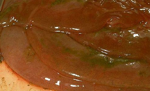
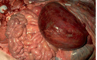
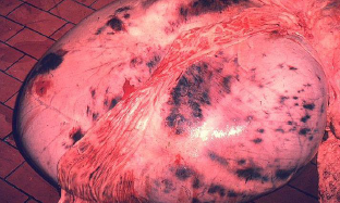
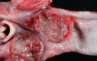
| Sheep liver with small intestinal carcinomas | |
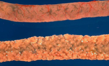
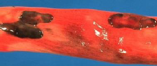
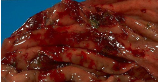
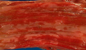
| hemorrhage in colon due to cyathostmonis | |
| Moniezia expasna | |
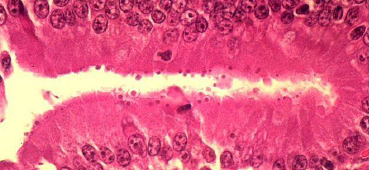
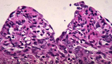
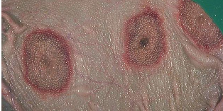
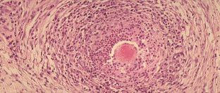
| Cryptosporidia on luminal surface of crypt | |
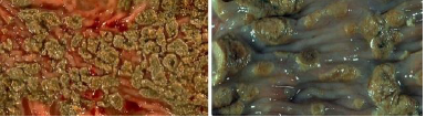
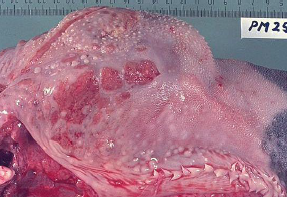
| Mulitfocal nodules due to coccidiosis in the S.I | |
| Congealed blood. PHE form of PPE (porcine proliferative enteritis) | |
| Necrotic mucosa NE form of PPE | |
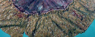
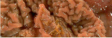
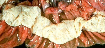
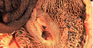
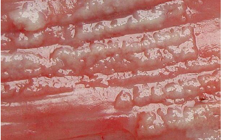
| Cerebriform appearance of thickened mucosa PIA form of PPE | |
| Cerebiform appearance PPE | |
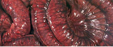
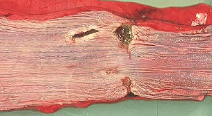
| Thickened ileum mucosa Johnes | |
| cerebiform pattern (left) Nodular lymhangitis (right Johnes | |
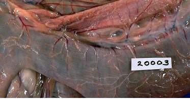
| prominent serosal lymphatics typical of Johnes | |
| Prominent lymphatics coursing from intestine to enlarged LN Johnes | |
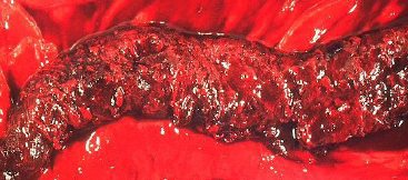
| Marked haemorrhagic gastrocenteritis due to yersiniosis in deer | |
| Button ulcers on colon mucosa Classical swine fever or salmonella | |
| A fibrinous pseudomembrane cow with salmonella | |
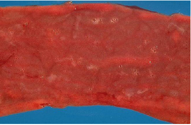
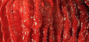
| catarrhal enteritis | |
| button ulcers in mucosa Salmonella or classical swine fever | |
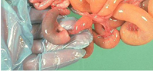
| Patchy hyperemia of the intestinal serosa with some fibrin strands Parvo puppy | |
| Red fluid intestinal contents in puppy with parvo | |
| Numerous small granulomas on the serosal surface of the gut FIP cat | |
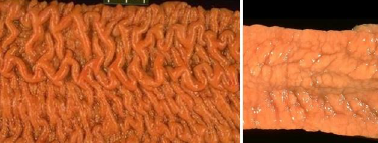
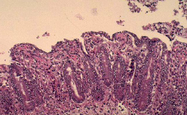
| Severe villus atrophy and hyperplasia of crypt epithelial cells Cornaviru enteritis | |
| Piglet with transmissible gastroenteritis Stunted villi covered squamous epithelial cells | |
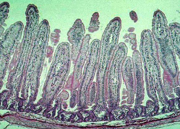
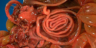
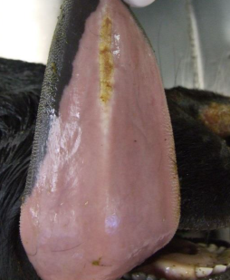
| normal mucosa | |
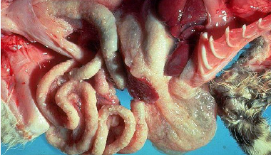
| haemomelsama ilei in the ileum Caused by migrating strongulus | |
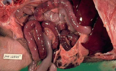
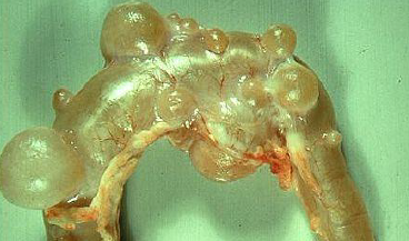
| Intestinal diverticulosis in a sheep | |
| Intussception | |
| Intussception | |
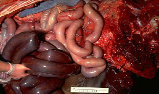
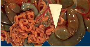
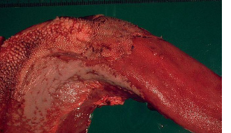
| caecum of horse following torsion | |
| Torsion and volvulus causing colic in a horse | |
| mesenteric vovulus | |
| Strangulation of a portion of bowel | |
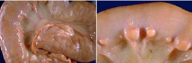
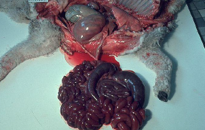
| Spiral colon atresia | |
| Small intestinal atresia | |
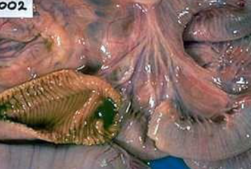
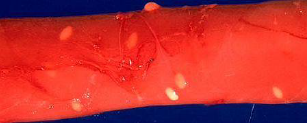
| haemonchus contortus in the abomasum | |
| Gastrophilus spp. in stomach of horse | |
| gastric pyloric ulcer in a dog | |
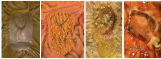
| pars oesophagia (left to right) normal, hyperkeratosis, early ulceration, total ulceration and haemorrhage | |
| Severe abomastitis due to ostertagiasis and secondary fusobacterium infection | |
| Acute abomastitis due to salmonellosis | |
| oedema of abomasal folds in case of chronic haemonchosis | |
| reddened areas of infarction | |
| Hyperaemia in acute abomastitis | |
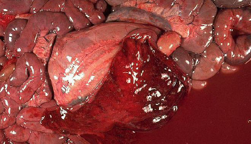
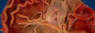
| volvulus of the abomasum in a calf | |
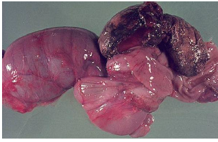
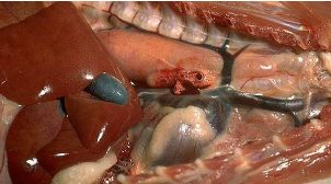
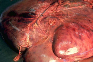
| gastric dilation and volvulus Congested spleen | |
| Wire embedded in reticulum Traumatic reticulopericarditis | |
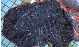
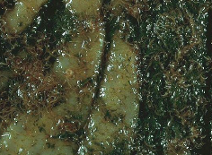
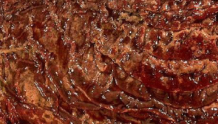
| Rumen mucosal infarcts due to zygomycosis | |
| serosal surface of rumen of a cow with numerous haemorrhagic infarcts due to zygomycosis secondary to acidosis | |
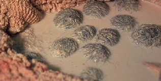
| bezoars in rumen | |
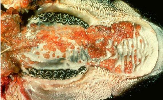
| sarcocystis gigantea | |
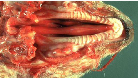
| megaoesphagus | |
| injury of oesophagus mucosa following pressure necrosis | |
| Superficial inflammation of oesophagous due to candidiasis (thrush) | |
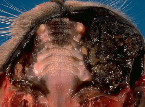
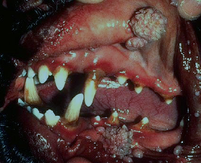
| Pyogranuloma in woddy tongue contains a club colony-aka sulphur granule | |
| eroded hard nodules bovine with woody tongue | |
| oral necrobacilliosis | |
| Severe bovine papular stomatitis | |
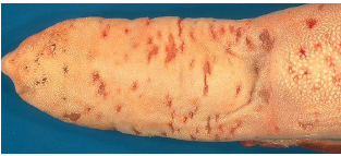
| Circular to horse shoe shapped papules calf with papular stomatitis | |
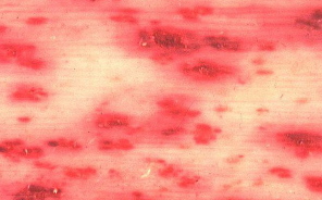
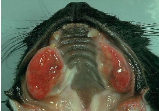
| petechaie on dental pad and erosions on lips of sheep with bluetongue | |
| Serosal haemorrhages MCF | |
| large coalescing erosions in the mucosa (MCF) | |
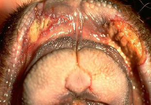
| severely eroded tongue (MCF) | |
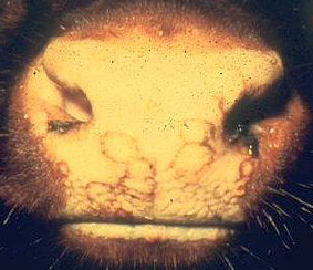
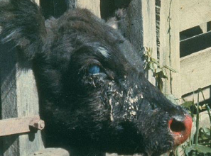
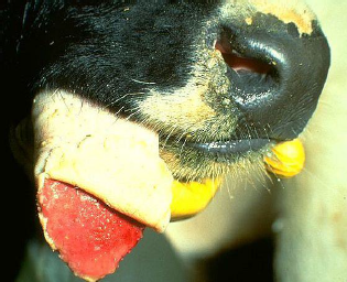
| Keratitis, occular discharge, inflammed nasolabium in MCF | |
| Oesophagus mucosa erosions from BVD or MD | |
| Erosions due to BVD or MD | |
| Enamel hypoplasia due to BVD | |
| FMD | |
| FMD | |
| oral melanoma | |
| fibrosarcoma | |
| SCC | |
| oral papilloma in dog | |
| photosensitization lesion | |
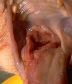
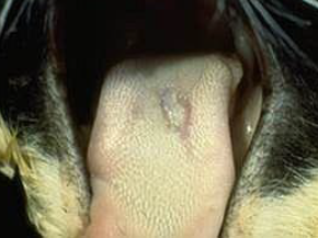
| glossitis | |
| ulcerative stomatitis | |
| Eosinipholic granuloma | |
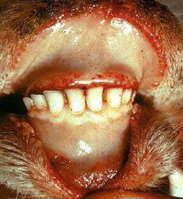
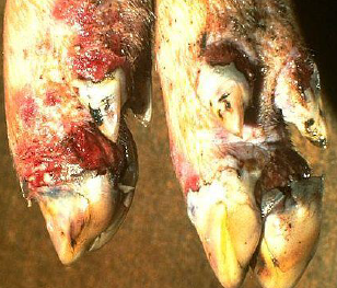
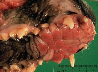
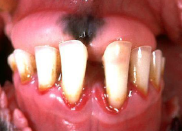
| Incisor peridontitis | |
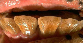
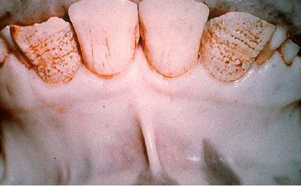
| hypoplasia of enamel | |
| cleft palate |
Want to create your own Flashcards for free with GoConqr? Learn more.