7277363
Description
Flashcards by Liam Musselbrook, updated more than 1 year ago
|
|
Created by Liam Musselbrook
over 7 years ago
|
|
| Question | Answer |
| What type of visual loss is caused by age-related macular degeneration (AMD)? | Progressive central visual loss |
| What are the risk factors for AMD? | Age Smoking Hypertension Hypercholesterolaemia UV exposure |
| What are the features of dry AMD? | Characterised by drusen - yellow round spots in Bruch's membrane Gradual deterioration of VA Central scotoma Focal RPE hyperpigmentation RPE (geographic) atrophy in advanced AMD |
| What are the features of wet AMD? | Characterised by choroidal neovascularisation Rapid vision loss and development of scotomas Metamorphosia (distortion of vision) Degeneration and localised detachment of RPE Fibrous scarring (end-stage) |
| Investigations for age-related macular degeneration | Optical coherence tomography: provide cross-sectional views of the macula Fluorescein angiography if neovascularisation is present |
| Management of age-related macular degeneration | General - refer for opthalmological assessment within a week, stop smoking, high dose of beta-carotene, vitamins C and E, and zinc Dry - no treatments Wet: - photocoagulation - photodynamic therapy - anti-vascular endothelial growth factor (anti-VEGF) treatments: intravitreal ranibizumab |
| What signs are seen in mild non-proliferative diabetic retinopathy (NPDR)? | 1 or more microaneurysm |
| What signs are seen in moderate non-proliferative diabetic retinopathy (NPDR)? | Microaneurysms Blot haemorrhages Hard exudates Cotton wool spots, venous beading/looping and intraretinal microvascular abnormalities (IRMA) less severe than in severe NPDR |
| What signs are seen in severe non-proliferative diabetic retinopathy (NPDR)? | Blot haemorrhages and microaneurysms in 4 quadrants Venous beading in at least 2 quadrants IRMA in at least 1 quadrant |
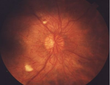
| Proliferative DR | Retinal neovascularisation - may lead to vitrous haemorrhage Fibrous tissue forming anterior to retinal disc More common in Type I DM, 50% blind in 5 years |
| What are the complications of proliferative DR? | Vitreous haemorrhage Rubeotic glaucoma Retinal fibrosis -> tractional retinal detachment |
| Diabetic maculopathy | Based on location rather than severity Anything is potentially serious Hard exudates and other 'background' Changes on macula Check visual acuity More common in Type II DM |
| Diabetic maculopathy: Focal | Leakage from capillaries in one part of the macula Leads to retinal thickening and surrounding exudates (circinates) |
| Diabetic maculopathy: Diffuse | Diffuse retinal oedema from dilated capillaries, may be associated with haemorrhages but rarely exudates |
| Diabetic maculopathy: Ischaemic | Closure of foveal capillary networks -> Diffuse oedema and dark haemorrhage Confirm with flourescein angiography |
| Management of diabetic retinopathy and maculopathy | Control of diabetes and risk factors Pan-retinal photocoagulation in PDR Focal/grid photocoagulation in maculopathy Anti-VEGF agents in combination with photocoagulation for maculopathy Vitrectomy if persistent vitreous haemorrhage |
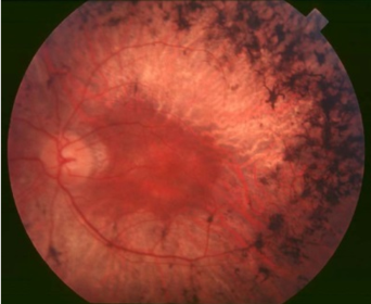
| What does this show? | Retinitis pigmentosa |
| What are the features of retinitis pigmentosa? | Night blindness Tunnel vision Fundoscopy: black bone spicule-shaped pigmentation in the peripheral retina |
| What does this show? | Proliferative retinopathy |
Want to create your own Flashcards for free with GoConqr? Learn more.