9258420
Description
Flashcards by Noah Bryan, updated more than 1 year ago
|
|
Created by Noah Bryan
almost 7 years ago
|
|
| Question | Answer |
| Practical Skills (x4) | Planning Implementing Analysis Evaluation |
| Planning | 1) Experimental Design, including to solve problems set in context of the question. 2) Identification of Variables that must be controlled (where appropriate) 3) Evaluation that an experimental method is appropriate to meet the expected outcomes |
| Implementation | 1) How to use a wide range of practical apparatus and techniques correctly 2) Appropriate units for measurements. 3) Presenting observations and data in an appropriate format. |
| Analysis | 1) Processing, analysing and interpreting qualitative and quantitative experimental results 2) Use of appropriate mathematical skills for analysis of quantitative data 3) Appropriate use of significant figures. d)Plotting and interpreting suitable graphs from experimental results, including (i) Selection and labelling of axes with appropriate scales, quantities and units (ii) Measurements of gradients and intercepts. |
| Evaluation | 1) How to evaluate results and draw conclusions 2) The identification of anomalies in experimental measurements 3) The limitations in experimental procedures 4) Precision and Accuracy of measurements and data, including margins of error, percentage errors and uncertainties in apparatus. 5) The refining of experimental design by suggestion of improvements to the procedures and apparatus. |
| Microscopy (PAG 1) | ... |
| Preparation and Examination of Microscope Slides (x4) | 1) Dry Mount 2) Wet Mount 3) Squash Slides 4) Smear Slides |
| Dry Mount | Solid specimens are viewed whole or cut into thin slices (sectioning). The specimen is placed in the centre of the slide and a cover slip is placed over the sample (ie. hair/dust - whole; muscle/plant tissue - sectioned) |
| Wet Mount | Specimens are suspended in a liquid such as water or immersion oil. A cover slip is placed on from angle. (ie. Aquatic organism) |
| Squash Slides | A wet mount is first prepared, then a lens tissue is used to gently press down on the cover slip. Depending on the material, potential damage to the cover slip may be avoided by squashing the sample between two microscope slide. (ie. soft tissue/ root tips - cell division) |
| Smear Slides | The edge of the slide is used to smear the sample, creating a thin, evenly distributed coating on another slide.A cover slip is then placed over the sample. (ie. blood cells in a droplet of blood) |
| Staining (PAG 1) | ... |
| Limitations of the light microscope: | Low Contrast - Most cells do not absorb a lot of light. Resolution is limited to the wavelength of light and diffraction of light as it passes through the sample. |
| What are stains used for? | Viewing transparent structures (ie. cytosol- aqueous interior of cells) Stains increase contrast as different components of the cell take up varying amounts of the stain - the increase in contrast allows components to become visible so they can be identified. |
| Preparation before staining | The sample is left to air dry. This is then heat-fixed by passing the slide over a flame. The specimens will adhere to the microscope slide and will take up stains. |
| Charged dyes | Crystal Violet/ Methylene Blue are positively charged dyes - these are attracted to the negatively charegd materials in the cytoplasm leading to staining of cell components Nigrosin/ Congo Red are negatively charged and are repelled by the negatively charged cytosol - hence these dyes remain outside of the cells. The cells have an increased contrast with the coloured background (aka. negative stain technique) |
| Differential Staining. Name Two Techniques | A technique used to distinguish between two types of organisms that would otherwise be hard to identify/ two components of a single organism within a tissue sample. Gram Staining & Acid-fast Technique |
| Gram Staining | Used to separate bacteria into two groups - Gram Positive/ Gram Negative. Crystal Violet is first applied to a bacterial specimen on a slide, then iodine, which fixes the dye. The slide is then washed with alcohol. The Gram Positive bacteria retain the crystal violet stain and appear blue. Gram Negative cells have thinner cell walls and so do not retain the dye. However safranin dye is used as a counterstain. These bacteria will then appear red. Gram Positive bacteria are susceptible to the antibiotic penicillin, which inhibits the formation of cell walls. Gram Negative bacteria are not susceptible to penicillin. |
| Acid-fast | Used to differentiate species of Mycobacterium from other bacteria. A lipid solvent is used to carry carbolfuchsin dye into the cells being studied. The cells are then washed with a dilute acid-alcohol solution. Mycobacterium are not affected by the acid-alcohol and retain the carbolfuchsin dye, which is bright red. Other bacteria lose the dye and are exposed to the methylene dye and appear blue. |
| Stages involved with fixing samples to stages. | 1) Fixing - Chemicals like formaldehyde are used to preserve specimens in a near natural state. 2) Sectioning - Specimens are dehydrated with alcohols and then placed in a mould with wax or resin to form a hard block. They can be slide thinly with a knife called a microtome. 3) Staining- Specimens are often treated with multiple stains to show different structures. 4) Mounting - The specimens are then secured to a microscope slide and a cover slip is placed on top. |
| Risk management: | Many of the dyes are toxic. |
| Scientific drawings (PAG 1) | A good scientific drawing will have: Title. Magnification stated. Sharp pencil used. White, unlined paper. As large as possible. Smooth, continuous lines. No shading. Clearly defined structures drawn. Correct Proportions. Labelled lines without arrow heads that are horizontal. |
| Ultrastructure of Prokaryotic and Eukaryotic Cells (Similarities and Differences) | ...(E=Eukaryotes/P=Prokaryotes)... |
| Nucleus | E |
| DNA | E=Linear P=Circular |
| DNA organisation | E= Associated with proteins called histones P= Proteins fold and condense DNA |
| Extra chromosomal DNA | E=Only present in certain organelle (ie. chloroplasts & mitochondria) P= Circular DNA called Plasmids |
| Organelles | E= Both membrane and non- membrane bound P= Non- membrane bound |
| Cell Walls | E=Chitin (fungi); cellulose (plants); not present in animals P= Peptidoglycan |
| Ribosomes | E=80s P=70s |
| Cytoskeleten | E=Complex P=Simple |
| Reproduction | E=Sexual/ asexual P= Binary Fission |
| Cell Type | E=Unicellular/Multicellular P= Unicellular |
| Cell-surface membrane | E=Present P=Present |
| You should be able to identify the following features of skeletal muscle (stained) | 1) Individual muscle fibres- long, thin, multinucleated fibres that are crossed with a regular pattern of fine red and white lines. 2) The highly structured arrangement of sarcomeres which appear as dark (A-bands) and light (I Bands) bands 3) Streaks of connective and adipose tissue 4) Capillaries running between fibres |
| ... | |
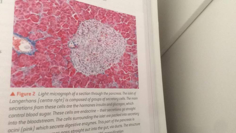
| Histology of the pancreas: Identify and examine stained sections of the pancreas to show the histology of the endocrine tissues | Endocrine =Islets of Langerhans - Large, spherical clusters - Produce and secrete hormones Exocrine = Pancreatic Acini - Small, berry shaped clusters - produce and secrete digestive enzymes. |
| ... | |
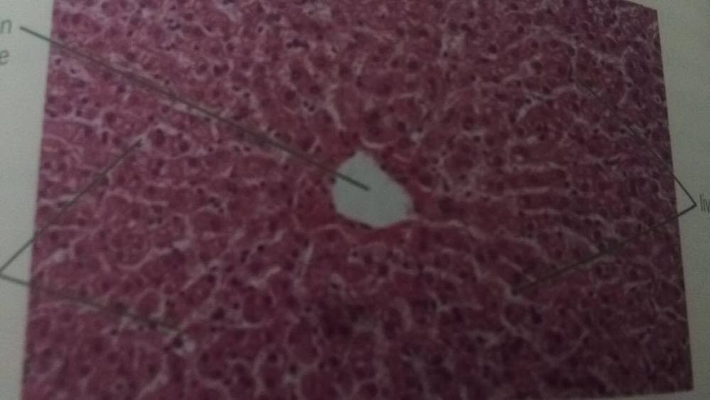
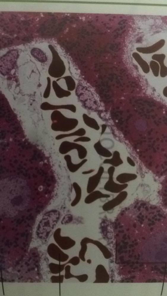
| Histology of the Liver | Low Mag: Liver Cells Central Vein and Lobule Sinusoids High Mag:Hepatocyte RBCs within sinusoids Kupffer Cells |
| ... | |
| ... | |
| Structures and functions of the components of the mammilian gaseous exchange system | 1) Trachea 2) Bronchus 3) Bronchioles 4) Alveoli |
| Trachea | Wide tube consisting of incomplete rings of strong, flexible cartilage, stopping a collapse. |
| Bronchus | They are similar in structure to the trachea but smaller. |
| Bronchioles | No cartilage rings. Contain smooth muscle - can contract and relax. This can affect the volume of air reaching the lungs. Lined with a thin layer of flattened epithelium. |
| Alveoli | Consists of a thin layer of flattened epithelial cells, some collagen and elastic fibres. |
| Dissection (PAG 2) | ... |
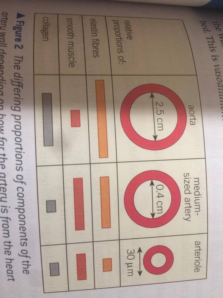
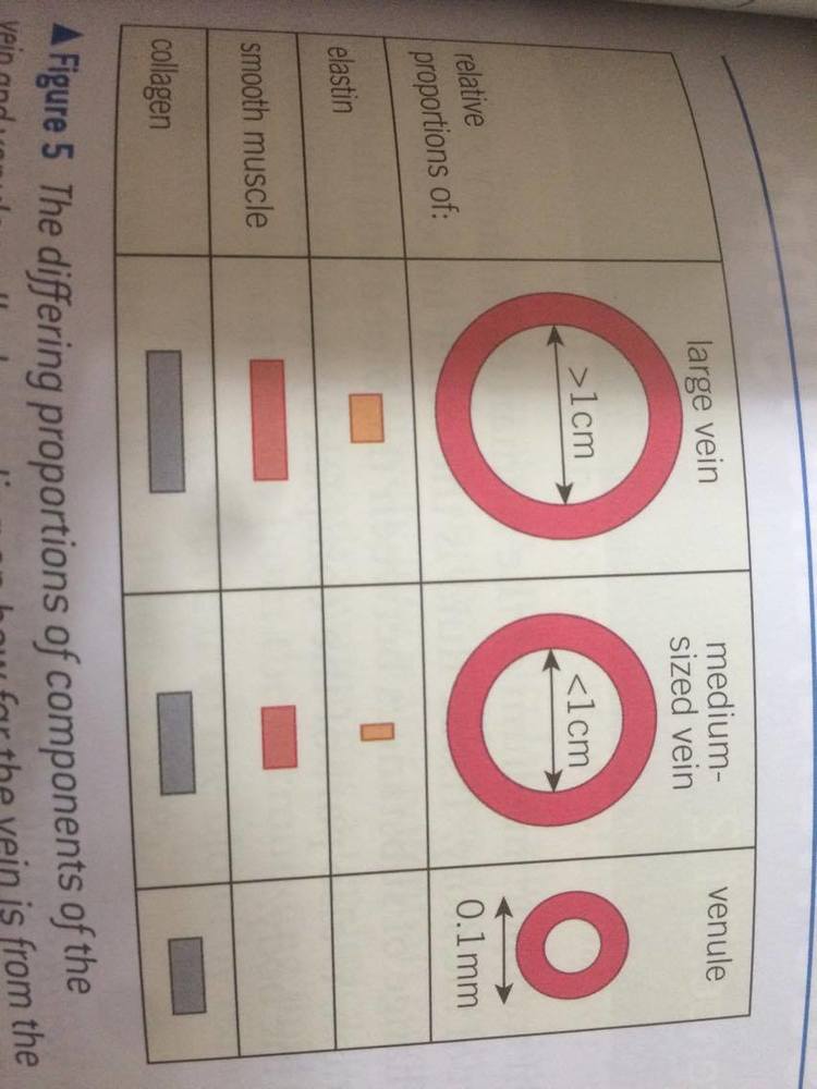
| Structure of the arteries, arterioles, capillaries, venules and veins. | ... |
| Arteries/Arterioles | |
| Capillaries | Diameter = 10 (x10^-6)m Very thin walls - about one epithelial cell thick - short diffusion distance. |
| Veins/ Venules | |
| The Heart | Be aware that when dissecting the mammalian heart supplied by the butcher, some components (like the aorta and the coronary artery) may be missing as they're removed before selling as many people don't want to eat these parts. |
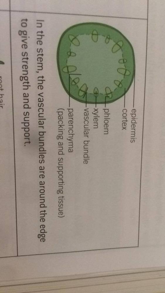
| Transport systems in dicotyledonous plants | Xylem and Phloem transport vessels form vascular bundles to transport water/ minerals/ solutes around the plant. |
| The vascular bundle arrangement inthe stems & reasons | |
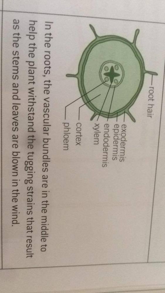
| The vascular bundle arrangement in in the roots & reasons | |
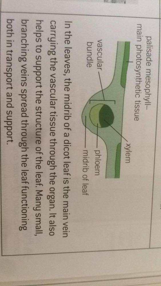
| The vascular bundle arrangement in in the dicot leaves & reasons | |
| The Kidney | Remove the perirenal fat. Slice the kidney open carefully to expose the internal structure. Dissecting the kidneys cannot show us the histology of the kidneys. To observe the structure of the individual nephons require hand-lens/stains. |
| Increase the visibility of nephons | Add a drop of H2O2, rapid effervescence will occur and after it's wiped off, the nephons will be easier to detect. |
Want to create your own Flashcards for free with GoConqr? Learn more.