1133201
Description
Mind Map by drmjmurphy, updated more than 1 year ago
|
|
Created by drmjmurphy
over 11 years ago
|
|
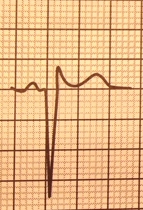
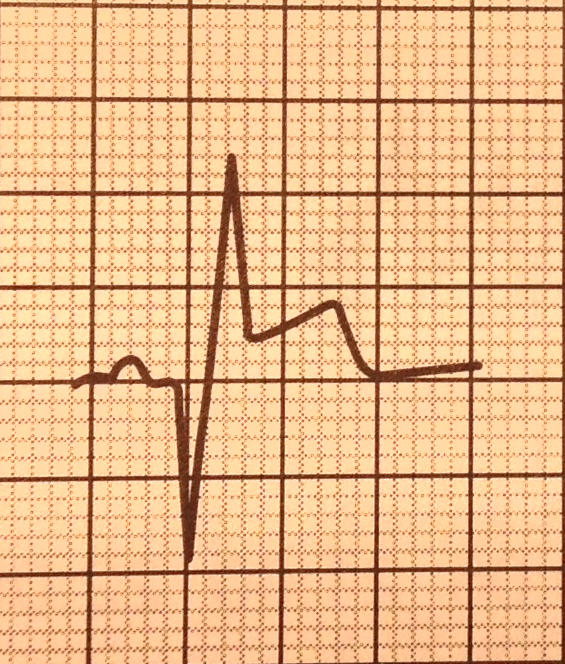
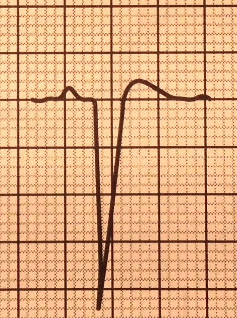
Abnormal Q waves
- Morphology
- QR Wave
- Qr Wave
- QS Wave
- Also due to LBBB,
LVH, cor pulmonale or
cardiomyopathy
- V1 to V3
- V1 to V3
- Also due to LBBB,
LVH, cor pulmonale or
cardiomyopathy
- qR Wave
- QR Wave
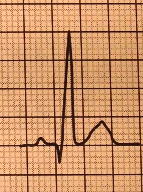
- Normal Q waves
- rapid depolarisation of thin septal wall
between two ventricles
- Maybe found in most leads
- very short and low amplitude
- Lead III, aVR, V1
- rapid depolarisation of thin septal wall
between two ventricles
- Location
- Lead V2 & V3
- Leads I, II, aVL, aVF, or
V4 to V6
- > or = 0.03 sec
- 2 contiguous leads
- > or = 0.03 sec
- Lead V4
- > 1mm deep or
at least 0.02 sec
or > Q wave in V5
- > 1mm deep or
at least 0.02 sec
or > Q wave in V5
- Lead aVL
- > 0.04 sec or > 50% amplitude of
QRS complex with upright P wave
- > 0.04 sec or > 50% amplitude of
QRS complex with upright P wave
- Lead III
- > or = 0.04 sec
- > or = 0.04 sec
- Lead V2 & V3
- Q Wave Equivalents
- tall R waves in leads V1 & V2
- Posterior infarction
- Posterior infarction
- Poor R wave progression
Annotations:
- localised R wave diminution
- localised R wave diminution
- reverse R wave progression
- R waves decrease in
amplitude from V1 to V4
- R waves decrease in
amplitude from V1 to V4
- tall R waves in leads V1 & V2
- dead or necrotic tissue act as an "electrical
window" transmitting depolarising forces (R
wave) from opposite position of heart
- Completely developed in
8-12 hours of occulsion
- 10% patients don't
develop until 3-11 days
after MI
- Completely developed in
8-12 hours of occulsion
Media attachments
Want to create your own Mind Maps for free with GoConqr? Learn more.