1135396
Description
Mind Map by Kristi Brogden, updated more than 1 year ago
|
|
Created by Kristi Brogden
over 11 years ago
|
|
Development of cortex
- From tube to laminae...
- How does the one-cell thick neural
tube turn into the multiple layered
cortices of the cerebrum and
cerebellum?
- Neural tube initially one cell thick
- Cells divide by nuclear translocation
- Marginal zone relatively cell- free
- Division ↑↑ after neuropores close
- Marginal zone relatively cell- free
- Cells divide by nuclear translocation
- Neural tube initially one cell thick
- What happens if it doesn't?
- Developmental abnormalities
- Lissencephaly
- Smooth brain
- Agyria
- Agyria
- Lissencephaly
- Migration abnormalities
- Developmental abnormalities
- How does the one-cell thick neural
tube turn into the multiple layered
cortices of the cerebrum and
cerebellum?
- Cerebral cortex
- Post-mitotic cells move
from ventricular zone to
form preplate
- Cortical plate
- Splits PP
- Forms many layers of adult cotex
- Splits PP
- Cortical plate
- Cajal-Retzius cells in
marginal zone
- 1st post-mitotic cells
- Disappear or change phenotype later
- Disappear or change phenotype later
- Cajal-retzius cells secrete reelin
- 3460 amino acids, ECM-like
- Reeler mice
- Preplate forms but cortical
layers malformed
- Parallels to lissencephaly
- Preplate forms but cortical
layers malformed
- Stops migrating cells?
- Unclear role
- Repellent / stop signal?
- Unclear role
- 3460 amino acids, ECM-like
- CR development regulated by Foxg1
- Foxg1 -/- has extra CR cells
- Foxg1 -/- has extra CR cells
- 1st post-mitotic cells
- Subplate
- Post-mitotic cells move
from ventricular zone to
form preplate
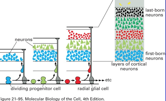
- Inside out development
- Using 3H – thymidine to find cell birthdays
- Cortical layers
- Subventricular zone appears
- Localised (Sanes) or widespread
- Also a germinal layer
- Localised (Sanes) or widespread
- White matter tracts and other layers form
- Subventricular zone appears
- Using 3H – thymidine to find cell birthdays
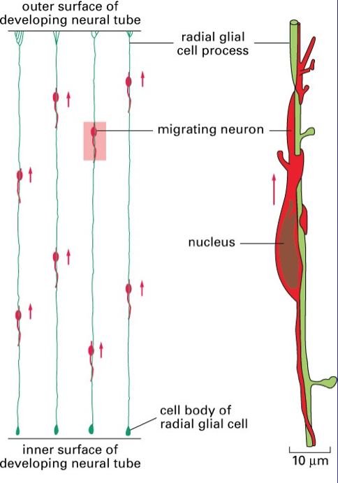
- Guiding migration
- Cell movement
is often
associated with
radial glia
- Radial glia have astrocyte markers
- Most
disappear
after
development
- Except bergmann glia
Annotations:
- What are they?
- Except bergmann glia
- Are stem cells
- Radial glia have astrocyte markers
- But some is
tangential (e.g.
cortical interneurons)
- Cell movement
is often
associated with
radial glia
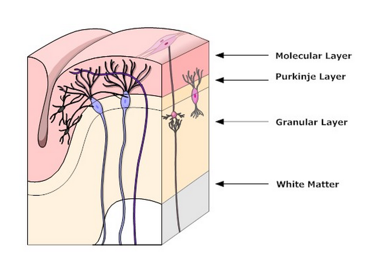
- Cerebellum
- Forms at the roof of the IVth ventricle
- Cortical region
- Cortical layers
- Cortical layers
- Central nuclei
- Deep cerebellar
- Deep cerebellar
- Roof of IVth very thin
- VZ is close to surface
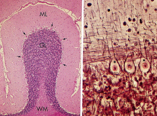
- Rhombic lip cells (germinal trigone) at the superior
and inferior edges of the medullary velum
Annotations:
- Whut?
- Superior rhombic lip cells form the external germinal layer
- VZ is close to surface
- Cortical region
- Forms at boundary of the mid and hindbrains
- Development
- Superior rhombic lip cells form the external germinal layer
- VZ produces all other cell types
- Inferior lip produces pontine nuclei and inferior olive
- VZ produces all other cell types
- The EGL produces granule cells (neurons)
- Granule cells migrate inwardly
- Cerebellar mass increases as granule cells ↑↑
- Folds appear
- Folds appear
- Cerebellar mass increases as granule cells ↑↑
- Granule cells migrate inwardly
- Superior rhombic lip cells form the external germinal layer
- Forms at the roof of the IVth ventricle
- Control of development
- Production of rhombic lip cells is
regulated by MATH-1 (transcription
factor)
- No MATH-1 no foliation, no IGL, no pontine nuclei
- Pontocerebellar hypoplasia
Annotations:
- Find a picture and research it a bit
- No MATH-1 no foliation, no IGL, no pontine nuclei
- Sonic HH released from Purkinje cells stimulates mitosis in EGL
- Reeler mice - disordered layers
- Reelin from EGL
- Other factors
- Reelin from EGL
- Production of rhombic lip cells is
regulated by MATH-1 (transcription
factor)
Media attachments
Want to create your own Mind Maps for free with GoConqr? Learn more.