3942578
Description
Mind Map by Jacob Shepherd, updated more than 1 year ago
|
|
Created by Jacob Shepherd
about 10 years ago
|
|
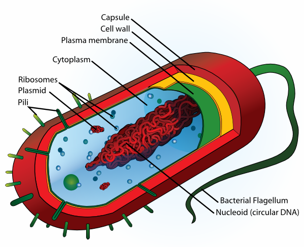
Prokaryotic Cells
- Its cells lack a nucleus
- The DNA is free floating in a circle
- The DNA is free floating in a circle
- e.g. bacteria cell
- Cell wall is made up of murein
- Cell wall is made up of murein
- Cell wall
- Functions
- Excludes substances
- Protects against
mechanical
damage and
osmotic lysis
- Excludes substances
- Functions
- Cell surface membrane
- This is inside
the cell wall
- It encloses the cytoplasm
- It encloses the cytoplasm
- Function
- Controls entry and exit of chemicals
- It has a permeable layer
- It has a permeable layer
- Controls entry and exit of chemicals
- This is inside
the cell wall
- Ribosomes
- Size
- 70S in prokaryotic cells
- 70S in prokaryotic cells
- Function
- Protein synthesis
- Protein synthesis
- Size
- Circular strand of DNA
- (DNA is still a double helix)
- It is not associated with proteins
- i.e. not wrapped around protein
- i.e. not wrapped around protein
- Found in central area
of the cell called the
nucleoid
- Function:
- Stores genetic
information for
replication of
bacterial cells
- Stores genetic
information for
replication of
bacterial cells
- (DNA is still a double helix)
- Microscopy
- Maginifcation
- This is how many times bigger the
image is than the actual object
- Magnification = size of image/size of real object
- This is how many times bigger the
image is than the actual object
- Resolution
- This is the minimum
distance apart two objects
can be in order to appear
as separate items
- The human eye has a
resolution of 0.2mm
- The light
microscope has a
resolving power of
0.2 micro metres
- The best electron
microscope has a
resolution of
0.1nm
- This is the minimum
distance apart two objects
can be in order to appear
as separate items
- The light microscope
- Specimens must be thin
sections of prepared tissue
that has been stained with
coloured pigments
- Or they can be small live
specimens like Daphnia
- Sections must be thin to allow
light to pass through and to
make sure you are only looking
at a single layer of cell
- They must be stained as
sections are often
transparent
- Or they can be small live
specimens like Daphnia
- A beam of light is shone
through the specimen through
a series of lenses resulting in a
magnified image
- Very little intracellular detail
can be seen
- The eyepiece graticule
- Usually around 10mm
long with 100 sub
divisions
- Stage micrometer
- This is usually 1mm long it goes
on the specimen stage
- This is usually 1mm long it goes
on the specimen stage
- Calibrating the
eyepiece graticule
- Because the scale remains
constant no matter what
magnification you're on, you
must change the eyepiece
graticule
- Because the scale remains
constant no matter what
magnification you're on, you
must change the eyepiece
graticule
- Usually around 10mm
long with 100 sub
divisions
- Specimens must be thin
sections of prepared tissue
that has been stained with
coloured pigments
- The electron microscope
- This uses a beam of electrons rather than light
- The electron beam has a shorter
wavelength than light which means
the resolving power is higher
- The electron beam has a shorter
wavelength than light which means
the resolving power is higher
- Transmission
electron microscope
(TEM)
- Ultra thin sections needed, must be
stained and heavy metals are needed
- Where electrons pass thought the
specimen the image appears bright
and when the electrons are
absorbed the image is dark
- A near vacuum is required
- Specimen must be dead
- Specimen must be dead
- 2D black and white images
- Ultra thin sections needed, must be
stained and heavy metals are needed
- Scanning Electron Microscope (SEM)
- Electrons do not penetrate the specimen
- The beam is passed to and
fro over the specimen
surface in a regular
pattern
- Electrons are scattered
depending on the contours
- Electrons are scattered
depending on the contours
- The beam is passed to and
fro over the specimen
surface in a regular
pattern
- Produces a 3D image
- Black and white
- Black and white
- Resolution is lower than that of the TEM
- Electrons do not penetrate the specimen
- This uses a beam of electrons rather than light
- Artefacts
- Due to all of the processes the specimens must
undergo under microscopes, images may contain
objects that shouldn't be there
- An artefact may be a break in a
membrane, empty spaces in cells
- In the TEM heavy metal stains are
used, so granular deposits may be
seen
- In the TEM heavy metal stains are
used, so granular deposits may be
seen
- It is therefore not possible to
be sure what we see on an
image is the actual natural
specimen
- Due to all of the processes the specimens must
undergo under microscopes, images may contain
objects that shouldn't be there
- Maginifcation
- Some prokaryotic cells have
- Plasmid
- Small circular
strand of DNA
- Function:
- Gives genes that aid survival
- e.g. they can reproduce
with antibiotic resistance
- e.g. they can reproduce
with antibiotic resistance
- Gives genes that aid survival
- Used
extensively as
vectors
- Small circular
strand of DNA
- Capsule
- This is mucilaginous
slime around the
outside of the cell wall
- Functions:
- It stops bacteria
being deteced
- This protected
them from other
cells
- This protected
them from other
cells
- It helps
bacteria stick
together for
protection
- It stops bacteria
being deteced
- This is mucilaginous
slime around the
outside of the cell wall
- Flagellum
- Function
- Used for locomotion
- Makes bacteria move
- Makes bacteria move
- Used for locomotion
- Function
- Plasmid
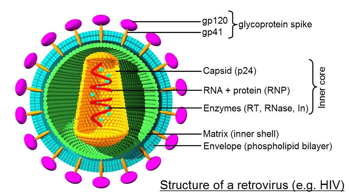
- Viruses
- This is an acellular,
non living particle
- Contains nucleic acids such as
DNA or RNA as genetic
material enclosed in a protein
coat or capsid
- Capsid or envelope
have attachment
proteins
- These allow the virus to
identify and attach to a host
cell
- These allow the virus to
identify and attach to a host
cell
- Capsid or envelope
have attachment
proteins
- Can only multiply inside a living host cell
- May be
surrounded by a
living host cell
- This is an acellular,
non living particle
Media attachments
Want to create your own Mind Maps for free with GoConqr? Learn more.