8604621
Description
Mind Map by leen abuafifeh, updated more than 1 year ago
|
|
Created by leen abuafifeh
almost 9 years ago
|
|
muscular dystophy
- Anatomy of the foot
- Bones
- talus, calcaneus, cuneiformes (3), cuboid, and navicular
- metatarsus (5): first, second, third, fourth, and fifth
metatarsal bone
- phalanges (14)
- talus, calcaneus, cuneiformes (3), cuboid, and navicular
- Joints
- Inferior
tibio-fibular joint
- Fibrous/syndysmosis
- Fibrous/syndysmosis
- ankle joint
- articulation
- mortise
- talus
- mortise
- movements
- dorsiflexion
- planterflexion
- dorsiflexion
- ligaments
- medial collateral
- tibionavicular
- tibiocalcaneal
- ant. and post. tibiotalar
- tibionavicular
- lateral collateral
- calcaneofibular
- ant. and post talofibular
- calcaneofibular
- post. tibiofibular
- medial collateral
- articulation
- Inferior
tibio-fibular joint
- Muscles
- 1st layer
- Abductor
hallucis
- Flexor digitorum
brevis
- Abductor digiti
minim
- Abductor
hallucis
- 2nd layer
- Quadratus
plantae
- tendon of FHL
- tendon of FDL
- tendon of FDL
- tendon of FHL
- Lumbricals
- Quadratus
plantae
- 3rd layer
- Flexor digiti minim
- Flexor hallucis brevis
- Adductor hallucis
- Adductor hallucis
- Flexor hallucis brevis
- Flexor digiti minim
- 4th layer
- Plantar
interossei
- Dorsal
interossei
- tendon of TP
- tendon of FL
- tendon of FL
- tendon of TP
- Plantar
interossei
- 1st layer
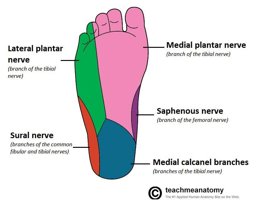
- Nerve
- post. tibial
- Lateral
Plantar
- the rest of
foot muscles
- the rest of
foot muscles
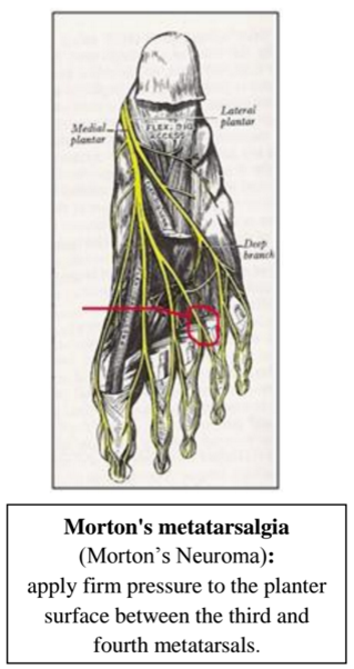
- Medial
Plantar
- 1) Abductor hallucis. 2)
Flexor digitorum brevis. 3)
Flexor hallucis brevis 4) First
lumbrical muscle
- 1) Abductor hallucis. 2)
Flexor digitorum brevis. 3)
Flexor hallucis brevis 4) First
lumbrical muscle
- Lateral
Plantar
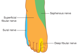
- cutaneus
- sole
- dorsum
- sole
- post. tibial
- blood supply
- sole
- posterior tibial artery
- Lateral
Plantar
Artery
- 1st digital artery
- anastomotic branch
- perforating branchs
- 1st planter metatarsal
- 2nd planter metatarsal
- 3rd planter metatarsal
- 4th planter metatarsal
- planter arch
- perforating branchs
- 1st digital artery
- Medial
Plantar
Artery
- deep branch
- superficial branch
- deep branch
- Lateral
Plantar
Artery
- posterior tibial artery
- dorsum
- anterior tibial artery
- Dorsalis Pedis Artery
- arcuate
- 4th dorsal metatarsal
- 5th dorsal metatarsal
- 5th dorsal metatarsal
- 3rd dorsal metatarsal
- 2nd dorsal metatarsal
- 4th dorsal metatarsal
- 1st dorsal metatarsal
- medial branch
- lateral branch
- medial branch
- deep planter
- medial and lateral tarsal
- arcuate
- Dorsalis Pedis Artery
- anterior tibial artery
- sole
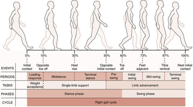
- Gait Cycle
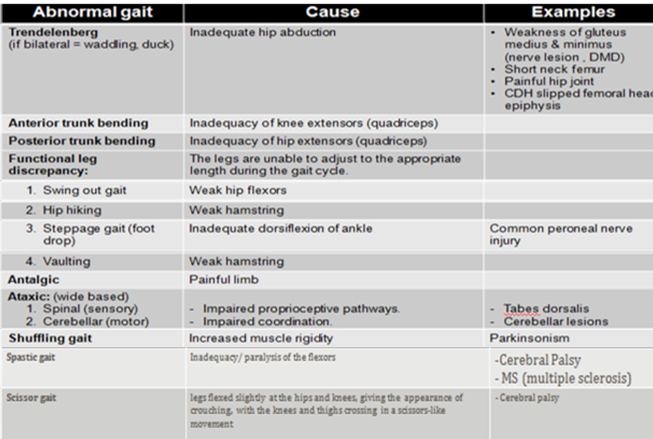
- Abnormalities in
- skeleton
- muscles
- nervous
system
- skeleton
- STRIDE
- from the initial contact of a
particular limb to the point
of initial contact of the SAME
limb and is equivalent to
one gait cycle
- from the initial contact of a
particular limb to the point
of initial contact of the SAME
limb and is equivalent to
one gait cycle
- STEP
- from initial contact of one limb to
the initial contact of the
contralateral limb. Therefore,
there are two steps in each stride
(or gait cycle).
- from initial contact of one limb to
the initial contact of the
contralateral limb. Therefore,
there are two steps in each stride
(or gait cycle).
- Abnormalities in
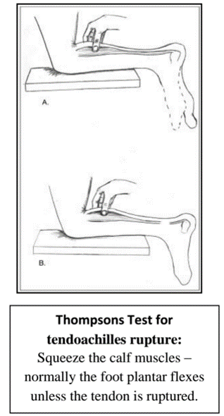
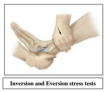
- Physical examination
- special tests
- special tests
- Bones
- Management
- STEROIDS
- creatine monohydrate
- STEROIDS
- Psychological aspects
- marginalization and
isolation
- hypochondriac
thoughts
- anxiety
- self-depreciation
- marginalization and
isolation
- TYPES
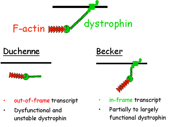
- Myotonic
Duchenne
Becker
Limb-girdle
Facioscapulohumeral
Congenital
Oculopharyngeal
Distal
Emery-Dreifuss
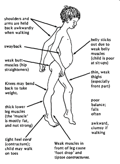
- Duchenne Muscular Dystrophy
- symptoms
- DMD is caused by an
absence of dystrophin, a
protein that helps keep
muscle cells intact
- Tests for DMD
- Creatin kinase test
- EMG
- Creatinine
- Dystrophin test
- MUSCLE BIOPSY
- function of the thyroid
- Creatin kinase test
- mode of
inheritance
- X-LINKED RESESSIVE
- X-LINKED RESESSIVE
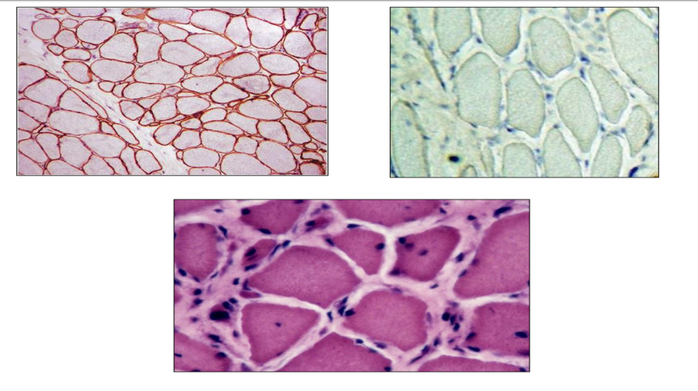
- MICROSCOPIC
- connective tissue
proliferation, scattered
degenerating and
regenerating myofibres.
- connective tissue
proliferation, scattered
degenerating and
regenerating myofibres.
- Complications
- Cardiomyopathy
- Congestive heart
failure
- Heart
arrhythmias
- Mental
impairment
- Cardiomyopathy
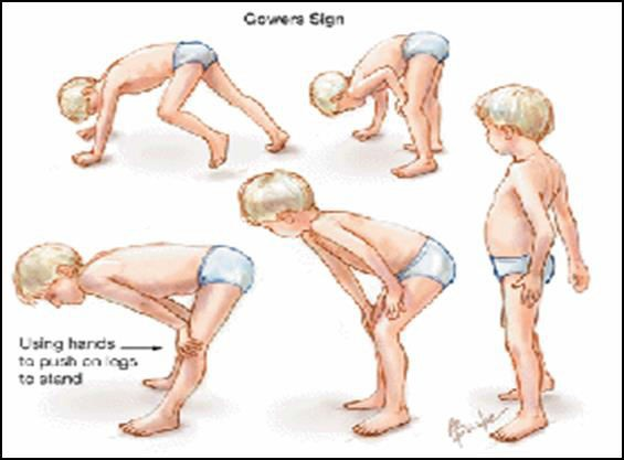
- GOWER'S SIGN
- symptoms
- Duchenne Muscular Dystrophy
- Myotonic
Duchenne
Becker
Limb-girdle
Facioscapulohumeral
Congenital
Oculopharyngeal
Distal
Emery-Dreifuss
Media attachments
Want to create your own Mind Maps for free with GoConqr? Learn more.