Description

|
|
Created by Erika Cobb
over 6 years ago
|
|
Page 1
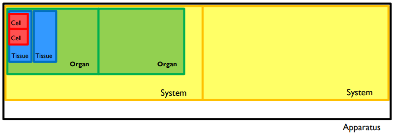
Definitions: Cell: A mass of protoplasm surrounded by a membrane containing a nucleus & some organelles The fundamental unit of life Tissue: An aggregation of cells & intercellular materials that have a specific function. The types of cells determine a tissue’s function. Organ: A collection of tissues joined in a structural unit to serve a common function System A group of physiologically or anatomically complementary organs or parts Apparatus: The complex structure of a particular organisation or system & use for specific purposes
Page 2
Epithelium: An aggregation of cells specialised for absorptive, secretory, excretory, protective, sensational and environmental exchange functions, resting on a membrane (extracellular matrix) Covering and Lining: Eg. Skin covering, duct lining Glandular or secretory Eg. Thyroid gland
Page 3
Embryological origins: Three embryological layers: ectoderm, mesoderm, endoderm. Epithelium may derive from any of these layers Ectoderm Forms skin and associated glands Form nervous system Lines oral, nasal and anal passages Mesoderm: Musculoskeletal and cardiovascular systems Mesothelium lining thoracic and abdominal cavities (pleura and peritoneum) Tubules, ducts and accessory glands of urogenital system Endoderm: Digestive and respiratory tracts, urinary bladder, liver, gall bladder, and pancreas Epithelial glands of the gut
Page 4
Characteristics of epithelium: Cell proliferation ◦ Permanent – cell division does not occur in adult life (eg. auditory cells) Stable – may still be able to replicate under certain conditions (eg. hepatic cells) Labile – renewable (eg. Intestinal cells, epithelial cells) Polarity Polar “top and bottom” Compartmentalise specific functions to specific areas Surfaces: Apical (top): Faces a lumen Basal (bottom): Faces extracellular matrix, consists of basal lamina and blood supply Lateral (lateral): adherent to adjacent cells Vascularity Epithelial sheets are avascular: no blood vessels on the epithelial side of the basement membrane Gain nutrients via diffusion only Glandular epithelia (ie. those with a secretory function) need to be located near a rich blood supply to take up and release materials and are VASCULAR Cell-cell adhesion Basement membrane Separates epithelial cells from underlying supportive structures Barrier function Secretion/excretion Interchange with the environment Protection Sensation
Page 5
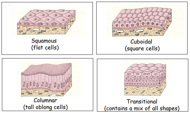
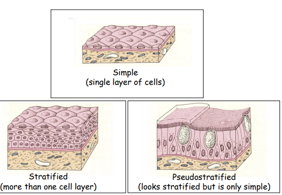
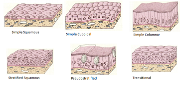
Types of epithelium shapes: Squamous: (flat) Cuboidal: (Square) Columnar (Tall, oblong) Transitional (Mix of shapes) Types of epithelium layers: Simple (single layer of cells) Stratified (more than one cell layer) Pseudostratified (Looks stratified but only simple)
Page 6
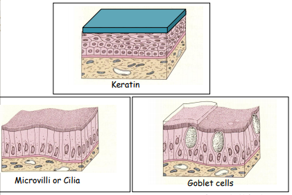
Specialisations: Keratin Protein formed by death of old skin cells Anchored to newer cells Strong waterproof barrier that protects underlying tissues and cells Microvilli or cilia Microvilli: Projection to increase surface are in absorption cells Cilia: internal structures (microtubules), sway with a metachronal rhythm, move substances over cells, located in respiratory tract Goblet cells Secrete mucous Modified columnar cells Protective function
Page 7
Common shapes: Simple squamous Nucleus usually bulges towards surface Lung tissue, blood vessels, peritoneal, pericardial, pleural membrane Simple cuboidal Height is similar to width Ducts of kidneys and exocrine glands, thyroid capsule of lens Simple columnar Same height and arranged in columns Nuclei usually at same level Eg. GIT, excocrine ducts, uterine tubes, endometrium Stratified squamous Provide resistance to wear and tear and form a protective barrier to underlying tissues Can be keratinised or non-keratinised Skin, oral cavity, oesophagus, anal canal, vagina Stratified cuboidal & columnar Two or more layers of cuboidal or columnar cells Stratified cuboidal: Ducts of mammary, salivary and sweat glands Stratified Columnar: Conjunctiva of eye, salivary gland, pharynx, male urethra Pseudostratified columnar Multilayered but every cell is attached to the base Not every cell reaches the free surface Nuclei appear at different levels Examples: upper respiratory tract, male reproductive tract Transitional Multilayered Cells transition from stratified to columnar Allow tissue to stretch Relaxed-several cells thick, stretched-only 2-3 cells thick Examples: renal calyces, ureters and urinary bladder
Want to create your own Notes for free with GoConqr? Learn more.