Description
|
|
Created by Cher Bachar
over 12 years ago
|
|
Page 1
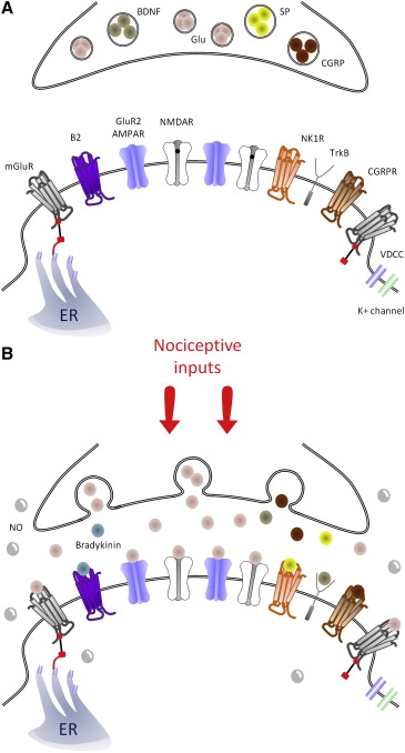
Figure 4. Central sensitization triggers: Schematic representation of key synaptic triggers of central sensitization. (A), Model of the synapse between the central terminal of a nociceptor and a lamina I neuron under control, basal conditions. mGluR receptors sit at the extremities of the synapse and are linked to the endoplasmic reticulum (ER). Note that NMDAR channels are blocked by Mg2+ in the pore (black dot). After a barrage of activity in the nociceptor (B), the primary afferent presynaptic terminal releases glutamate that binds to AMPAR, NMDAR (now without Mg2+), and mGluR, as well as substance P, CGRP, and BDNF, which bind to NK1, CGRP1, and TrkB receptors, respectively. B2 receptors are also activated by spinally produced bradykinin. NO is produced by several cell types in the spinal cord and can act presynaptically and postsynaptically.
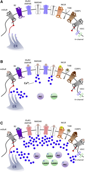
Figure 5. Sources of Ca2+ in the synapse of nociceptive neurons for inducing central sensitization. (A), Model of a nociceptor–dorsal horn neuron synapse under control, nonactivated conditions. After nociceptor input (B), activation of NMDAR and mGluR result in a rapid increase of [Ca2+]i that activates PKC and CaMKII, 2 major effectors of central sensitization. (C), Representation of a synapse during peripheral inflammation-induced central sensitization, where there is a shift from GluR2/3 to GluR1-containing AMPARs that enables, along with voltage-dependent calcium channels and NMDAR, entry of Ca2+and which, together with activation of the G-coupled MGluR, NK1, B2, and CGRP1 receptors, which release intracellular Ca2+stores, recruits PKC and CaMKII, strengthening the excitatory synapse.
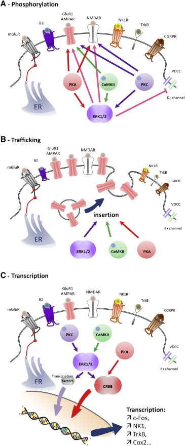
Figure 6. Contribution of PKC, CaMKII, PKA, and ERK activation to central sensitization. (A), Phosphorylation by PKC, CaMKII, PKA, and ERK cause changes in the threshold and activation kinetics of NMDA and AMPA receptors, boosting synaptic efficacy. ERK also produces a decrease in K+ currents through phosphorylation of Kv4.2 channels, increasing membrane excitability. (B), PKA, CaMKII, and ERK promote recruitment of GluR1-containing AMPAR to the membrane from vesicles stored under the synapse. (C), Transcriptional changes mediated by activation of CREB and other transcription factors driving expression of genes including c-Fos, NK1, TrkB, and Cox-2, to produce a long-lasting strengthening of the synapse.
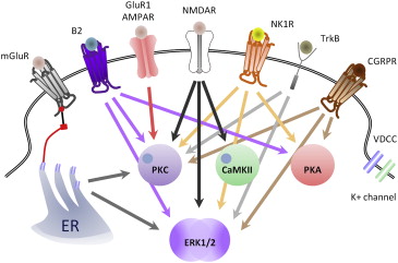
Figure 8. Key intracellular pathways contributing to the generation of central sensitization. NMDAR activation causes activation of PKC, CaMKII, and ERK (black arrows); GluR1-containing AMPAR activate PKC (red arrow); NK1 and CGRP1 receptors activate PKC, PKA, and ERK (orange and brown arrows, respectively); TrkB s activates of PKC and ERK (purple arrows); and mGluR, via release of Ca2+ from microsomal stores, activates PKC and ERK (gray arrows). Note that most of the triggers of central sensitization: Activation of NMDAR, mGluR, TrkB, NK1, CGRP1, or B2 converge to activate ERK.
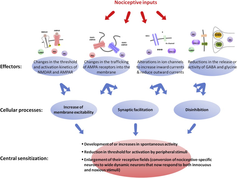
Figure 9. Multiple cellular processes lead to central sensitization. Central sensitization is not defined by activation of a single molecular pathway but rather represents the altered functional status of nociceptive neurons. During central sensitization, these neurons display 1 or all of the following: i, development of or an increase in spontaneous activity; ii, reduction in threshold for activation; and iii, enlargement of nociceptive neuron receptive fields. These characteristics can be produced by several different cellular processes including increases in membrane excitability, a facilitation of synaptic strength, and decreases in inhibitory transmission (disinhibition). Similarly, these mechanisms can be driven by different molecular effectors including PKA, PKC, CaMKII, and ERK1/2. These kinases participate in changes in the threshold and activation kinetics of NMDA and AMPA receptors and in their trafficking to the membrane, cause alterations in ion channels that increase inward currents and reduce outward currents, and reduce the release or activity of GABA and glycine.
Triggers
Signalling- induction
Signalling-maintenance
intracellular pathways summary
Multiple cellular pathways
Want to create your own Notes for free with GoConqr? Learn more.