
Flashcards
Aug 14 lecture
Flashcards do conjunto (60)
- 1Gross AnatomyStudy of structures visible to the unaided eye
- 2Topographical anatomyAnatomy of one part of the body in relation to another
- 3Applied Anatomyapplied to disease
- 4Microscopic anatomystudy of tissue with a microscope
- 5Ultra Structural Anatomystudy of tissues with electron microscope
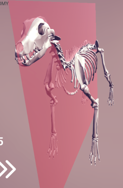
- 6What type of plane?Median
- 7What is a sagittal plane?Any plane parallel to the median plane
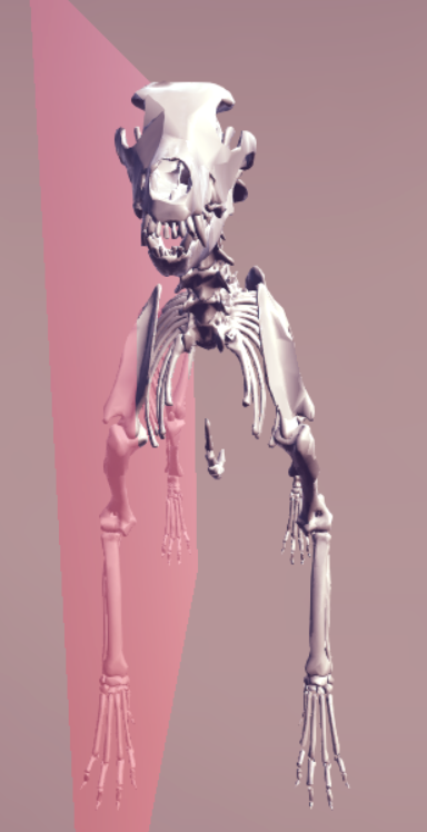
- 8What type of plane?Dorsal plane (any level that is perpendicular to the median plane
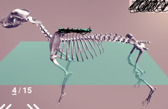
- 9What type of plane?Transverse plane
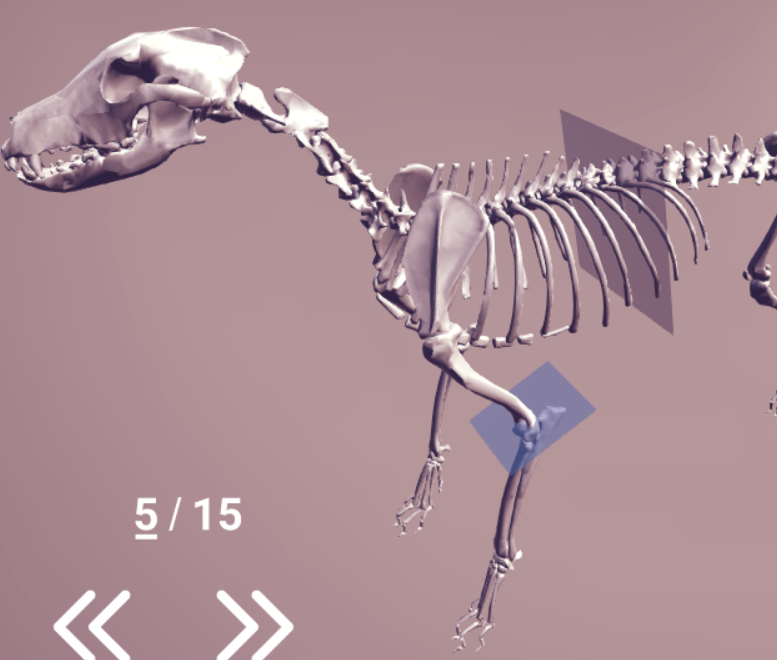
- 10Cranial vs CaudalCloser to head=cranial
Closer to tail=caudal
Can also be used to directly compare parts (ie the forelimb cranial to the hind limb)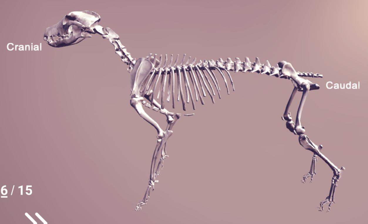
- 11Where is rostral vs caudal used?The head region
- 12Rostral vs caudalRostral= toward the nose (nostral)
Caudal= toward the tail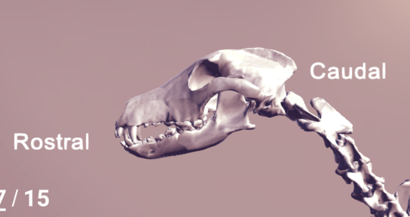
- 13Dorsal vs ventralDorsal=closer to top
Ventral=closer to bottom (think V)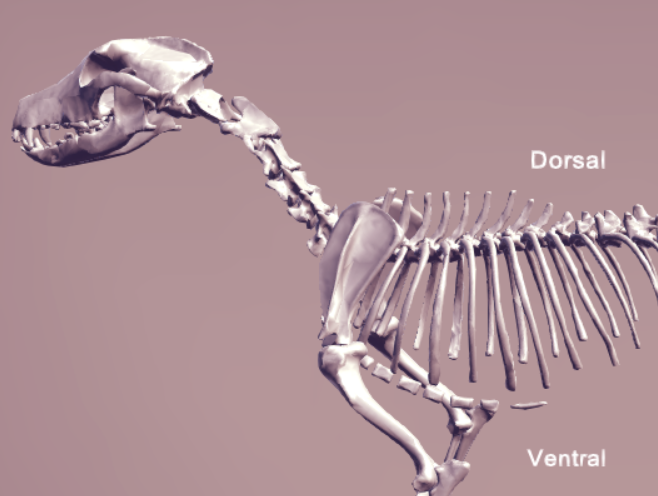
- 14Axial vs AbaxialAxial= Facing or towards the central line or axis
Abaxial= facing away or farther from the central line or axis - 15Medial vs LateralMedial- closer to median plane
Lateral- farther away from median plane - 16Proximal vs distalProximal- closer to the trunk (body)
Distal- farther from the trunk - 17Which three terms are used distal to the carpal bones?Dorsal and Palmar/Plantar
- 18Dorsal and Palmar and PlantarDorsal- top
Palmar- bottom (palm) forelimb
Plantar- bottom (palm) hindlimb - 19Superficial vs deepSuperficial- near the surface of the body
deep- near the center (inside the trunk) of the body - 205 general functions of bones1) Provide form and support
2) Protect soft tissues (i.e lungs)
3) act as levers to facilitate locomotion
4) have a role in blood cell formation
5) maintain mineral homeostasis - 21Two classes of skeleton and what they includeAxial skeleton- forms and supports the body trunk
Appendicular skeleton- forms the limbs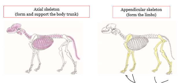
- 227 classes of bones1) Long bones
2) Irregular bones
3) Short bones
4) flat bones
5) sesamoid (round) bones
6) pneumatic bones
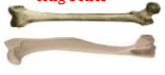
7) Heterotropic or splanchnic bones - 23What class of bone?Long
- 24What class of bone?Irregular
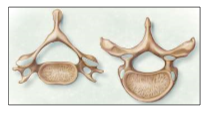
- 25What class of bone?Short
- 26What class of bone?Flat
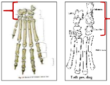
- 27Form and function of sesamoid (round) bonessmall, nodular, embedded within tendons and adjacent to joints where the tendons are compressed
- 28Pneumatic bonesair filled bones
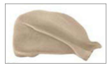
- 29Heterotrophic or splanchnic bones (3 types)-os penis (dog)
-os cordis (cattle)
os rostrum (pigs) - 30#2? What is it composed of?Diaphysis, composed of compact bone
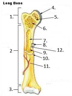
- 31What structure is the diaphysis covered by?periosteum
- 32#1? Composed of what?Epiphysis, made up of spongy bone covered by compact bone
- 33#3/#4Articular cartilage (AKA hyaline cartilage)
- 34#5, function?Epiphyseal line, thin strip of bone marking the fusion of epiphysis to diaphysis
- 35What is the difference between epiphyscal growth plate and the epiphyseal lineArea actively growing in bones is called epiphyscal growth plate. It is replaced by the epiphyseal line when the bone stops growing
- 36#6Spongy bone
- 37#7?Medullary cavity
- 38Where is metaphysis on a long bone and what is it made of?Metaphysis is part of the diaphysis that borders the epiphyseal plate/line and consists of spongy bone
- 39Blue?Articular cartilage (hyaline cartilage)
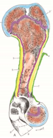
- 40Light purple (top)Epiphyseal plate or line
- 41Orangish pink?Medullary cavity/spongy bone
- 42Green (outer covering)Periosteum
- 43Yellow? (middle)Diaphysis (compact bone)
- 44Dark purple (inner)Endosteum
- 45Almost mature animals have an open or closed growth plate?Closed
- 46number and locations of ossification centers in long bones(3), one diaphyseal center (middle of the bone) and two epiphyseal centers (top and bottom)
- 47Number of ossification centers in a short boneone
- 48Which two bone classifications have variable numbers of ossification centers?Flat and irregular
- 49Where does the nutrient artery attach?in the middle of the diaphysis
- 50What is the nutrient foramen?A gap in the bone that allows arteries through
- 51Three properties of muscles that allow them to produce movement1) Contractility
2) Excitability
3) Extensibility - 52Why do muscles differ in shape?Based on the amount of work they do (shortening depends on fiber length, power depends on cross sectional area)
- 53Two ways muscles attach to bones and their characteristicsTendons- discrete band of tissue
Aponeurosis- flat, wide, thin sheet of fiberous connective tissue - 542 Different muscle attachments and their characteristicsOrigin- more fixed (least movable), usually proximal attachment
Insertion- more movable, usually distal attachment - 55Muscle bellyfleshy contractile part of muscle
- 56RetinaculumBand of dense regular tissue than binds down muscle tendons as they pass over bone surfaces
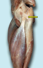
- 57When is the retinaculum generally broad?When it passes over many tendons (ex dorsal aspect of carpus)
- 58Function of synovial bursa?Permits frictionless movement of a tendon over bone
- 59Differences between synovial bursa and tendon sheath?Same function, tendon sheath (4) normally lays under a retinaculum (3) and wraps completely around the tendon (1)
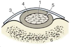
- 60How does the synovial sheath relate to the tendon sheathThe synovial sheath is one of two layers that surrounds a tendon sheath