
Mapa mental
Visual system 1
Conteúdo do mapa
Visual system 1
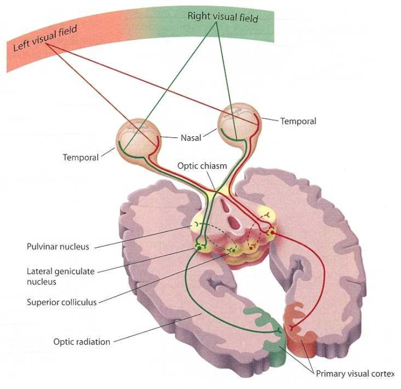
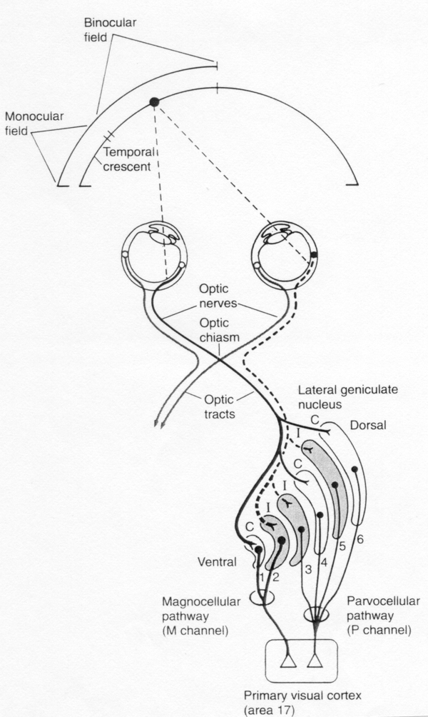
Visual pathways
- Right hemifield activates left brain - and vice versa
- The main (but not only) function of the retina is image acquisition
- LGN preprocesses the visual information
- Main processing of information happens in the visual cortex
Retinal mosaic
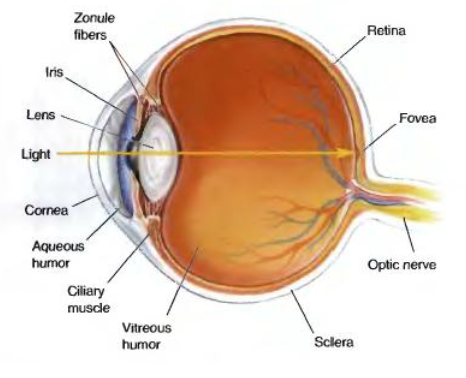
- Pupil regulates the amount of light that falls on the retina
- Lens focuses image on the fovea
- Fovea is the part of the retina with the highest visual acuity
- The rest of the retina has smaller acuity and contains primarily rods
- Fovea is the part of the retina with the highest visual acuity
Laminar retinal organisation
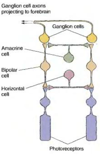
- Retina consists of :
- 3 layers of neurons
- Feedforward neurons
- Photoreceptors
- Photoreceptor synapses
- Have special “ribbon synapses” able to keep vesicle release permanently
- Do not spike (i.e. have “graded potentials”)
- Release glutamate
- Therefore, when the light goes up, the release rate goes down and vice versa.
- Photoreceptor synapses
- Bipolar cells
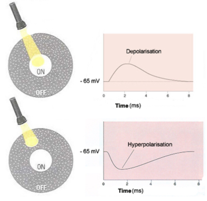
- Centre cell organization of receptive field
- Receptive field is an area in the retina which when illuminated activates a visual neuron
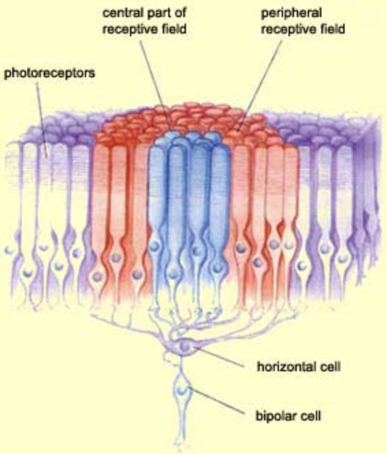
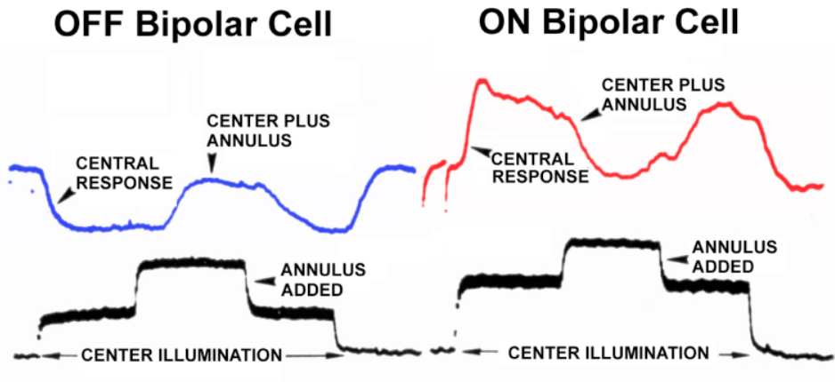
- Many visual neurons have centre-surround organisation of the RF, meaning that illumination of the centre and the surround leads to responses in opposite polarities
- Mechanism
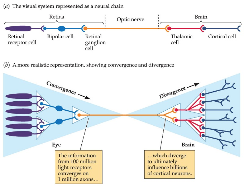
- Many photoreceptors converge to 1 bipolar cell
- Some activate directly (centre) while others activate via horizontal cells(surround) and therefore cause surround photoreceptors to depolarise
- Many photoreceptors converge to 1 bipolar cell
- Some bipolar cells hyperpolarise while others depolarise in response to light
- Why do ON cells depolarise?
- Sign inverting synapse in On bipolar cell
- Photoreceptors release glutamate
- OFF cells express ionotropic glutamate receptor
- Excitatory
- ON cells express metabotropic glutamate receptor
- Inhibitory
- Sign inverting synapse in On bipolar cell
- Why do ON cells depolarise?
- Stratification of bipolar cells in the IPL
- Like many other neurons, BC terminate in different sublayers of the IPLS
- Recall, that this is very precisely regulated during development
- Centre cell organization of receptive field
- Ganglion cells
- Also send their dendrites to different layers of the IPL
- Dendritic trees
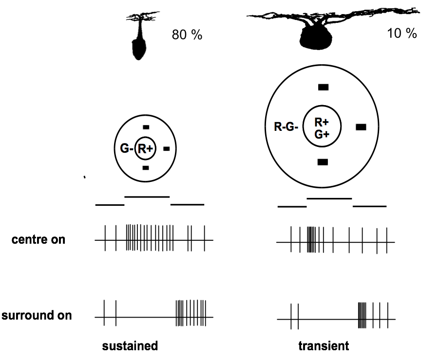
- Ganglion cells have diverse but distinct morphology of the dendritic trees
- Note that some have very small but very dense dendritic trees
- Some cells have larger and diffuse dendritic trees
- How does this determine function?
- FIND OUT
- Receptive field
- Centre surround orginization
- Illumination of the whole receptive field does not activate ganglion cells
- Appears that they are designed to respond to differences in illumination that occur within the receptive field
- Illumination of the whole receptive field does not activate ganglion cells
- Centre surround orginization
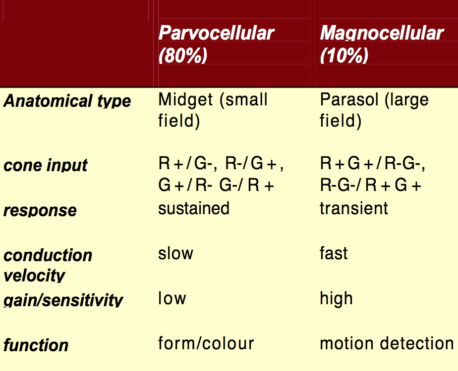
- Two major classes
- Parvocellular
- Magnocellular
- In LGN
- Axons from each eye remain segregated
- P- and M-Ganglion Cells remain segregated
- Photoreceptors
- Feedback neurons
- Horizontal cells
- Amacrine cells
- Feedforward neurons
- 2 layers of synapses
- Inner plexiform layer
- Outer plexiform layer
- 3 layers of neurons
- Retina consists of :
Convergence, divergence and information bottleneck
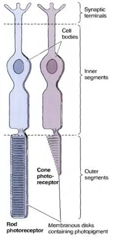
Rods and Cones
- Rods
- Active at dim light
- Cones
- Active at bright light
- Phototransduction happens in the outer segment
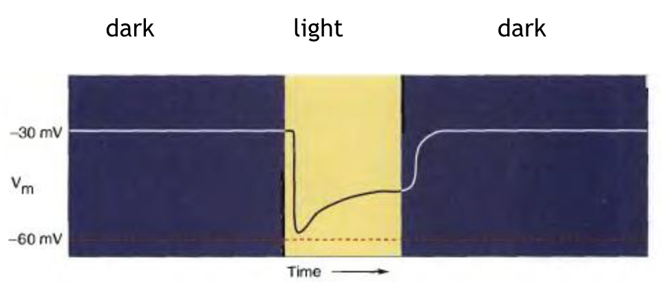
- Photoreceptors respond to light flashes by hyperpolarisation (decrease in membrane potential)
- Rods
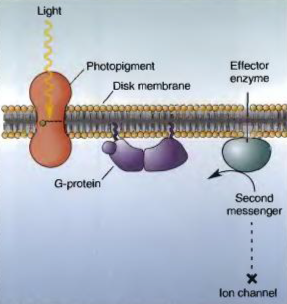
Phototransduction cascade
- Stimulus
- Light
- Receptor activation
- Change in protein conformation
- G-protein response
- Binds GTP
- Second messenger
- cGMP
- Change
- Decrease second messenger
- Ion channel
- cGMP gated channel
- Response
- Decrease na+ conductance
- Stimulus
Retina
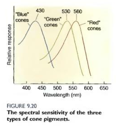
- Colour processing
- All rods have same spectral sensitivity
- Cones
- Red
- Blue
- Much less of these in the fovea
- Green
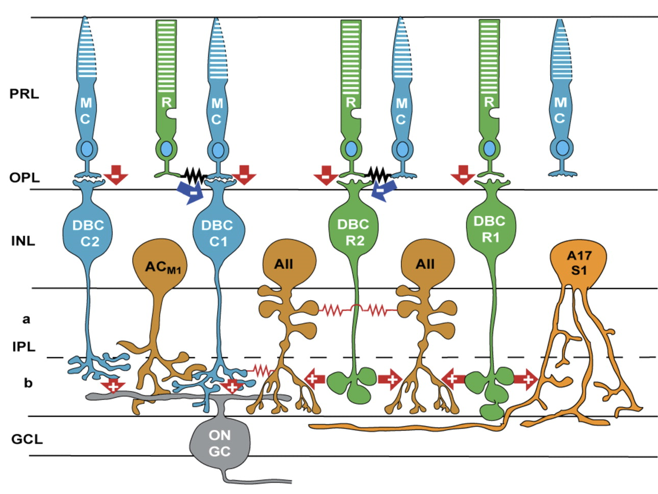
- Rod pathways
- No direct rod-ganglion cell pathway
- Rod bipolar cells activate special class of amacrine cell
- AII form gap junctions with ON cone bipolar cell and inhibitory (glycinergic) synapse with OFF ganglion cell
- Colour processing
Imagens e arquivos do mapa
Informações
- Descrição
- Undergraduate BMS236 Building Nervous Systems (Visual system) Mapa Mental sobre Visual system 1, criado por Kristi Brogden em 05-08-2014.
- Criado por
- Kristi Brogden
- Visualizações
- 19
- Avaliação