
Mapa mental
Apocrine Gland

Conteúdo do mapa
Apocrine Gland
release their products by “decapitation,”
- process by which membrane-bound cytoplasm from the apical surface of the cells
- buds off into the lumen of the duct and is secreted
- entails membrane budding of the apical cytoplasm.
- buds off into the lumen of the duct and is secreted
- process by which membrane-bound cytoplasm from the apical surface of the cells
They are made up of 2 primary parts
- coiled secretory structure
- is made up of
- a single layer of cuboidal cells
- a single layer of columnar cells
- is made up of
- accompanying straight duct
- coiled secretory structure
originate from the hair germ
- generally enter the pilosebaceous follicle at its infundibulum
are a subtype of exocrine secretory glands
- They are found in many locations
- anogenital region
- areolae
- primarily in the axillae
- periumbilical region
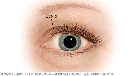
- Eyelids
- external auditory canals
- They are found in many locations
The ductal portion is made up
- a double layer of basophilic cells
- with a periluminal eosinophilic cuticle
- a double layer of basophilic cells
There are 2 main types of secretory glands
- endocrine glands
- exocrine glands
- remain connected with the surface epithelium
- secrete their products through ducts
- They are subclassified
- holocrine
- as merocrine (or eccrine)
- apocrine
- remain connected with the surface epithelium
nonfunctional before puberty
- they grow
- commence secretion.
believed to be an evolutionary remnant of an odorous organ of animals.
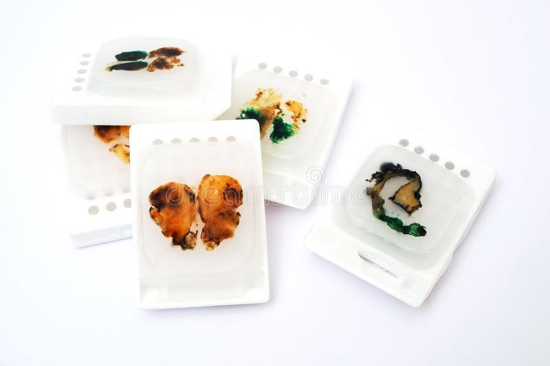
Tissue Preparation
- can be obtained from patients with osmidrosis
- the glands will be maintained in an active state
- can be cut into small pieces
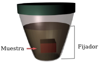
- fixed with 70% ethanol for 12 hours at 4 C
- embedded in paraffin
- Staining
- can be visualized with hematoxylin-eosin staining
- reveal eosinophilic cytoplasm within the secretory cells of apocrine glands
- Periodic Acid Schiff (PAS)
- allows for visualization of diastase-resistant mitochondria-like granules within the secretory portion of the gland
- also contain iron and can be visualized with acid-fast stains.
- allows for visualization of diastase-resistant mitochondria-like granules within the secretory portion of the gland
- can be visualized with hematoxylin-eosin staining
- Staining
- embedded in paraffin
- fixed with 70% ethanol for 12 hours at 4 C
- can be obtained from patients with osmidrosis
Microscopy
- reveals the decapitation of the luminal portion of the secretory cells
- the luminal portion of the cells is lined with microvilli
- reveals the decapitation of the luminal portion of the secretory cells
Imagens e arquivos do mapa






Informações
- Descrição
- Undécimo Histologia Mapa Mental sobre Apocrine Gland, criado por Andrea Mendez em 09-08-2021.
- Criado por
- Andrea Mendez
- Visualizações
- 3
- Avaliação
Mais sobre Glandular Epithelium and Types of Glands