
Mapa mental
Visual system 3
Conteúdo do mapa
Visual system 3
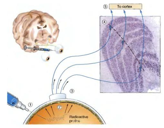
Lateral geniculate nucleus
- Thalamic relay station
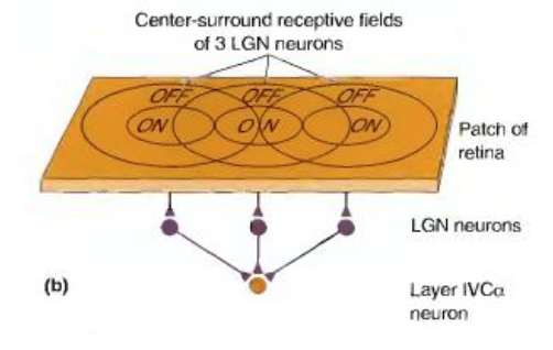
- Ganglion cell axons make 1:1 connections with LGN projection neurons
- Receptive fields of LGN neurons are similar to the receptive fields of retinal ganglion cells
- But:
- 60% of synaptic input from cortex (“back propagation”)
- Local interneurons in LGN
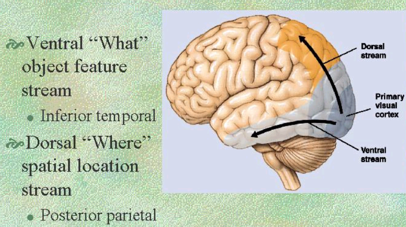
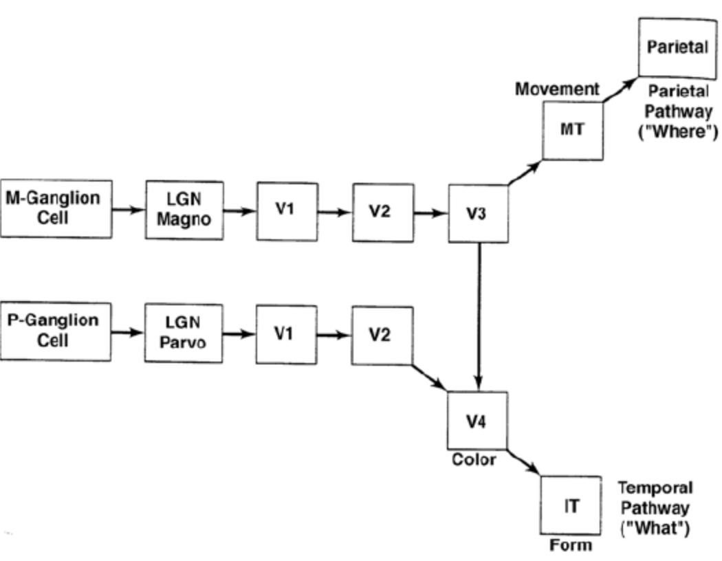
- The human and monkey results indicate two visual pathways
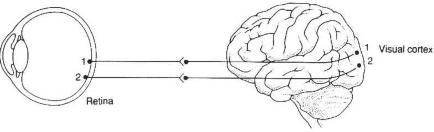
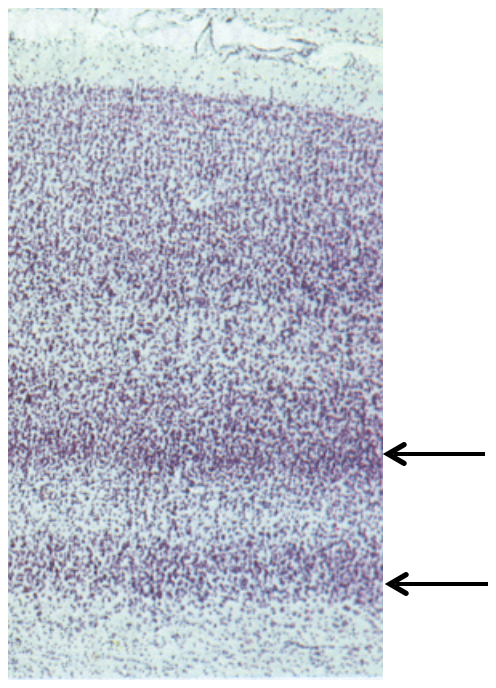
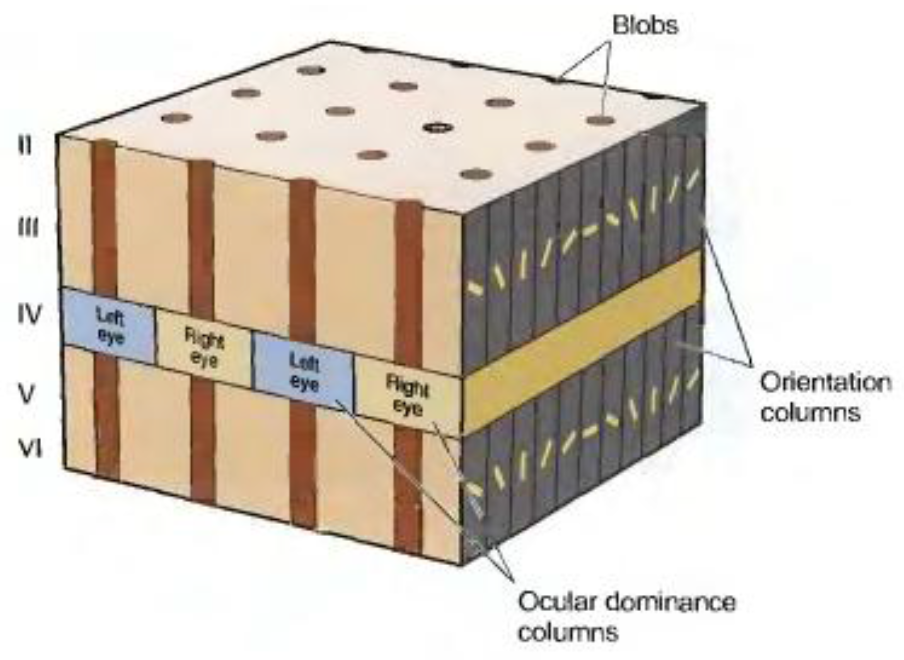
- Neurons in the V1 cortex form retinotopic maps
- Occular dominance columns in the visual cortex
- Each column receives input from either ispilateral or contralateral eye
- Occular dominance columns in the visual cortex
- Neurons in the V1 cortex form retinotopic maps
- Ganglion cell axons make 1:1 connections with LGN projection neurons
- Thalamic relay station
Simple and complex cells
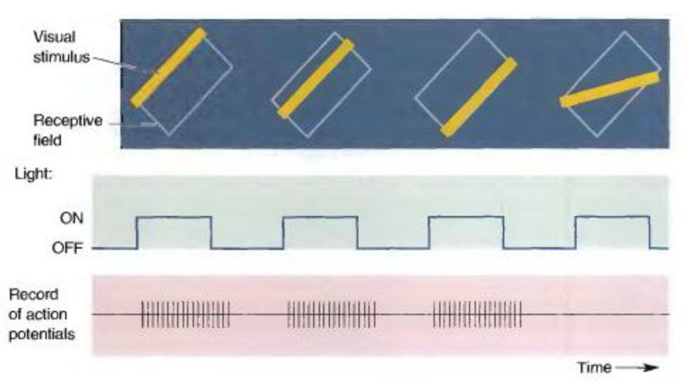
- Simple cells
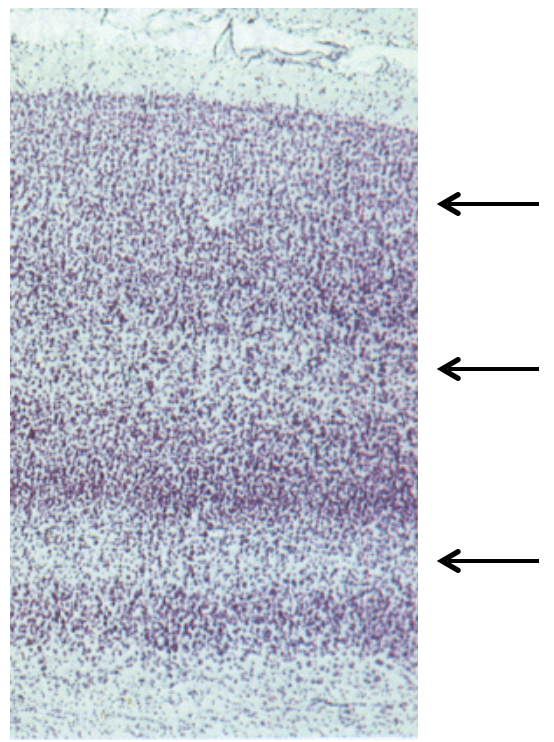
- Localised in layers 4 and 6
- Probable circuit mechanism of receptive field
- See bears neuroscience p328
- Complex cells
- Receptive field
- i.e. respond to a bar in certain orientation presented anywhere in the receptive field
- circuit basis
- Complex cells collect information from many simple cells with similar orientation
- Localised in layers 2,3 and 5
- 'Hypercomplex' = end stopped complex cell
- Receptive field
- Simple cells
Receptive fields downstream of L1
- Increase complexity
- Difficult to study
- 2 approaches
- Guess
- “Spike triggered average” and the like
- Both approaches have limitations
- 2 approaches
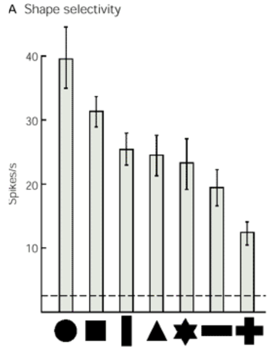
V4 neurons respond to presentation of simple shapes
- Response of the same neuron to presentation of different shapes
- Complexity taken into extreme "grandmother cell" or "jennifer anniston neuron"
- Recording from humans find that 1 or a few neurons recognise a particular object
Object recognition
- Orientation invariance
- Scale invariance
- Lesions in IT cortex decrease ability to recognise objects
- Mainly studied by modelling
- Hierarchical models of object recognition inspired by cortical processing
- IT and higher areas perform categorisation
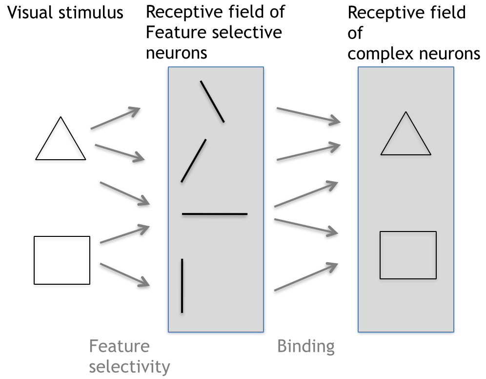
- Shape selective neurons converge to form object selective neurons
- Feature selective neurons converge to form shape selective neurons (V2-V4?)
- Each small part of the retina contains a set of feature selective neurons
- Features = edges?
- Each small part of the retina contains a set of feature selective neurons
- Feature selective neurons converge to form shape selective neurons (V2-V4?)
- Shape selective neurons converge to form object selective neurons
- Problems
- Still poor in scale- and orientation invariance
- Do not take into account feedbacks from higher cortical areas (“top-down regulation”)
- Need experimental validation
- Difficult because would need to record pre- and post-synaptic responses
- Discussed in lecture 5
- Difficult because would need to record pre- and post-synaptic responses
- IT and higher areas perform categorisation
Imagens e arquivos do mapa
Informações
- Descrição
- Undergraduate BMS236 Building Nervous Systems (Visual system) Mapa Mental sobre Visual system 3, criado por Kristi Brogden em 05-08-2014.
- Criado por
- Kristi Brogden
- Visualizações
- 11
- Avaliação