
Mapa mental
Skeletal muscle structure
Mind map: Skeletal muscle structure
Conteúdo do mapa
Skeletal muscle structure
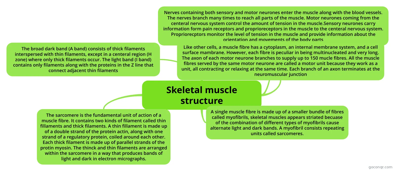
Nerves containing both sensory and motor neurones enter the muscle along with the blood vessels. The nerves branch many times to reach all parts of the muscle. Motor neurones coming from the centeral nervous system control the amount of tension in the muscle.Sensory neurones carry information form pain receptors and proprioreceptors in the muscle to the centeral nervous system. Propriorecptors monitor the level of tension in the muscle and provide information about the orientation and movements of the body parts
Like other cells, a muscle fibre has a cytoplasm, an internal membrane system, and a cell surface mambrane. However, each fibre is peculiar in being multinucleated and very long. The axon of each motor neurone branches to supply up to 150 mucle fibres. All the muscle fibres served by the same motor neurone are called a motor unit because they work as a unit, all contracting or relaxing at the same time. Each branch of an axon terminates at the neuromuscular junction
A single muscle fibre is made up of a smaller bundle of fibres called myofibrils, skeletal muscles appears striated becuase of the combination of different types of myofibrils cause alternate light and dark bands. A myofibril consists repeating units called sarcomeres.
The sarcomere is the fundamental unit of action of a muscle fibre. It contains two kinds of filament called thin fillaments and thick filaments. A thin fillament is made up of a double strand of the protein actin, along with one strand of a regulatory protein, coiled around each other. Each thick filament is made up of parallel strands of the protin myosin. The thinck and thin filaments are arranged within the sarcomere in a way that produces bands of light and dark in electron micrographs.
The broad dark band (A band) consists of thick filaments interspersed with thin filaments, except in a centeral region (H zone) where only thick filaments occur. The light band (I band) contains only filaments along with the proteins in the Z line that connect adjacent thin filaments
Informações
- Descrição
- A-Levels Biology 5 (Nerves and Muscles) Mapa Mental sobre Skeletal muscle structure, criado por harry_bygraves em 14-06-2013.
- Criado por
- harry_bygraves
- Visualizações
- 145
- Tags
- Avaliação