
Mapa mental
Action Potentials (L6-12)

Conteúdo do mapa
Action Potentials (L6-12)
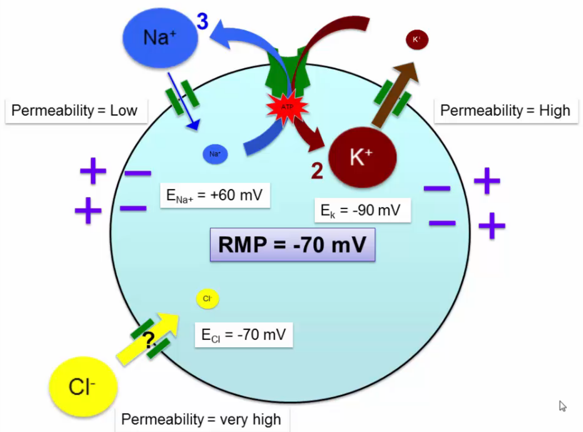
Resting Membrane Potential
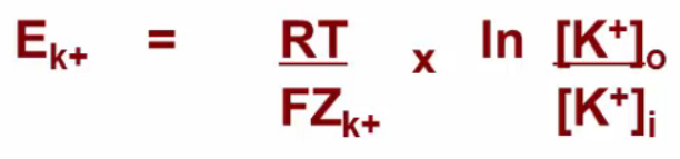
- Nernst Equation for equilibrium potential
- Ek = -90mV
- Means you need to apply -90mV to the cell to keep K from following concentration gradient out
- ENa = +60mV
- ECl = -70mV
- Ek = -90mV
- Goldman Equation for RMP combines all EP
- Typically -70mV
- So K diffuses out, Na diffuses in, no net Cl diffusion
- Typically -70mV
- Nernst Equation for equilibrium potential
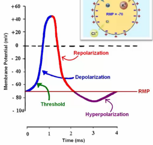
Action Potential
- @ threshold -55mV
- Na+ voltage gated channel opens, depolarizing membrane to +45mV
- K+ voltage gated channel opens (0.5ms delay), repolarizing membrane to -70mV
- Hyperpolarizes below -70 as it reequilibriates
- positive feedback loop
- Inactivation gate closes, enters refractory period
- Tetrodoxin blocks Na+ VG channel
- Reaches threshold but no depolarization
- Greyanotoxin blocks Na+ inactivation gate
- Resists repolarization
- Tetraethylammonium (TEA) & Dendrotoxin inhibits K+ VG channel preventing repolarization
- Local anaesthetics act on the Na+ activation gate
- K+ voltage gated channel opens (0.5ms delay), repolarizing membrane to -70mV
- Cell maintains homeostasis up to -55mV
- Na/K pump
- K leaks out of cell
- Cl leaks into cell
- Depolarizing subthreshold = EPSP
- Hyperpolarizing subthreshold = IPSP
- Na+ voltage gated channel opens, depolarizing membrane to +45mV
- Neurons 1-2ms, Heart 300ms
- @ threshold -55mV
AP Propagation
- Local + charge inside axon is attracted to proximal - charge
- Initially can move any direction but once directionality is established refractory period keeps it moving forward
- Myelin prevents ion leakage, speeding up AP
- AP channels at Nodes of Ranvier
- Thicker axons = faster AP propagation
- MS demyelinates axons
- AP channels at Nodes of Ranvier
- Local + charge inside axon is attracted to proximal - charge
Skeletal Muscle
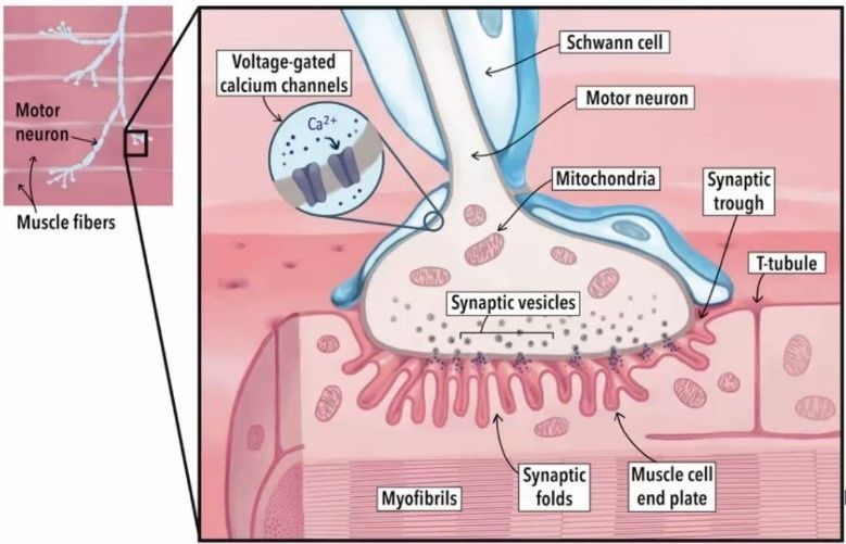
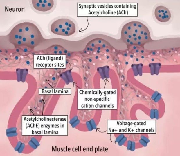
- Neuromuscular Junction
- AP in alpha-motor neuron
- Depolarization of neuron terminal
- Voltage-gated Ca channels open, Ca enters terminal
- Acetylcholine (Ach) vesicles in terminal fuse to membrane, releasing Ach
- Ach diffuses to muscle folded endplates
- 2 Ach bind to Ach-Nicotinic (n1) receptors, opening channel for K and Na to pass through
- Muscle membrane depolarizes, opening VG channels
- AP propagates through muscle from endplate
- Acetylcholinesterase breaks down Ach, closing channels
- AP spreads over sarcolemma and down T-tubules
- DHP receptor on T-tubule changes conformation
- Ryanodine receptor/Ca release channel on SR opens, Ca diffuses to thin filaments (fast)
- Ca binds to troponin C, pulls tropomyosin off actin's myosin binding sites and myosin binds to actin forming crossbridges
- Myosin head power stroke
- Sarcomere shortens
- Ca pumped back into SR by Ca-ATPase (slow)
- Tropomyosin covers Actin's myosin sites again
- Muscle relaxes (slow)
- Tropomyosin covers Actin's myosin sites again
- Ca pumped back into SR by Ca-ATPase (slow)
- Actin
- Actin
- Actin + Myosin + ADP + Pi
- Actin
- Myosin + ATP
- Myosin + ADP + Pi
- Actin + Myosin + ADP + Pi
- Actin + Myosin
- Actin + Myosin + ADP + Pi
- 1. ATP Hydrolyzed. Rate limiting step
- 2. AP on muscle cell releases Ca to expose myosin binding site on Actin
- 3. Crossbridge formation. Pi ejected, triggering power stroke. ADP released after power stroke
- 4. Crossbridge remains until new ATP binds to Myosin, releasing actin
- Continues until ATP runs out (Rigor mortis) or Ca is gone (pumped back into SR)
- 4. Crossbridge remains until new ATP binds to Myosin, releasing actin
- 3. Crossbridge formation. Pi ejected, triggering power stroke. ADP released after power stroke
- 2. AP on muscle cell releases Ca to expose myosin binding site on Actin
- Myosin + ADP + Pi
- Sarcomere shortens
- Myosin head power stroke
- Ca binds to troponin C, pulls tropomyosin off actin's myosin binding sites and myosin binds to actin forming crossbridges
- Ryanodine receptor/Ca release channel on SR opens, Ca diffuses to thin filaments (fast)
- DHP receptor on T-tubule changes conformation
- AP propagates through muscle from endplate
- Muscle membrane depolarizes, opening VG channels
- 2 Ach bind to Ach-Nicotinic (n1) receptors, opening channel for K and Na to pass through
- Ach diffuses to muscle folded endplates
- Acetylcholine (Ach) vesicles in terminal fuse to membrane, releasing Ach
- Voltage-gated Ca channels open, Ca enters terminal
- Depolarization of neuron terminal
- 1 AP in neuron = 1 AP in muscle
- Botulin blocks release of Ach, leads to muscular paralysis
- Nerve gas blocks acetylcholinesterase, keeping channels open and muscles contracted
- Myasthenia Gravis autoimmune disease that blocks Ach receptors. Treated with Neostigmine - competes with antibodies for Ach binding sites
- AP in alpha-motor neuron
- Fascicles --> Muscle cells --> Myofibrils --> Myofilaments - thin and thick
- Membrane = Sarcolemma
- T-tubules = sarcolemma inside the muscle
- Sarcoplasmic Reticulum (SR) between, Ca reservoir
- T-tubules = sarcolemma inside the muscle
- Thick filaments
- Myosin
- 2 long chains forming tail, arm, hinge, and 1 head
- Head contains 2 light protein chains - binding ATP and Actin
- Titin
- Holds thick filaments in place, prevents overstretching
- M-line
- Down middle of thick filaments, holding them in place
- Myosin
- Thin filaments
- G-Actin
- 2 alpha-helices, myosin binding site
- Tropomyosin
- alpha helix that covers myosin binding sites
- Troponin
- Holds tropomyosin over myosin binding site. Has a Ca binding site that releases the tropomyosin
- Z-lines
- Connects thin filaments keeping them in place
- Sarcomere is distance between Z-lines
- G-Actin
- Membrane = Sarcolemma
- Motor units
- 1 alphamotor neuron and all the muscles it innervates
- each muscle has 1 neuron, but multiple muscles per neuron
- Small diameter = small # of muscles
- each muscle has 1 neuron, but multiple muscles per neuron
- More motor units = stronger twitch
- Varying AP frequency can cause multiple twitches that sum without time for the muscle to relax
- No relax = complete tetanic contraction ~30 Hz
- Varying AP frequency can cause multiple twitches that sum without time for the muscle to relax
- 1 alphamotor neuron and all the muscles it innervates
- Muscle fiber types
- Type I - slow-oxidative
- Marathoners
- Low myosin ATPase. 100ms contraction
- High myoglobin (dark)
- Small motoneurons
- Recruited first
- Small motoneurons
- High myoglobin (dark)
- Low myosin ATPase. 100ms contraction
- Marathoners
- Type IIa - fast-oxidative
- Type IIb - fast-glycolytic
- Sprinters
- High myosin ATPase. 10ms contraction
- Glycolysis - easily fatigued
- Low myoglobin (light)
- Large motoneurons
- Recruited last
- Large motoneurons
- Low myoglobin (light)
- Glycolysis - easily fatigued
- High myosin ATPase. 10ms contraction
- Sprinters
- Type I - slow-oxidative
- Neuromuscular Junction
Imagens e arquivos do mapa
Informações
- Descrição
- Mapa Mental sobre Action Potentials (L6-12), criado por J J em 14-11-2018.
- Criado por
- J J
- Visualizações
- 0
- Avaliação