
Mapa mental
The Upper Limb
Conteúdo do mapa
The Upper Limb
Muscles
- Pectoral and Shoulder Regions
- Serratus Anterior
- - rotates the scapula = allows arm to be raised above 90 degrees - long thoracic nerve = when damaged "winging of the scapula" (long thoracic nerve palsy) is produced as serratus anterior is paralysed - thought to occur from traction injuries where upper limb is stretched violently
- Pectoralis Major
- - adducts and medially rotates upper limb - draws scapula anteroinferiorly - lateral and medial pectoral nerves
- Pectoralis Minor
- - stabilises the scapula by drawing it anteroinferiorly against the thoracic wall - medial pectoral nerve
- Subclavius
- - anchors and depresses the clavicle - nerve to subclavius
- Intrinsic
- Subscapularis
- - subscapular fossa - lesser tubercle - upper and lower subscapular nerves - medially rotates the arm
- Rotator Cuff
- - gives glenohumeral joint a lot of additional stability - pull humeral head into glenoid fossa - repetitive use of shoulder joint --> rotator cuff tendonitis - Swimmers shoulder = overuse of supraspinatus muscle - causes inflammation and degenerative changes in the tendon itself
- Supraspinatus
- - supraspinatus fossa - greater tubercle of humerus - suprascapular nerve - abducts arm first 15 degrees - assists deltoid after that
- Infraspinatus
- - infraspinatous fossa - greater tubercle - suprascapular nerve - laterally rotates arm
- Teres Minor
- - posterior surface of scapula - greater tubercle of humerus - axillary nerve - laterally rotates the arm
- Deltoid
- - originates from scapula and clavicle - attaches to deltoid tuberosity on the lateral surface of humerus - axillary nerve - anterior fibers flex arm at the shoulder - posterior fibres extend arm at shoulder - middle fibres major abductor of the arm - takes over from supraspinatus
- Teres Major
- - forms inferior border of quadrangular space - originates from posterior surface of the inferior angle of the scapula - attaches to intertubercular groove of the humerus - lower subscapular nerve - adducts at shoulder and medially rotates the arm
- Subscapularis
- Extrinsic
- - originate from torso and attach to bones of the shoulder
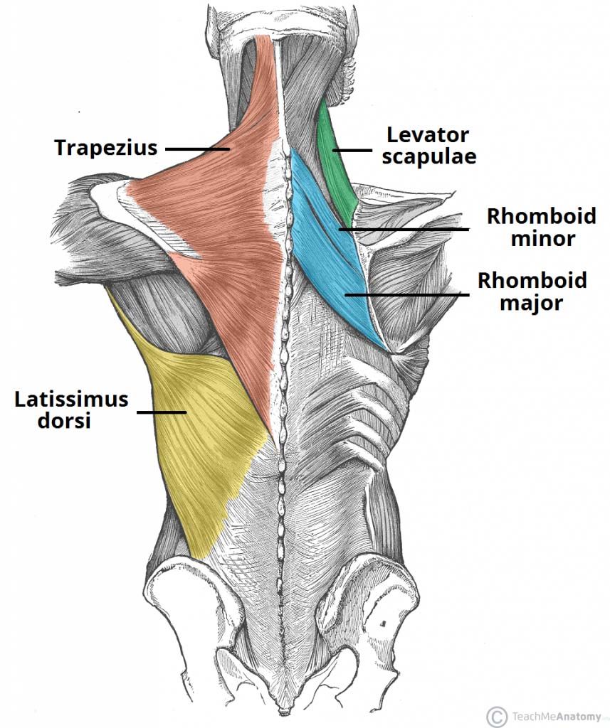
- Trapezius
- - originates from skull - fibres attach to clavicle, acromion and scapula spine - accessory nerve - proprioceptor fibres from C3 and C4 spinal nerves - upper fibres elevate scapula and rotates it during abduction of the arm - middle fibres retract scapula - lower fibres full scapula inferiorly
- Latissimus Dorsi
- - thoracodorsal nerve - extends, adducts and medially rotates the upper limb
- Rhomboid Major
- - attaches to medial border of the scapula, between the scapula spine and inferior angle - dorsal scapular nerve - retracts and rotates scapula
- Rhomboid Minor
- - attaches to medial border of scapula, at level of the spine of scapula - dorsal scapula nerve - retracts and rotates the scapula
- Levator Scapulae
- - attaches to medial border of the scapula - dorsal scapular nerve - elevates the scapula
- Serratus Anterior
- Arm
- Anterior
- Biceps brachii
- - 2 headed - gives of the bicipital aponeurosis - supraglenoid tubercle of scapula = long head - coracoid process of scapula = short head - inserts into radial tuberosity and fascia of the forearm via bicipital aponeurosis - supination of the forearm - rupture of long head = popeye sign- flexes arm at elbow and at shoulder- musculocutaneous nerve
- Brachialis
- - medial and lateral surfaces of humeral shaft - inserts into ulna tuberosity - flexion at elbow - musculocutaneous nerve with contributions from radial nerve
- Coracobrachialis
- - coracoid process - flexion of arm at shoulder - weak adduction - musculocutaneous nerve
- Biceps brachii
- Posterior
- Triceps brachii
- - 3 headed - long head = infraglenoid tubercle - lateral head = humerus superior to radial groove - medial head = inferior to radial groove - all insert into olecranon of ulna - extension of arm at elbow - radial nerve
- Triceps brachii
- Anterior
- Forearm
- Posterior
- Superficial
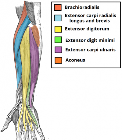
- Brachioradialis
- - flexes at elbow - radial nerve
- Extensor Carpi Radialis Longus
- - supracondylar ridge - extends and abducts wrist -radial nerve
- Extensor Carpi Radialis Brevis
- - lateral epicondyle - extends and abducts wrist - radial nerve
- Extensor Digitorum
- - lateral epicondyle - extends medial 4 fingers at the MCP and IP joints - radial nerve
- Extensor Digiti Minimi
- - lateral epicondyle - extends little fingers, contributes to extension at wrist - radial nerve (deep branch)
- Extensor Carpi Ulnaris
- - lateral epicondyle - extension and adduction of wrist - radial nerve (deep branch)
- Aconeus
- - lateral epicondyle - extends and stabilises elbow joint - abducts ulna during pronation of the forearm - radial nerve
- Brachioradialis
- Deep
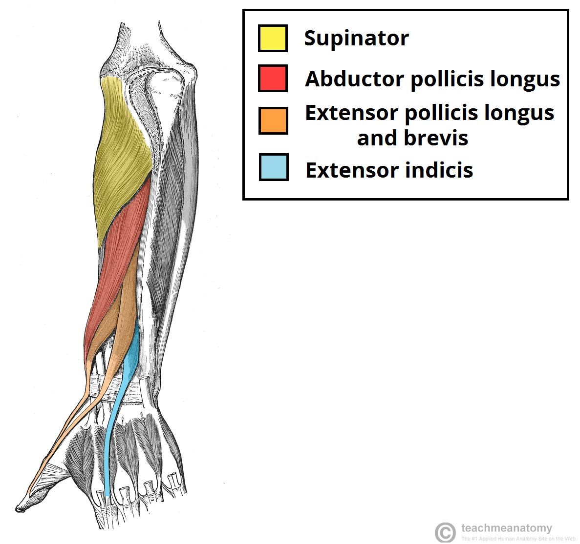
- Supinator
- - lateral epicondyle and posterior surface of ulna - supinates forearm - radial nerve (deep branch)
- Abductor Pollicis Longus
- - interosseous membrane and adjacent posterior surfaces of radius and ulna - abducts thumb - radial nerve (posterior interosseous branch)
- Extensor Pollicis Brevis
- - posterior surface of ulna and interosseous membrane - extends at MCP and CM joints - radial nerve (posterior interosseous branch)
- Extensor Pollicis Longus
- - posterior surface of ulna and interosseous membrane - extends all joints of the thumb - radial nerve (posterior interosseous branch)
- Extensor Indices
- - posterior surface of ulna and interosseous membrane - extends index finger - radial nerve (posterior interosseous branch)
- Supinator
- Superficial
- Anterior
- Superficial
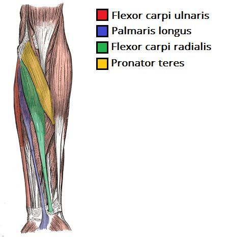
- Flexor Carpi Radialis
- - medial epicondyle - flexion and abduction at wrist - median nerve
- Palmaris longus
- - flexion and abduction of wrist - median nerve - medial epicondyle
- Flexor carpi ulnaris
- - ulnar nerve - flexion and adduction at wrist - medial epicondyle
- Pronator Teres
- - 2 attachments = medial epicondyle and coronoid process of ulna - pronation of forearm - median nerve
- Flexor Carpi Radialis
- Intermediate
- Flexor Digitorum Superficialis
- - medial epicondyle and radius - flexes MCP joints and PIP joints - flexes at wrist - median nerve
- Flexor Digitorum Superficialis
- Deep
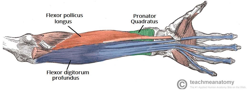
- Flexor Digitorum Profundus
- - ulna and associated interosseous membrane - only muscle that can flex DIP joints of fingers - also flexes MCP joiints and at wrist - medial half = ulnar nerve - lateral half = median nerve
- Pronator Quadratus
- - anterior surface of ulna - pronates forearm - median nerve (anterior interosseous branch)
- Flexor Pollicis Longus
- - lies laterally to FDP - anterior surface of radius and surrounding interosseous membrane - flexes IP joint and MCP join of thumb - median nerve (anterior interosseous branch)
- Flexor Digitorum Profundus
- Superficial
- Posterior
- Hand
- Pectoral and Shoulder Regions
Brachial Plexus
- Median n.
- Musculocutaneous n.
- Ulnar n.
- Radial n.
- Axillary n.
Bones
- Shoulder Area
- Scapula
- Clavicle
- Function - attaches upper limb to trunk - protects underlying neurovascular structures supplying - transmits force from upper limb to axial skeleton Bony landmarks- sternal ended has large facet for articulation with the manubrium of the sternum- costoclavicular ligament attaches to inferior surface of sternal end- conoid tubercle where the conoid ligament attaches- trapezoid line where the trapezoid ligament attaches Clinical RelevanceFracture: most common is junction of the medial 2/3 and lateral 1/3. Suprascapular nerves (medial, intermedial and lateral) can be damaged by upwards movement of the medial part of the the fracture = waiter's tip
- Arm
- Humerus
- Forearm
- Ulna
- Radius
- Hand
- Shoulder Area
Vasculature
Clinical Examination
- Shoulder
- Wrist
- Hand
- Systems Review
Imagens e arquivos do mapa
Informações
- Descrição
- Mapa Mental sobre The Upper Limb, criado por Michelle Davis em 15-08-2017.
- Criado por
- Michelle Davis
- Visualizações
- 4
- Avaliação