
Notas
Sem título
Conteúdo das notas (3)
Página 1
surgery anatomy notes : hernia Surface Anatomy
surface anatomy :
The linea alba:
The linea alba is a shallow groove that runs vertically in the median plane from
the xiphoid to the pubis .components of the rectus sheath intersect at the linea alba.
Planes and Regions:
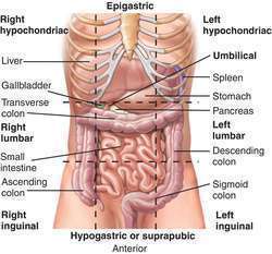
The anterior abdominal wall is divided into 9 regions separated by several
planes and lines.
Te subcostal plane (horizontal) : passes through the inferior margins of the
10th costal cartilages at the level of the third lumbar vertebra.
The transpyloric plane passes through the L1 vertebra, being half the distance
between the pubis and the jugular notch. The plane passes through several
important abdominal landmarks useful for radiology: pylorus of the stomach
(variable), fundus of gallbladder, neck, and body of the pancreas, hila of kidneys,
first part of the duodenum, and origin of the superior mesenteric artery.
Te midclavicular lines (vertical) are the 2 planes that pass from the midpoint
of the clavicle to the midpoint of the inguinal ligament on each side.
Página 2
surgery anatomy notes :(breast)
axilla anatomy
Borders
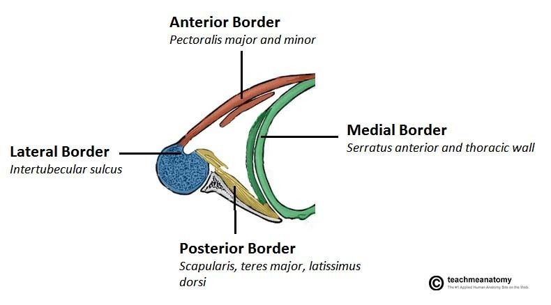
The overall 3D shape of the axilla looks slightly like a pyramid. It consists of four sides, an open apex and base:
Apex – also known as the axillary inlet, it is formed by lateral border of the first rib, superior border of scapula, and the posterior border of the clavicle.
Lateral wall – formed by intertubercular groove of the humerus.
Medial wall – consists of the serratus anterior and the thoracic wall (ribs and intercostal muscles).
Anterior wall – contains the pectoralis major and the underlying pectoralis minor and the subclavius muscles.
Posterior wall – formed by the subscapularis, teres major and latissimus dorsi.
contents:
Axillary artery (and branches): first part: superior thoracic artery
second part: thoracoacromial and lateral thoracic
third part: posterior and anterior humeral circumflex and subscapular (gives 2 branches:1) thoracodorsal 2) circumflex scapular)
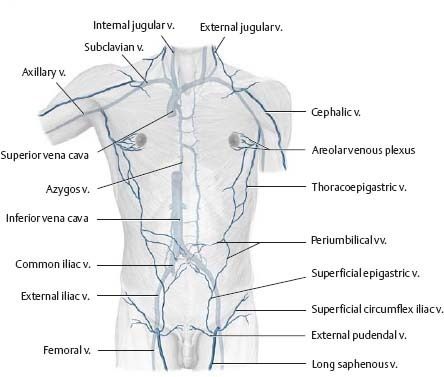
Mondor's disease occur in :-lateral thoracic vein
- and thoracopigastric disease (runs along the lateral aspect of the trunk between the superficial epigastric vein below and the lateral thoracic vein above and establishes an important communication between the femoral vein and axillary vein.)
Página 3
Informações
- Descrição
- Notas sobre Sem título, criado por reem alawnaa em 03-04-2019.
- Criado por
- reem alawnaa
- Visualizações
- 5
- Avaliação