
Quiz
Gastrointestinal pathology :)
Este quiz tem tipos de pergunta que abrem no player de sempre.
Perguntas do quiz (119)
1A middle-aged man with longstanding Barrett’s esophagus is referred for specialist review after he reports increasing dysphagia over the last 4 months. He has noted that it initially only affected solids but now struggles with liquids as well. What is the next most important investigation to aid diagnosis?
- ABlood investigation for tumour markers
- BBarium swallow
- CCT
- DPET
- EEndoscopy
2What is the first line maintenance treatment for Ulcerative Collitis?
- AMesalazine
- BCiclosporin
- CPrednisolone
- DInfliximab
- EAzathioprine
3What do parietal cells produce?
- Apepsinogen
- Bsomatostatin
- Cgastrin and histamine
- Dhydrochloric acid and intrinsic factor
- Ehistamine only
4An elderly man with a history of alcohol abuse goes to ED with hematemesis for 1 day. He has no abdominal or chest pain. His eyes are reddened and he admits to drinking heavily the night before he vomited several times. What may be the reason for this?
- AOesophageal varices
- BLung Cancer
- CAchalasia
- DPerforated gastric ulcer
- EMallory-Weiss tear
5A 20-year-old man presents to the ED with fever and severe right lower quadrant (RLQ) pain for 1 day. Prior to this episode, he reports 2 months of crampy abdominal pain, generalized malaise, a 5kg weight loss, and occasional bloody diarrhoea. On physical exam, the patient appears uncomfortable and has a tender mass is in the RLQ. An abdominal CT scan demonstrates normal appendix and inflammation of the distal ileum and several areas of the colon without rectal involvement. Which of the following is the most likely diagnosis?
- ACrohn's Disease
- BUlcerative Colitis
- CAppendicitis
- DDiverticulitis
- EPseudomembranous colitis
6An older woman arrives at ED complaining of abdominal pain. She states that the pain began as a vague aching but has migrated to the left side of her abdomen. She had a fever over the past 3 days and just not feeling like herself. She also complains of being more constipated than usual. Your abdominal exam reveals normal bowel sounds, mild distension, and tenderness over the left lower quadrant. What is most likely the reason for this
- AColorectal cancer
- BDiverticulitis
- CAppendicitis
- DCrohn’s disease
- EDuodenal ulcer
7What is the blood supply to the superior rectum?
- Aceliac artery
- Bsuperior mesenteric artery
- Cinferior mesenteric artery
- Dinternal iliac artery
- Eexternal iliac artery
8A elderly man attends your clinic with a three-month history of weight loss of approximately 9 kg despite a normal appetite. A full blood count reveals that his hemoglobin is 9.0 g/dL (previous hemoglobin was 13.5 g/dL one year ago) and the MCV is 71 fL.
The abdominal examination is unremarkable and per rectum exam is unremarkable. The patient states that he has normal bowel habits and has been feeling quite tired lately. What is the most appropriate management?- AReassure and discharge
- BPrescribe iron tablet supplementation
- Chave an abdominal ultrasound
- Dhave an abdominal x-ray
- Ehave an upper and lower GI endoscopy
9A middle aged woman has a four-month history of diarrhoea and bloating. Associated symptoms include lethargy and weight loss.
Full blood count reveals:
hemoglobin of 9.3 d/gL (13.8 to 17.2)
MCV 70 fL. (77-95)
What other investigation allow proper patient diagnosis?- AAnti-mitochondrial antibodies
- BAnti-smooth muscle antibodies
- CAnti-nuclear antibodies
- DAnti-tissue transglutaminase antibodies
- EAnti-neutrophil cytoplasmic antibodies
10You see an elderly man with an acute episode of hematemesis, who was admitted the night before and is awaiting an upper GI endoscopy. You are asked on the ward round about the common causes of upper GI bleeding. From the list below, which of the following is the most common cause of upper GI bleeding?
- AMallory–Weiss tear
- BPeptic ulcers
- CEsophageal varices
- DDrug induced
- ESporadic malignancy
11You see a middle aged woman with Crohn’s disease. Due to a severe attack of Crohn’s which failed to respond to medical therapy, she had a small bowel resection. Your registrar tells you that she is at risk of developing vitamin B12 deficiency as a result of her surgery. Which part of the bowel is responsible for the absorption of vitamin B12?
- AAscending colon
- BTerminal ileum
- CDuodenum
- DJejunum
- EProximal Ileum
12You see a middle aged man who was admitted for an elective upper GI endoscopy due to longstanding GORD which has failed to improve on antacids and PPIs. Your registrar suspects that this patient may have Barrett’s oesophagus and asks you to define what this is:
- AMetaplasia of the squamous epithelium of the lower third of the esophagus to columnar epithelium
- BMetaplasia of the columnar epithelium of the upper third of the oesophagus to squamous epithelium
- CMetaplasia of the squamous epithelium of the middle third of the oesophagus to columnar epithelium
- DMetaplasia of the squamous epithelium of the upper third of the oesophagus to columnar epithelium
- EMetaplasia of the columnar epithelium of the lower third of the oesophagus to squamous epithelium
13Which of the following is the most common cause of duodenal ulcers?
- ANSAIDs
- BAlcohol Abuse
- CHelicobacter pylori
- DChronic corticosteroid therapy
- EZollinger-Ellison syndrome
14A middle-aged lorry driver presents to the GP with chest pain that usually occurs after having a meal. He has no major past medical history and he takes ibuprofen regularly. He is otherwise fit and healthy. What is the most important next step in management?
- AUrgent referral to endoscopy.
- BRoutine referral to endoscopy.
- CPrescribe omeprazole, a PPI.
- DPerform a H. pylori urea test.
- EPerform a 12-lead ECG
15You see a middle aged lorry driver, who presents to you with a three-month history of heartburn after meals which has not been settling with antacids and PPIs. You suspect that the patient has a hiatus hernia. The most appropriate investigation for diagnosing a hiatus hernia is:
- ACT scan
- BChest x-ray
- CBarium meal
- DUltrasound
- EUpper GI endoscopy
16A middle-aged woman is referred for upper gastrointestinal (GI) endoscopy following a four-month history of epigastric pain despite treatment with antacids and proton pump inhibitors (PPIs). The results demonstrate a duodenal ulcer coupled with a positive campylobacter-like organism (CLO) test. The patient has no past medical history and has no known drug allergies. What is the most appropriate treatment?
- ASeven-day course of twice daily omeprazole 20 mg, 1 g amoxicillin and 500 mg clarithromycin
- BSeven-day course of twice daily omeprazole 20 mg
- CSeven-day course of twice daily omeprazole 20 mg and 1 g amoxicillin
- DSeven-day course of twice daily omeprazole 20 mg and 500 mg clarithromycin
- ESeven-day course of twice daily 1 g amoxicillin and 500 mg clarithromycin
17You see a middle aged man with a three-month history of epigastric dull abdominal pain. The pain is worse in the mornings and is better after meals. There is no history of weight loss and the patient’s bowel habits are normal. His abdomen is soft and experiences moderate discomfort on palpation of the epigastric region. What is the most likely diagnosis?
- AGastric ulcer
- BGastro-oesophageal reflux disease (GORD)
- CGastritis
- DGastric carcinoma
- EDuodenal ulcer
18You see an elderly man who comes to ED with epigastric pain. The pain started 3 days ago and today his stools color has changed to a ‘tarry-black’ color. Associated symptoms include nausea and lethargy.
The patient is alert and orientated with a pulse rate of 99 and blood pressure of 98/69, respiratory rate of 18, oxygen saturations of 98 per cent on room air and temperature of 37.2°C.
The abdomen is soft with marked tenderness in the epigastric region and bowel sounds are present. The rectum is empty, on PR examination, with some traces of melaena. The patient has been started on high flow oxygen and has been given some oral analgesia. The most appropriate next step in managing this patient is:- AECG
- BIntravenous omeprazole
- CRequest an erect chest x-ray
- DKeep nil by mouth and arrange endoscopy
- EIntravenous cannulation and fluids
19As you palpate the right upper quadrant (RUQ) of a middle aged woman’s abdomen, you notice that she stops her inspiration for a brief moment. The patient states that over the last 2 days she gets pain in her RUQ that radiates to her back shortly after eating.
Her vitals are temperature 38°C, heart rate (HR) 95, blood pressure (BP) 130/75, respiratory rate (RR) 16. What is the initial diagnostic modality of choice for this disorder?- APlain film radiograph
- BCT scan
- CUltrasound
- DMRI
- EERCP
20A middle-aged woman at ED complains of pain in her RUQ that is steady but gets worse with eating over the past 2 days. The pain also radiates to the right side of her mid-back. No vomiting. Her only medication is an oral contraceptive.
Her BP is 140/75 mm Hg, HR is 80 beats per minute, temperature is 37°C, and RR is 16 breaths per minute. Laboratory tests are within normal limits. What is the most likely diagnosis?- ACholangitis
- BPancreatitis
- CCholecystitis
- DPeptic ulcer disease
- EBiliary colic
21A 50-year-old man presents to the ED complaining of abrupt onset of epigastric pain that radiates to his back. He describes the pain as constant and associated with nausea and vomiting. The pain improves mildly if he leans forward. He has a 20-pack-year smoking history and consumes a 6-pack of beer daily for more than 5 years. His BP is 150/80 mm Hg, HR is 98 beats per minute, temperature is 37.8°C, and his RR is 18 breaths per minute. He is tender to palpation in the epigastric area. His WBC count is normal and lipase 520 U/L (0-160 U/L). What is the most likely diagnosis?
- ACholecystitis
- BBowel perforation
- CMesenteric ischemia
- DAbdominal aortic aneurysm
- EPancreatitis
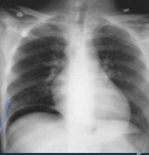
22A 55-year-old man presents to the ED complaining of mild diffuse abdominal pain. The pain began upon waking up and is associated with some nausea. He denies fever, vomiting, diarrhoea, and rectal bleeding. His BP is 143/71 mm Hg, HR is 87 beats per minute, temperature is 36°C, and RR is 16 breaths per minute. His abdomen is tense but only mildly tender. You order baseline labs. His chest radiograph is seen below. Which of the following is the most likely diagnosis?
- A
- BAscending cholangitis
- CAcute pulmonary edema
- DRuptured aneurysm
- EPancreatitis
- FPerforation with pneumoperitoneum
- A
23A 33-year-old man presents to the ED complaining of lower abdominal pain. He states that last night he ate pizza with pepperoni for dinner, went to sleep, and awoke several hours later with a vague, periumbilical pain. The pain has been constant since its onset and has migrated to his lower abdomen more on the right than left. Since eating the pizza he has no appetite. He is nauseated and vomited twice in the ED. His BP is 125/70 mm Hg, HR is 88 beats per minute, temperature is 38°C, and his RR is 16 breaths per minute. On exam, his RLQ is tender to palpation. What is the most likely diagnosis?
- ACholangitis
- BAppendicitis
- CDiverticulitis
- DPancreatitis
- EAcute cholecystitis
24Patient presents with painless jaundice and palpable RUQ mass. Which of the following is the most likely diagnosis
- ACholecystitis
- BCholangitis
- CAbdominal aortic aneurysm
- DCrohn’s disease
- EGall bladder carcinoma
25A 71-year-old obese man is brought to the ED complaining of constant left middle quadrant abdominal pain with radiation into his back. His past medical history is significant for hypertension, peripheral vascular disease, and kidney stones. He smokes a pack of cigarettes daily. His BP is 145/80 mm Hg, HR is 90 beats per minute, temperature is 37.2°C, and RR is 16 breaths per minute. Abdominal exam shows a pulsatile abdominal mass. An abdominal radiograph reveals normal loops of bowel. Which of the following is the most likely diagnosis?
- ABiliary colic
- BDiverticulitis
- CPancreatitis
- DSmall bowel obstruction
- EAbdominal aortic aneurysm
26A 78-year-old woman is brought to the ED by EMS complaining of vomiting and abdominal pain that began during the night. EMS reports that her BP is 90/50 mm Hg, HR is 110 beats per minute, temperature is 38°C, and RR is 18 breaths per minute. After giving her a 500 mL bolus of normal saline, her BP is 115/70 mm Hg. During the exam, you notice that her face and chest appear jaundiced. What is the most likely diagnosis?
- ACholecystitis
- BAcute hepatitis
- CCholangitis
- DPancreatic cancer
- EBowel obstruction
27What is NOT true regarding GI anatomy?
- AThe foregut is from the distal (abdominal) esophagus to hepatopancreatic ampulla of the duodenum (2nd part of the duodenum.)
- BThe mesentery includes the liver, gallbladder, pancreas, and spleen
- CMidgut ends at 2/3rds along the transverse colon.
- DThe Hindgut ends at the anus.
28The GI wall, from the [blank_start]lower[blank_end] [blank_start]espohagus[blank_end] to the [blank_start]anus[blank_end], has the same 4 layers (from outermost to innermost)
1. [blank_start]Serosa[blank_end]
2. [blank_start]Muscularis[blank_end]
3. [blank_start]Submucosa[blank_end]
4. [blank_start]Mucosa[blank_end]- Alower
- Bespohagus
- Canus
- DSerosa
- EMuscularis
- FSubmucosa
- GMucosa
29The foregut is supplied by the [blank_start]celiac[blank_end] [blank_start]trunk[blank_end], which comes off the aorta at level [blank_start]T12[blank_end].
The midgut is supplied by the [blank_start]superior[blank_end] [blank_start]mesenteric[blank_end] artery, which comes off the aorta at level [blank_start]L1[blank_end].
The hindgut is supplied by the [blank_start]inferior[blank_end] [blank_start]mesenteric[blank_end] artery, which comes off the aorta at level [blank_start]L3[blank_end].- Aceliac
- Btrunk
- CT12
- Dsuperior
- Emesenteric
- FL1
- Ginferior
- Hmesenteric
- IL3
30The GI tract lymphatics and vasculature is about the same.
- AVerdadeiro
- BFalso
31Which is NOT a major lymph structures involved with abdominal drainage?
- ACeliac nodes
- BSuperior Mesenteric Aortic/Caval nodes
- CInferior Mesenteric Aortic/Caval nodes
- DRenal nodes
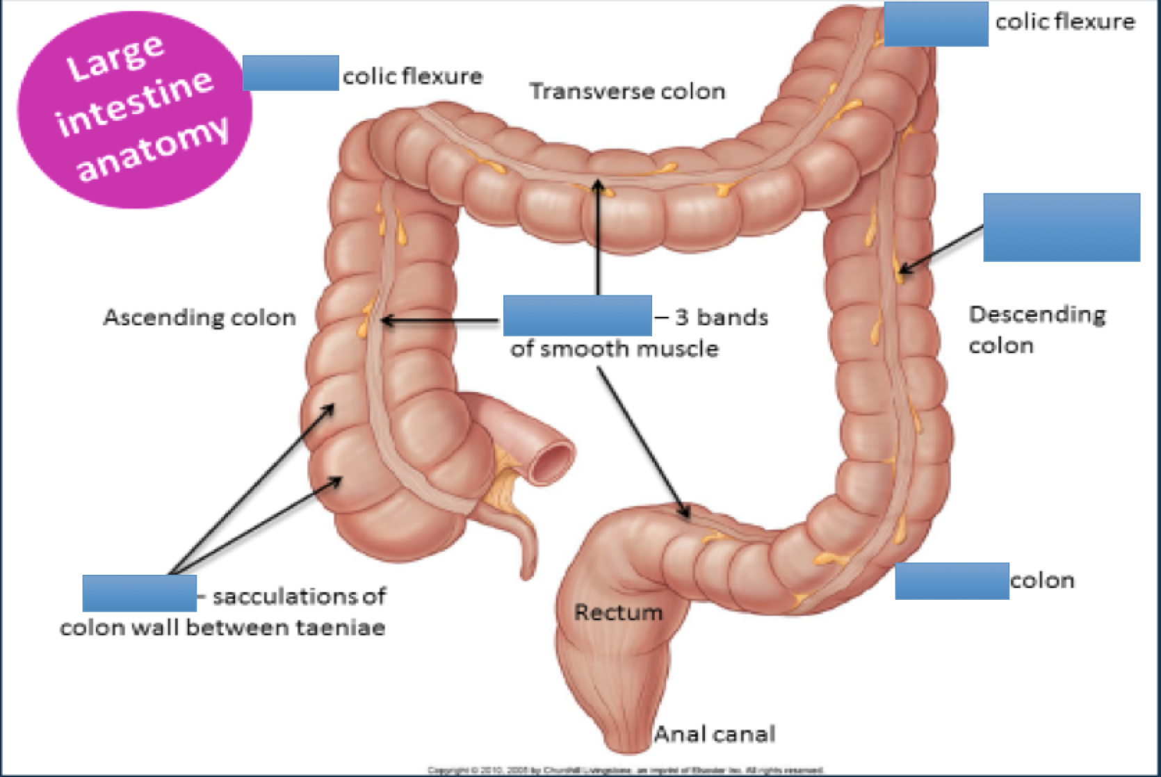
32Label the parts.
- Aright
- Bleft
- Chastura
- Dtenie coli
- Esigmoid
- Fomental appendices
33Large Intestine Blood Supply:
[blank_start]Superior[blank_end] rectal artery is a branch of the [blank_start]inferior[blank_end] [blank_start]mesenteric[blank_end] artery.
However, the [blank_start]middle[blank_end] rectal and [blank_start]inferior[blank_end] rectal arterys are both branches of [blank_start]internal[blank_end] [blank_start]iliac[blank_end] artery.- Ainferior
- Bmesenteric
- Cinternal
- Diliac
- ESuperior
- Fmiddle
- Ginferior
34What is true regarding the esophagus' histology?
- Amade of nonkeratinized squamous epithelium
- Bupper skeletal sphincter
- Clower smooth sphincter
- Dupper 1/3 skeletal muscle
- Emiddle 1/3 skeletal and smooth muscle
- Flower 1/3 smooth muscle
- Gcyclic mucus secretions
- Hupper 1/3rd squamous cells
35Pharyngeal Pouch ([blank_start]Zenker[blank_end] diverticulum) is an out-pouching (diverticulum) that develops from the [blank_start]posterior[blank_end] wall of the [blank_start]pharynx[blank_end] just prior to the [blank_start]esophagus[blank_end].
- Aposterior
- Bpharynx
- Cesophagus
- DZenker
36What is true regarding Zenker diverticulum?
- Amanagement: surgery
- Binvestigation: barium swallow
- Cmanagement: corticosteroids
- Ddysphagia
- Ehalitosis
- Fstomach gurgling upon palpatation
- Gconstipation
- Hdiarrea
37What is true regarding achalasia?
- Aaffects esophageal motility
- Blower esophageal sphincter unable to relax
- Cgradual dysphagia to solids and liquids
- Dweight loss
- Eform of acid reflux--> damages esophageal cells
- Fvomiting
- Gprolonged dysphagia
- Hdrugs trtment: nitrates
- Idrugs trtment: caclium channel blockers
- Jinvestiagtion: manometry and endoscopy
38[blank_start]Gastroesophageal[blank_end] [blank_start]Reflux[blank_end] [blank_start]Disease[blank_end] (GERD/GORD) is a condition in which the stomach contents are regurgitated back the esophagus, usually because the [blank_start]lower[blank_end] [blank_start]esophageal[blank_end] sphincter is unable to fully close after swallowing.
- AGastroesophageal
- BReflux
- CDisease
- Desophageal
- Elower
39What is true regarding GORD?
- Adyspepsia is main symptom
- Bpreferable investigation via endoscopy
- Cmanage via lifestyle changes
- Dmanage via proton pump inhibitors
- Emanage via gastrin inhibitors
- Frisk factors = smoking, alcohol, and obesity
- Grisk factor = chocolate
- Hrisk factor= pregnancy
- Ipreferable investigation via blood tests
- Jpreferable investigation via sputum culture
40A Mallory-Weiss Tear is when there is bleeding from a laceration in the [blank_start]mucosa[blank_end] at the junction of the [blank_start]stomach[blank_end] and [blank_start]esophagus[blank_end]. This is usually caused by severe [blank_start]vomiting[blank_end], such as from [blank_start]alcoholism[blank_end], bulimia, or food [blank_start]poisoning[blank_end].
The signaling symptom is painful hematemesis. It is diagnosed by endoscopy.- Amucosa
- Bstomach
- Cesophagus
- Dvomiting
- Ealcoholism
- Fpoisoning
41Label the diagram showing the GI venous drainage. (the colors are just to differentiate between the various vessels)
- Ainferior mesenteric vein
- Bsuperior mesenteric vein
- Chepatic portal vein
- Dsplenic vein
- Esplenic vein
42The liver is supplied by the hepatic portal vein (which is made up of the [blank_start]splenic[blank_end] vein and the [blank_start]superior[blank_end] [blank_start]mesenteric[blank_end] vein) and the hepatic artery.
[blank_start]75[blank_end]% of blood going to the liver is from the hepatic portal vein while [blank_start]25[blank_end]% is from the hepatic artery.
However, [blank_start]oxygenated[blank_end] blood is split ~50:50 between the portal vein and hepatic arteries.- Asplenic
- Bmesenteric
- Csuperior
- D75
- E25
- Foxygenated
43Which of these does NOT directly come off of the celiac trunk?
- Asplenic artery
- Bcommon hepatic artery
- Cleft gastric artery
- Dproper hepatic artery
44The common hepatic artery becomes the proper hepatic artery after the gastroduodenal artery branch.
- AVerdadeiro
- BFalso
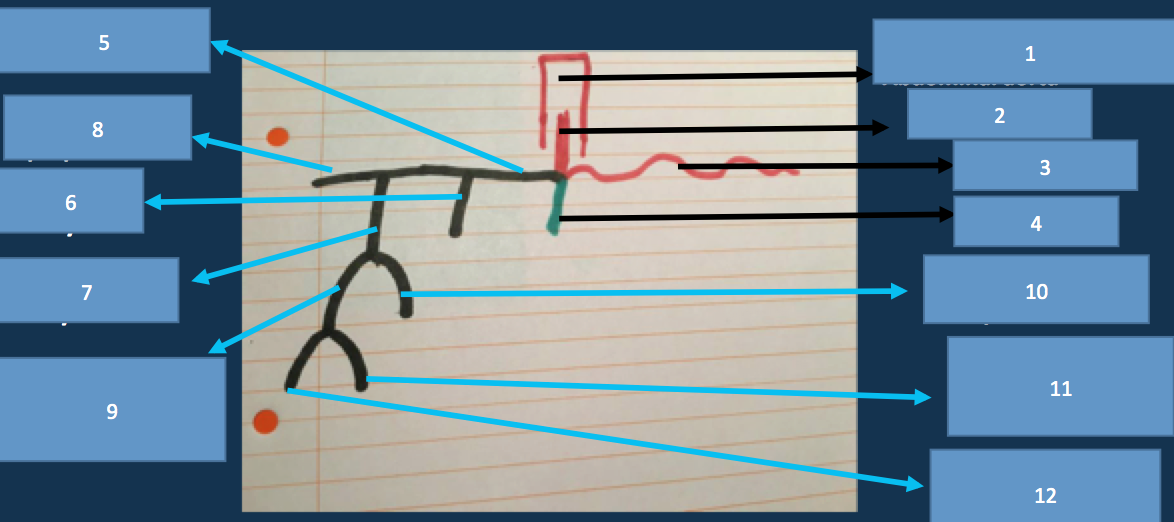
45Label the parts of the GI arterial vasculature. (max amnt words for any answr is 3)
- Aabdominal aorta
- Bceliac artery
- Csplenic artery
- Dleft gastric artery
- ESuperior Posterior Pancreatoduodenal
- FSuperior Anterior Pancreatoduodenal
- GSuperior pancreatoduodenal artery
- HRight gastroepiploic artery
- Icommon hepatic artery
- JHepatic artery proper
- KRight gastric artery
- LGastroduodenal artery
46Which comes first in the development of a carcinoma?
- Ametaplasia
- Bdysplasia
47[blank_start]Metaplasia[blank_end] is the reversible change of one cell type to another
[blank_start]Dysplasia[blank_end] is abnormal changes in the cell and disorganized [blank_start]growth[blank_end]
Carcinoma is the eventual transformation into malignant [blank_start]neoplastic[blank_end] cells.- AMetaplasia
- Bgrowth
- CDysplasia
- Dneoplastic
48[blank_start]Barrett’s[blank_end] [blank_start]Esophagus[blank_end] is a pre-malignant condition of the lower esophagus. This happens when the [blank_start]squamous[blank_end] cells go through metaplasia and become [blank_start]columnar[blank_end] cells. This change usually happens as defence against an [blank_start]acidic[blank_end] environment, which may be due to conditions like [blank_start]GERD[blank_end]. This condition puts the patient of increased risk of esophageal [blank_start]adenocarcinoma[blank_end].
- ABarrett’s
- BEsophagus
- Csquamous
- Dcolumnar
- Eacidic
- FGERD, GORD
- Gadenocarcinoma
49The management of Barret's Esophagus is to do a [blank_start]biopsy[blank_end].
Premalignant changes are indicative of high-grade [blank_start]dysplasia[blank_end]. The treatment for this in younger and more fit individuals is an [blank_start]esophageal[blank_end] [blank_start]resection[blank_end]. For others, it's [blank_start]mucosal[blank_end] [blank_start]ablation[blank_end].
If there are no premalignant channges, the patient will have regular [blank_start]endoscopy[blank_end], biopsy, and other anti-reflux measures, including long term [blank_start]proton[blank_end] [blank_start]pump[blank_end] [blank_start]inhibitors[blank_end] (drugs).- Aendoscopy
- Bproton
- Cpump
- Dinhibitors
- Edysplasia
- Fesophageal
- Gresection
- Hablation
- Imucosal
- Jbiopsy
50What is true regarding esophageal carcinoma?
- Asymptom: vomiting
- Bsymptom: weight loss
- Csymptom: anorexia
- Dpresentation: progressive dysphagia
- Einvestigation: chest xray
- Finvestigation: urgent endoscopy + biopsy
- Ginvestigation: CT or MRI chest and upper abdomen
- Hsymptom: hoarse voice
- Isymptom: excessive acid reflux
- Jinvestigation: neck ultrasound
51The stomach has 2 muscle layers: inner oblique and outer longitudinal
- AVerdadeiro
- BFalso
52The stomach is lined by simple squamous epithelial cells
- AVerdadeiro
- BFalso
53What controls the secretion of gastric acid?
- Athe parasympathetic nervous system
- Bgastrin
- Chistamine
- DPGE2
- EPGI2
- Fpepsinogen concentration
- Ggut bacteria
54Prostaglandins increase acid secretion
- AVerdadeiro
- BFalso
55A side effect of NSAIDs is peptic ulcers.
- AVerdadeiro
- BFalso
56Which of these are considered GI drugs?
- Aproton pump inhibitors
- Bhistamine receptors
- Csynthetic prostaglandins
- Dantacids
- Eamoxicillin
- Fmetronidazole
- Gclarithromycin
- Hcalcium channel blockers
- Ibeta blockers
- Jpepto bismol
57Fill in which drugs go into which parts of the cycle to help with acid control:
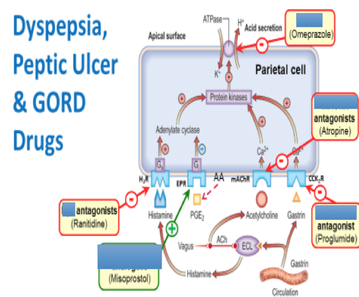
- Ahistamine
- Bprostaglandin analogues
- Cmuscarinic acetylcholine
- Dproton pump inhibitors
- Egastrin receptor
58What is not a sign of dyspepsia?
- Aepigastric pain
- Bbloating
- Cheartburn
- Dburning sensation during defecation
59Worrying symptoms of dyspepsia:
A [blank_start]anemia[blank_end]
L loss of weight
A [blank_start]anorexia[blank_end]
R recent [blank_start]progressive[blank_end] [blank_start]symptoms[blank_end]
M [blank_start]melena[blank_end] (tarry feces) and [blank_start]hematemesis[blank_end]
S wallowing difficulty- Aanemia
- Banorexia
- Cprogressive
- Dsymptoms
- Emelena
- Fhematemesis
60What are ways to manage dyspepsia?
- Asimple antacids for 4 weeks
- Btest for H. Pylori and eliminate if possible
- CProton pump inhibitors for 4 weeks
- Durgent endoscopy
- Ebiopsy
- Fblood works
61Label the different types of hiatus hernias
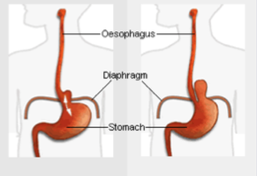
- Asliding
- Brolling
62Barium swallow via fluoroscopy is how one is able to diagnose a hiatus hernia.
- AVerdadeiro
- BFalso
63What is true regarding peptic ulcers?
- Ainclude duodenal ulcers
- Binclude gastric ulcers
- Ccomplication: hemorrhage
- Dcomplication: ulcer perforation
- Ecomplication: malignancy (tumor)
- Finclude jejunal ulcers
- Ggastric ulcers > common than duodenal ulcers
64What is true regarding duodenal ulcers?
- Aepigastric tenderness
- Bepigastric pain before meals
- Crelief via milk
- Depigastric pain better at night
- Erelief via lemon soda
65Gastric ulcers:
- happens mainly in [blank_start]elderly[blank_end]
- [blank_start]epigastric[blank_end] pain (dependent on [blank_start]meals[blank_end] and can be relieved by antacids)
- weight gain or loss- Aelderly
- Bepigastric
- Cmeals
66What is this sign and what is indicative of?
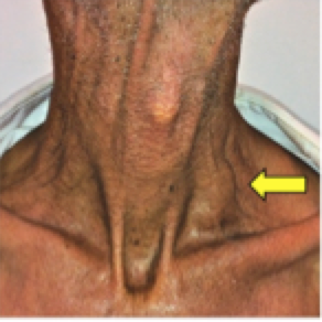
- AVirchow's node
- Bgastric cancer
67What is true regarding zollinger-ellison syndrome?
- Agastrinoma--> peptic ulcer
- Busually in duodenum
- Cusually in pancreas
- Dinvestigation: endoscopy
- Einvestigation: very high fasting and then serum gastrin level investigation
- Finvestigation: biopsy
- Gmanagement: surgery
- Hmanagement: proton pump inhibitors
- Imanagement: antacids
- Jusually in ileum
68iron is absorbed in the jejunum.
- AVerdadeiro
- BFalso
69[blank_start]Celiac[blank_end] [blank_start]disease[blank_end] is an inflammation of the proximal small intestine mucosa in response to dietary gluten.
It is Hypersensitivity [blank_start]IV[blank_end]. It is a disease of the small intestines in which [blank_start]prolamin[blank_end] [blank_start]intolerance[blank_end] causes villous atrophy and [blank_start]malabsorption[blank_end].- ACeliac
- Bdisease
- CIV
- Dprolamin
- Eintolerance
- Fmalabsorption
70What are symptoms of Celiac's Disease?
- Adiarrhea
- Bmalaise
- Cabdominal discomfort
- Diron deficiency anemia
- Eweight loss
- Fsteatorrhea
- Gweariness
- Hconcentrated pain
- Ibursts of energy
71Celiac disease makes you at more risk of colon cancer and lymphoma.
- AVerdadeiro
- BFalso
72Crohn's Disease treatment is a gluten free diet and necessary vitamin supplements.
- AVerdadeiro
- BFalso
73Which of the following is not a way to diagnose Celiac's disease?
- Aduodenal biopsy via endoscopy
- Bblood test: abnormally high levels of IgA endomysial
- Cblood test: if anti-tissue transglutaminase antibodies are found
- Dabdominal x-ray
74What is true regarding diverticulitis?
- Aonly in relation to the large intestine
- Binflammation of colon's diverticulae
- Cmore common in elderly patients
- Dmore common in those chronically constipated
- Emore common in women
- Fmore common in those with acute constipation
- Gcommonly associated with anal fissures
75Which are symptoms of diverticulitis?
- Alower left quadrant pain
- Blower left quadrant tenderness
- Cfever
- Dtenesmsus
- Ediarrhea
- Fconstipation
76How can you investigate for diverticulitis?
- Abarium enema
- Bcolonoscopy
- CCT colonogram
- Drectal biopsy
77Treatment of diverticulitis:
mild:
at home with [blank_start]paracetamol[blank_end], fluids, and [blank_start]oral[blank_end] [blank_start]antibiotics[blank_end].
severe or ill patients:
admission to hospital (cannot manage [blank_start]pain[blank_end], problems with [blank_start]dehydration[blank_end], [blank_start]frail[blank_end])- Aparacetamol
- Bantibiotics
- Coral
- Dpain
- Edehydration
- Ffrail
78What are these two colon related conditions?
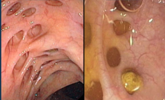
- Adiverticulitis
- Bdiverticulosis
79Which conditions make up inflammatory bowel disease?
- ACrohn's disease
- BCeliac's disease
- CUlcerative Colitis
- DDiverticulitis
- EPeptic Ulcers
80[blank_start]Crohn’s[blank_end] is a chronic inflammatory bowel disease affecting all layers of the GI tract ([blank_start]transmural[blank_end]). Its key characteristic is [blank_start]skip[blank_end] [blank_start]lesions[blank_end], and on a colonscopy there is a “[blank_start]cobblestone[blank_end] [blank_start]appearance[blank_end]." ANY part of the GI tract but most commonly the [blank_start]terminal[blank_end] [blank_start]ileum[blank_end] (can cause [blank_start]vitamin[blank_end] [blank_start]B12[blank_end] deficiency).
- ACrohn’s, Crohn's Disease
- Btransmural
- Cskip
- Dlesions
- Ecobblestone
- Fappearance
- Gterminal
- Hileum
- IB12
- Jvitamin
81What are symptoms of Crohn's Disease?
- Adiarrhea
- Babdominal pain
- Cright lower quadrant mass
- Dperianal disease
- Emouth ulcers
- Fextra-intestinal manifestations
- Gpale and bulky stools
- Hright lower quadrant pain
82What is not an investigation for Crohn's Disease?
- Astool sample
- Bcolonscopy and biopsy
- Cendoscopy and biopsy
- DBlood: CRP/ESR
- EBlood: FBC
- FBlood: LFTs
- GBlood: B12
- HBlood: ferritin
- IBarium swallow
83Management of Crohn Disease attacks:
Mild attacks: [blank_start]Prednisolone[blank_end]
Severe attacks: IV [blank_start]steroids[blank_end], nil by mouth, and IV [blank_start]hydration[blank_end].- APrednisolone
- Bsteroids
- Chydration
84Label the parts of the biliary tree?
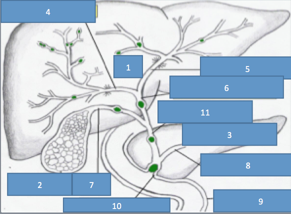
- Aliver
- Bright hepatic tree
- Cleft hepatic duct
- Dcommon hepatic duct
- Ecommon bile duct
- Fpancreatic
- Gpancreatic duct
- Hduodenum
- Isphincter of Oddi
- Jcystic duct
- Kgallbladder
85Bile is produced by [blank_start]hepatocytes[blank_end] and is stored in the [blank_start]gallbladder[blank_end]. Bile [blank_start]emulsifies[blank_end] the fat and allows fat absorption in [blank_start]enterocytes[blank_end]. An example of fat soluble vitamins: [blank_start]ADEK[blank_end]
- Ahepatocytes
- Bgallbladder
- Cemulsifies
- Denterocytes
- EADEK
86An [blank_start]enterogastrone[blank_end] is any hormone secreted by the [blank_start]duodenum[blank_end] mucosa in response to dietary lipids that inhibits the caudal (or "forward, analward") motion of the contents of [blank_start]chyme[blank_end].
- Aenterogastrone
- Bduodenum
- Cchyme
87Relapsing inflammatory disorder of [blank_start]colonic[blank_end] mucosa which usually begins in the [blank_start]rectum[blank_end] and doesn’t spread past the [blank_start]ileocaecal[blank_end] valve is ulcerative colitis. This inflammation is not [blank_start]transmural[blank_end] and is [blank_start]continous[blank_end].
- Acolonic
- Brectum
- Cileocaecal
- Dtransmural
- Econtinous
88What is true regarding ulcerative colitis?
- Abloody diarrhea
- Babdominal pain
- Ctenesmus
- Dbathroom urgency
- Emalaise
- Fweight loss
- Gextra-intestinal features
- Hpale and bulky stool
- Idehydration
89Which is not a way to investigate for ulcerative colitis?
- AFBC
- Bliver function tests
- Curea and electrolytes profile
- Derythrocyte sedimentation rate
- Ec reactive protein
- Fferritin
- GB12
- Hfolate
- Ihistamine
90You can investigat for ulcerative colitis via sigmoidscopy or colonoscopy.
- AVerdadeiro
- BFalso
91Management of Ulcerative Colitis:
[blank_start]Mesalazine[blank_end] (also known as 5-aminosalicylate) and steroids (such as [blank_start]prednisolone[blank_end]) to induce remission.
Surgery ([blank_start]colectomy[blank_end] for those non-responsive to drugs)
Mesalazine to maintain remission.- AMesalazine
- Bcolectomy
- Cprednisolone
92Pancreatic secretions are controlled by the [blank_start]parasympathetic[blank_end] nervous system.
[blank_start]Secretin[blank_end] in response to H+ in the duodenum causes the pancreas to secrete [blank_start]bicarbonate[blank_end].
In response to [blank_start]fats[blank_end] and [blank_start]amino[blank_end] [blank_start]acids[blank_end] in the duodenum, the pancreas releases CCK cholecystokinin (CCK). CCK also causes [blank_start]gallbladder[blank_end] contractions.
These proteolytic enzymes are released inactively as [blank_start]zymogens[blank_end] to protect the pancreatic tissue from being destroyed.- Aparasympathetic
- BSecretin
- Cbicarbonate
- Dgallbladder
- Efats
- Famino
- Gacids
- Hzymogens
93Which of these are pancreatic enzymes?
- Atrypsinogen
- Bchymotripsinogen
- Cprocarboxypeptidase
- Dpancreatic amylase
- Epancreatic lipase
- Fgastrin
- Gpepsinogen
94pancreatic enzymes:
trypsin, chymotrypsin, carboxypeptidase: act on [blank_start]proteins[blank_end]
pancreatic lipase acts on [blank_start]fat[blank_end]
pancreatic amylase acts on [blank_start]carbohydrates[blank_end]
ribonucleoase/deoyribonuclease acts on nucleic acids- Aproteins
- Bfat
- Ccarbohydrates
95What are examples of tumor markers?
- ACEA
- Balpha fetoprotein (aFP)
- CCA 1999
- Dbeta HCG
- ECa 125
- FBRCA 2
- GCa 187
96Tumor suppressor genes cause tumors to actually progress.
- AVerdadeiro
- BFalso
97p53 is a tumor suppressor gene that is important because it can trigger apoptosis.
- AVerdadeiro
- BFalso
98What is the condition that describes a type of pain related to the gallbladder that occurs when a gallstone temporarily obstructs the cystic duct and the gallbladder contracts?
- ABiliary Colic
- BCholangitis
- CCholecystitis
- DCourvoisier's syndrome
99What is true regarding biliary colic?
- Agoldstone investigation: ultrasound of right upper quadrant
- Bmanage: analgesia
- Cmanage: rehydrate
- Dmedication given via orally
- Emanage: elective cholecystectomy
- Fnausea and vomiting
- Gindigestion symptoms
- Hpain after a fatty meal
- Iright upper quadrant pain which radiates
- Jacid reflux
100[blank_start]Acute[blank_end] cholecystitis:
Due to stone impaction in the gallbladder neck
Symptoms:
Continuous [blank_start]RUQ[blank_end] pain or [blank_start]Epigastric[blank_end] pain which radiates to the right shoulder
[blank_start]vomiting[blank_end]
fever
Investigation: [blank_start]Ultrasound[blank_end]
Treatment: Nil by mouth: analgesia; antibiotics; and [blank_start]cholecystectomy[blank_end]- AAcute
- BRUQ
- CEpigastric
- Dvomiting
- EUltrasound
- Fcholecystectomy
101[blank_start]Chronic[blank_end] cholecystitis:
Stones cause chronic inflammation of gallbladder +/- [blank_start]colic[blank_end] inflammation
Symptoms:
Abdomen discomfort
[blank_start]distension[blank_end]
nausea
[blank_start]flatulence[blank_end]
fat intolerance
Investigation: [blank_start]Ultrasound[blank_end]
Treatment: [blank_start]cholecystectomy[blank_end]- AChronic
- Bcolic
- Cdistension
- DUltrasound
- Echolecystectomy
- Fflatulence
102Courvoisier's law is that the possible malignancy of the [blank_start]gallbladder[blank_end]/[blank_start]pancreas[blank_end], such as swelling, is not probably due to [blank_start]gallstones[blank_end].
- Agallbladder
- Bpancreas
- Cgallstones
103What is not true regarding cholangitis?
- Ainflammation of the common bile duct due to gall stones obstruction
- Binvestigation: ERCP
- CCharcot's triad
- Dinvestigation: ultrasound
104Pancreatitis is due to injuries that lead to autodigestion of the pancreas by its own [blank_start]enzymes[blank_end].
[blank_start]Acute[blank_end] pancreatitis is characterized by reversible pancreatic [blank_start]parenchymal[blank_end] injury associated with inflammation. [blank_start]Biliary[blank_end] [blank_start]tract[blank_end] disease and alcoholism account for approximately 80% of cases of [blank_start]acute[blank_end] pancreatitis in Western countries.
[blank_start]Chronic[blank_end] pancreatitis is defined as prolonged inflammation of the pancreas associated with irreversible destruction of [blank_start]exocrine[blank_end] parenchyma, [blank_start]fibrosis[blank_end], and, in the late stages, the destruction of [blank_start]endocrine[blank_end] parenchyma. The most common cause of [blank_start]chronic[blank_end] pancreatitis by far is long-term [blank_start]alcohol[blank_end] abuse- Aenzymes
- BAcute
- CChronic
- Dparenchymal
- Etract
- FBiliary
- Gacute
- Hchronic
- Ialcohol
- Jexocrine
- Kendocrine
- Lfibrosis
105What is NOT true regarding pseudocyst?
- Amost common complication of pancreatitis
- Bis the accumulation n of fluid within and around the pancreatic tissue.
- Ctrtment: drainage of fluid into stomach's lumen
- Dinvestigation: type II MRI
106Which of these can cause chronic pancreatitis?
- Aalcohol
- Bcystic fibrosis
- Chemochromastosis
- Dpancreatic duct obstruction
- Erenal failure
- Fliver disease
- Gobesity
107Ultrasound and CT to check for pancreatic calcifications is how to investigate for chronic pancreatitis.
- AVerdadeiro
- BFalso
108What are symptoms of chronic pancreatitis?
- Aepigastric pain radiating to the back
- Bepigastric pain is less when sitting forward
- Cepigastric pain is less with hot water bottles
- Dbloating
- Eweight loss
- Fbrittle diabetes
- Gsteatorrhea
- Hmanage: lipase and fat soluble vitamins
- Imanage: low fat diet
- Jmanage: surgery
109CAUSES for acute pancreatitis:
GET SMASHED:
[blank_start]Gallstones[blank_end] (38%)
[blank_start]Ethanol[blank_end] (35%)
[blank_start]Trauma[blank_end] (1.5%)
[blank_start]Steroids[blank_end]
[blank_start]Mumps[blank_end]
[blank_start]Autoimmune[blank_end]
Scorpion venom
Hyperlipidaemia/[blank_start]hypothermia[blank_end]/ hypercalcaemia
[blank_start]ERCP[blank_end] and emboli
Drugs.- AGallstones
- BEthanol
- CTrauma
- DSteroids
- EAutoimmune
- FMumps
- Ghypothermia
- HERCP
110What are the signs and symptoms of acute pancfdatitis?
- Atachycardia
- Bfever
- Cjaundice
- DCullen's sign
- EGray Turner's sign
- Fgradual epigastric pain
- Gsevere epigastric man
- Hvomiting
- Icentral abdominal pain which radiates to the back
- Jinvestigation: dropped serum levels of amylase
111Which sign is which?
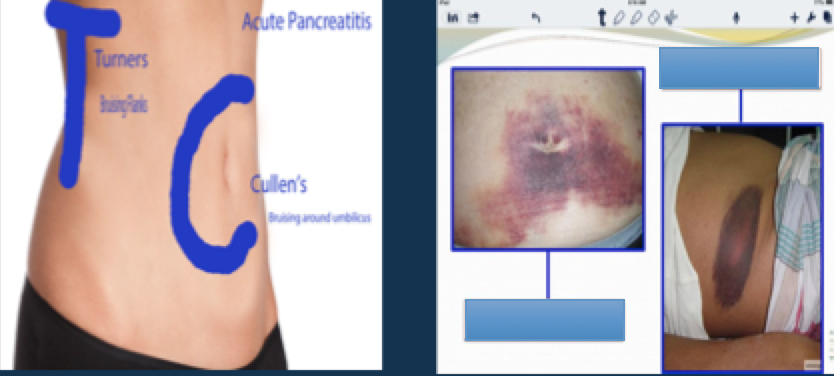
- Acullen's sign
- Bturner's sign
112Serum lipase is the most reliable investigating factor for pancreatitis.
- AVerdadeiro
- BFalso
113Management of acute pancreatitis:
Nil by mouth: [blank_start]analgesia[blank_end]
[blank_start]ERCP[blank_end] + [blank_start]gallstone[blank_end] [blank_start]removal[blank_end] may be needed if progressive jaundice.- Aanalgesia
- BERCP
- Cgallstone
- Dremoval
114Those at an increased risk of developing cancer often are born with one defective copy of a [blank_start]tumor[blank_end] [blank_start]suppressor[blank_end] gene.
- Atumor
- Bsuppressor
115An oncogene is when a proto-oncogene becomes mutated.
- AVerdadeiro
- BFalso
116Oncogenes cause uncontrolled tumor growth.
- AVerdadeiro
- BFalso
117Appendicitis:
presentation:
[blank_start]Fever[blank_end]
RLQ pain or [blank_start]umbulical[blank_end] pain
nausea
vomiting
KEY: loss of appetite
investigation: [blank_start]Xray[blank_end]- AXray, x-ray
- Bumbulical
- CFever
118How does ischemic bowel NOT present?
- Asevere abdominal pain
- Btachycardia + hypotension
- Craised leukocyte count
- Dfever intervals
119Murphy's sign is for acute cholecystitis.
Murphy's sign is that on palpation of the RUQ, patient stops inspiration for a moment.- AVerdadeiro
- BFalso
Informações
- Descrição
- Content courtesy of Dayo's Mat 2 lecture. credits to him :)
- Autor
- Usuário eliminado
- Idioma
- English
- Visualizações
- 30
- Histórico
- Criado em 10 mar 2018 · atualizado em 13 mar 2018
- Etiquetas
- Avaliação
- Ainda sem avaliações Avalie este quiz