
Flashcards
RCA Practical 5
Flashcards do conjunto (77)
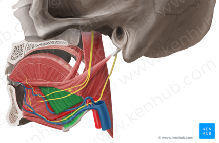
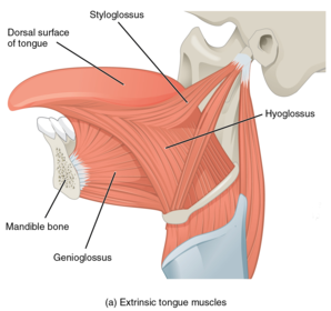
- 1Name Origin: Insertion: Function: Innervation:Hyoglossus:
Origin: Hyoid bone
Insertion: Side of the tongue
Function: Retracts the tongue as well as makes the dorsum more convex
Innervation: Hypoglossal nerve (cranial nerve 12)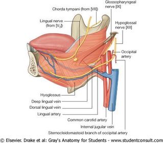
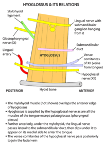
- 2Name Structures deep (3): Structures superficial (6):Hyoglossus
Deep: Lingual artery and vein, glossopharyngeal nerve (IX)
Lateral: Sublingual gland, portion of the submandibular gland, submandibular duct, lingual nerve, hypoglossal nerve and venae comitantes of 7 (veins from tongue).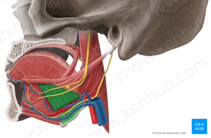
- 3Name Origin: Insertion: Function: Innervation:Genioglossus:
Origin: Superior mental spine of the mandible
Insertion: Floor of the tongue and body of the hyoid bone
Function: Protrusion and depression of the tongue
Innervation: Hypoglossal nerve (cranial nerve 12)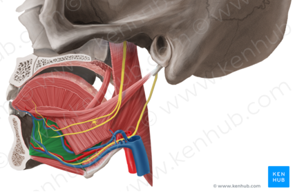
- 4Name Composition: Relation to hyoglossus: Drainage:Submandibular gland
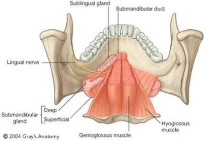
Composition: Consists of two part with respect to the mylohyoid; superficial and deep.
Relation to hyoglossus: Lateral/superficial
Drainage: The submandibular duct opens into the sublingual caruncle/papillae; a small prominence on either side of the frenulum of the tongue demarcating the opening.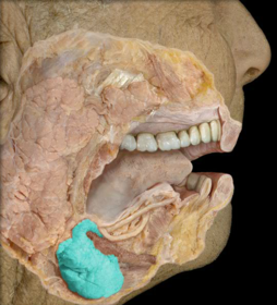
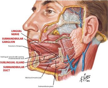
- 5Name Relation to hyoglossus: Position: Drainage:Sublingual gland:
Relation to hyoglossus: Lateral/superficial.
The sublingual gland lies anterior to the submandibular gland, inferior to the tongue beneath the mucous membrane of the floor of the mouth forming the sublingual folds.
The sublingual gland has two points of drainage; many of the small ducts coalesce to form the major sublingual duct/duct of Bartholin which will join the submandibular. The other remaining ducts will open into an elevated crest in the mucous membrane of the mouth called the sublingual fold, either side of the lingual frenulum.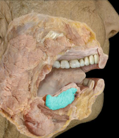
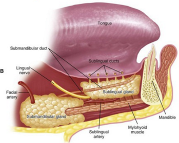
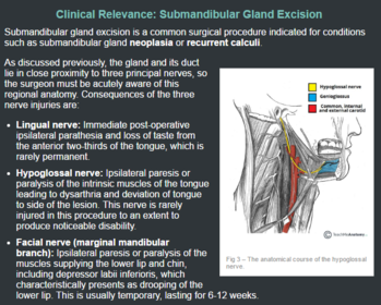
- 6Should be considered during operation on the submandibular gland to treat neoplasia or recurrent calculi:Both the gland and duct share intimate anatomical relations with nerves; lingual nerve begins laterally to the duct and wraps under to travel anteromedially. The hypoglossal nerve lies deep to the submandibular gland. The marginal mandibular branch of the facial nerve curves inferior to the submandibular gland.
- 7Name all: Deciduous and permanent dentition numbers:
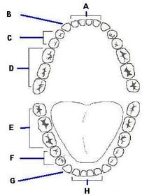
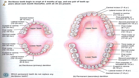
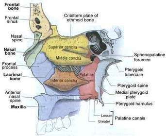
- 8Main bones of the lateral nasal wall: Conchae: Function of conchae:Lateral wall: Formed by the maxilla with contributions from ethmoid, palatine and lacrimal bones. It is comprised of three curved shelves of bone, the superior, middle (two process of ethmoid bone) and inferior conchae (separate bone).
Function: The conchae increase the surface area of contact between tissues of the lateral wall and inspired air.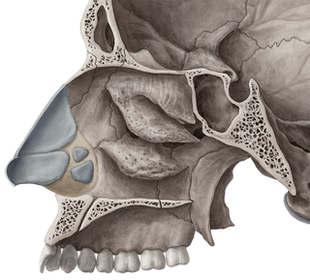
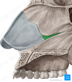
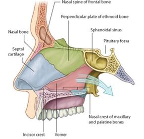
- 9Main bones of the nasal septum:The nasal septum (medial wall of the nasal cavity): Is comprised of the septal nasal cartilage anteriorly, the vomer and perpendicular plate of the ethmoid bone posteriorly.
- 10Name Bone of origin: Passage below:Superior concha
Bone of origin: Ethmoid
Passage below: Superior meatus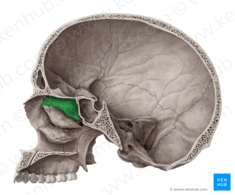
- 11Name Bone of origin: Passage below:Middle nasal concha
Bone of origin: Ethmoid
Passage below: Middle meatus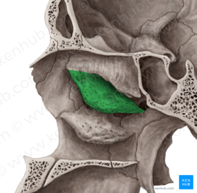
- 12Name Bone of origin: Passage below:Inferior meatus
Bone of origin: It's own bone
Passage below: Inferior meatus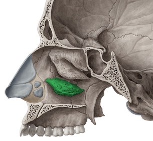
- 13Describe the arterial supply to the lateral nasal wall:Terminal branches of the maxillary and facial arteries, and ethmoidal branches of the ophthalmic artery.
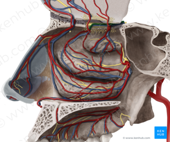
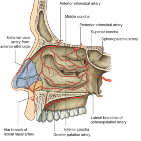
- 14Describe the venous drainage of the lateral nasal wall:Venous drainage: Follow the arteries.
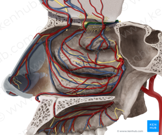
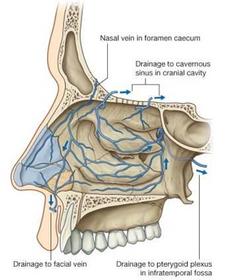
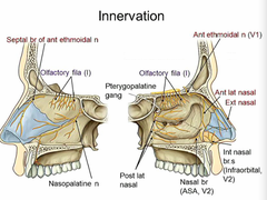
- 15Describe the innervation of the lateral nasal wall and nasal septum:Innervation: Olfactory nerve (CN 1), ethmoidal branches of the nasociliary nerve from the ophthalmic division of trigeminal (V1), and nasal nerve branches of maxillary division of trigeminal (V2).
Innervation of the anterior nasal septum is also via the nasal nerve branches of the maxillary division of trigeminal (V2).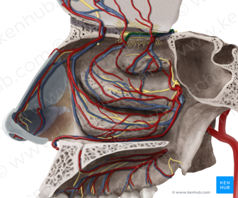
- 16Name 1, 2, 3 and 41: Frontal sinus
2: Ethmoidal sinus (anterior, middle, posterior)
3: Sphenoid sinus
4: Maxillary sinus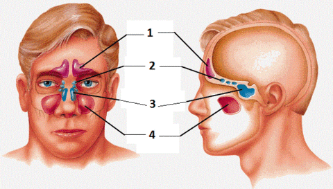
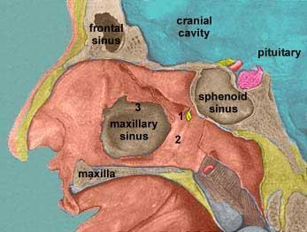
- 17Name sinuses 1-7 List sinuses which drain into 1. Superior meatus 2. Middle meatus 3. Inferior meatusDrainage of air sinuses:
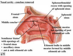
Superior meatus: The posterior ethmoid air cells drain into this.
Middle meatus: Of importance here, we have the uncinate process which lies anterior to the semilunar hiatus. The frontal sinus, anterior ethmoidal air cells and maxillary sinus. Then we have the ethmoid bulla, the large bulge seen, and on this we have the opening of the middle ethmoidal air cells.
Inferior meatus: The nasolacrimal duct into which tears drain into opens here.
Sphenoethmoidal recess: this is where the sphenoid sinus drains into.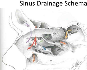
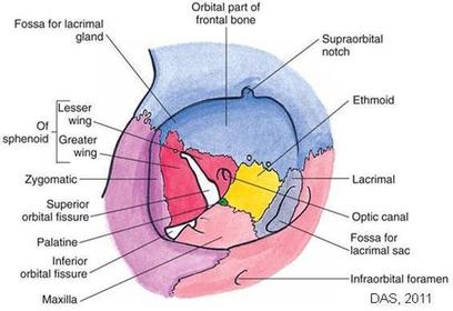
- 18Name the bones forming the 4 walls of the orbit and the apex:The roof: Mainly the orbital plate of frontal bone, near the apex lesser wing of sphenoid.
Medial wall: Mainly ethmoid with contributions from frontal, lacrimal, and sphenoid. Anteriorly indented by lacrimal groove.
Floor: Mainly by maxilla, partly by zygomatic bone. Floor is partly separated from lateral wall by the inferior orbital fissure.
Lateral wall: Zygomatic and greater wing of sphenoid. Partly separated from roof by superior orbital fissure (communicates with the middle cranial fossa).
Apex: Is at the optic canal, medial to superior orbital fissure.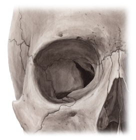
- 19Name all1. Lesser wing of sphenoid
2. Greater wing of sphenoid
3. Palatine bone
4. Ethmoid
5. Lacrimal
6. Nasal bone
7. Maxillary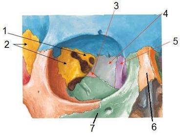
- 20Name C and describe the covering externally and internallyEyelid:
Externally covered by thin skin
Internally covered by conjuctiva which is then reflected onto the eyeball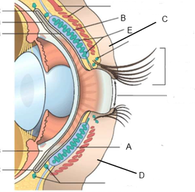
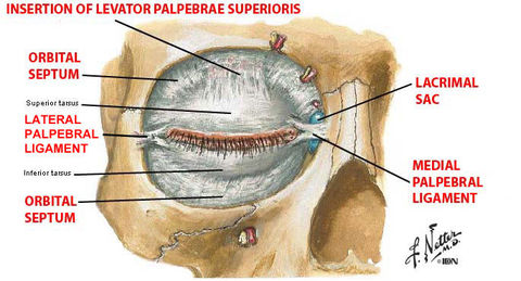
- 21Name A and B Describe them and their function:A: Superior tarsus
B: Inferior tarsus
Tarsal plates are dense fibrous tissue bands that strengthen the eyelid.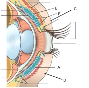
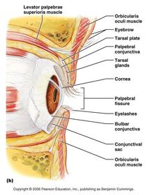
- 22Name E Describe their location and function:Tarsal glands
Location: Embedded within the tarsal plates
Function: Secrete fluid to lubricate the eyelids to prevent them sticking together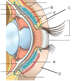
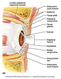
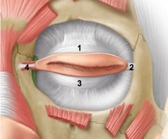
- 23Describe the attachments of the eyelid identifying the numbers:Medial palpebral ligament (4) connects the eyelids to the medial margin of the orbit and lateral palpebral ligament (2) to the lateral margin.
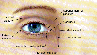
- 24Identify 1 Describe its location and drainage:Lacrimal gland
Location: In a fossa in the lateral part of the roof of the orbit
Drainage: By about a dozen ducts onto the conjuctiva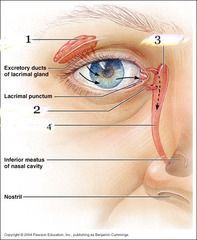
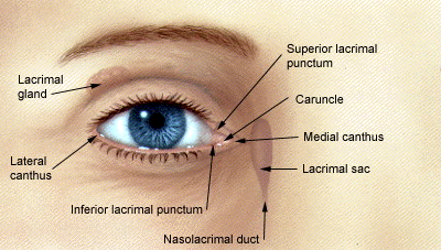
- 25Name 3 Describe its location and drainage identifying the numbers:Lacrimal sac
Location: In the lacrimal groove, which indents the anterior part of the medial wall of the orbit
Drainage: Receives fluid from the eye via lacrimal canaliculi (labelled 3). The lacrimal sac then drains into the nasolacrimal duct which it is continuous with. The nasolacrimal duct (labelled 4) will open into the inferior meatus on the lateral wall of the nasal cavity.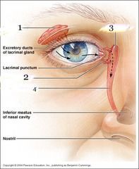
- 26Name Origin: Insertion: Function: Innervation:Levator palpebrae superioris:
Origin: Roof of the orbit close to apex
Insertion: Tarsal plate and skin of upper eyelid
Function: Elevates upper eyelid
Innervation: Oculomotor nerve (cranial nerve 3)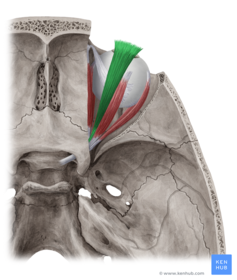
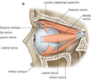
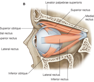
- 27Name Origin: Insertion: Function: Innervation:Superior rectus:
Origin: Common tendinous ring at the apex of the orbit
Insertion: Superior surface of eyeball
Function: Elevates the eyeball
Innervation: Oculomotor nerve (cranial nerve 3)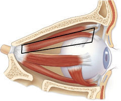
- 28Name Origin: Insertion: Function: Innervation:Inferior rectus:
Origin: Common tendinous ring at the apex of the orbit
Insertion: Inferior surface of the eyeball
Function: Depresses the eyeball
Innervation: Oculomotor nerve (cranial nerve 3)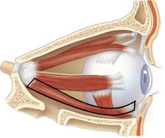
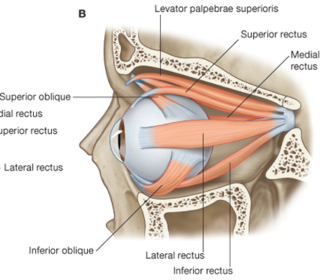
- 29Name Origin: Insertion: Function: Innervation:Medial rectus:
Origin: Common tendinous ring at the apex of the orbit
Insertion: Medial surface of the eyeball
Function: Adduct the eyeball
Innervation: Oculomotor nerve (cranial nerve 3)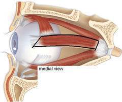
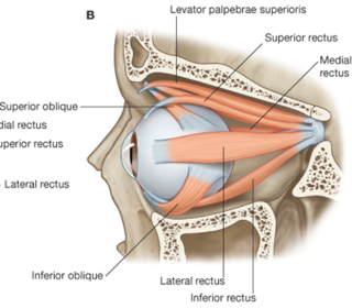
- 30Name Origin: Insertion: Function: Innervation:Lateral rectus:
Origin: Common tendinous ring at the apex of the orbit
Insertion: Lateral surface of the eyeball
Function: Abducts the eyeball
Innervation: Abducens nerve (cranial nerve 6)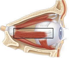
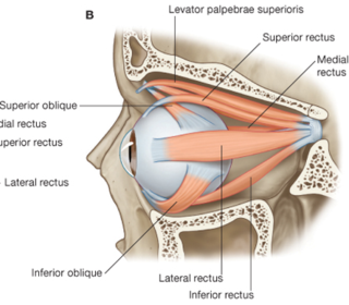
- 31Name Origin: Insertion: Function: Innervation:Superior oblique:
Origin: Body of sphenoid
Insertion: Tendon passes through a fibrous ring (trochlea) at a superomedial angle to the orbital wall, changes direction and inserts into sclera deep to superior rectus muscle
Function: Abducts and depresses eyeball
Innervation: Trochlear nerve (cranial nerve 4)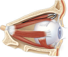
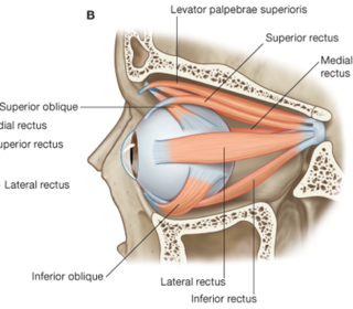
- 32Name Origin: Insertion: Function: Innervation:Inferior oblique:
Origin: Anterior part of floor of orbit
Insertion: Sclera deep to the lateral rectus muscle
Function: Abducts and elevates the eyeball
Innervation: Oculomotor nerve (cranial nerve 3)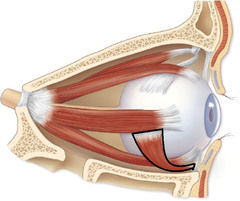
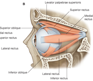
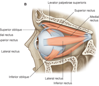
- 33Name all1. Superior oblique
2. Medial rectus
3. Superior rectus
4. Common tendinous ring
5. Inferior rectus
6. Inferior oblique
7. Lateral rectus
8. Levator palpebrae superioris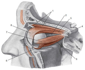
- 34Name 4 Origin: Supplies:Lacrimal nerve:
Origin: Ophthalmic division of trigeminal (V1)
Supplies: Innervates the lacrimal gland as well as the lateral skin and conjunctiva of the upper eyelid.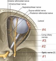
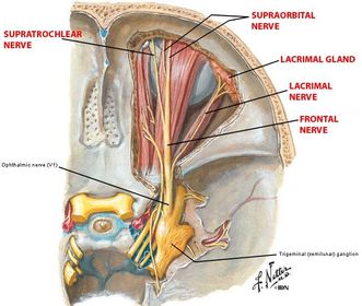
- 35Name 3 Origin: Terminal branches:Frontal nerve
Origin: Ophthalmic division of trigeminal (V1)
Terminal branches: Supratrochlear and supraorbital nerves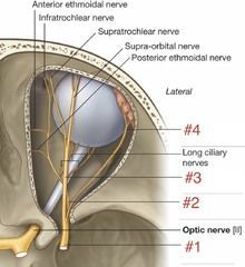
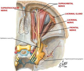
- 36Name 7 Origin: Course: Supplies:Supraorbital nerve
Origin: One of the two terminal branches of the frontal nerve originating from ophthalmic (V1)
Course: Exits the orbit via the supraorbital foramen and emerges onto the face travelling superiorly
Supplies: The skin and conjunctiva of the upper eyelid, and the skin of the forehead and scalp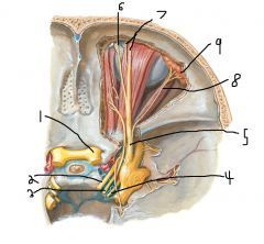
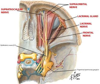
- 37Name 6 Origin: Course: Supplies:Supratrochlear nerve
Origin: From the frontal nerve which originates from ophthalmic (V1)
Course: Exits the orbit between the trochlea (fibrous ring of superior oblique) and supraorbital foramen
Supplies: Skin and conjunctiva of upper eyelid, and skin of forehead.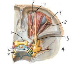
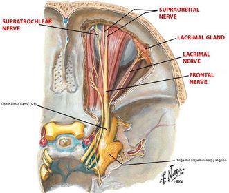
- 38Name 5 Origin: Course: Branches:Ophthalmic division of trigeminal (V1)
Origin: V1 division of trigeminal
Course: Enters orbit via superior orbital fissure
Branches: Lacrimal nerve, frontal nerve and nasociliary nerve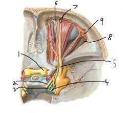
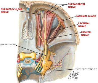
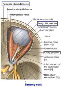
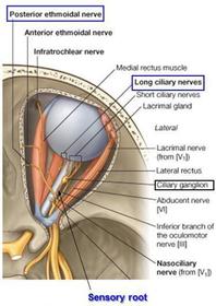
- 39Name Origin: Course: Branches:Nasociliary nerve
Origin: From the ophthalmic division of the trigeminal (V1)
Course: Passes forward between the cone of muscles and gives of the following branches
Branches: Anterior and posterior ethmoidal nerves, long ciliary nerves and infratrochlear nerve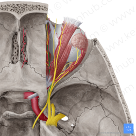
- 40Name Origin: Course: Supplies:Long ciliary nerves
Origin: From the nasociliary nerve which originates from the ophthalmic division of trigeminal (V1)
Course: Forward to the eyeball
Supplies: Carries sensory fibres to the cornea and postganglionic sympathetic fibres from superior cervical ganglion (T1 origin) to the dilator pupillae muscles.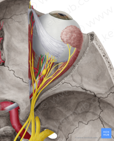
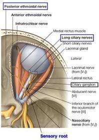
- 41Name Origin: Course: Supplies:Posterior ethmoidal nerve
Origin: Nasociliary nerve which originates from the ophthalmic division of trigeminal (V1)
Course: Enters the posterior ethmoidal sinus via the respective foramen in the medial wall of the orbit
Supplies: Sensory to posterior ethmoidal sinus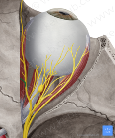
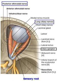
- 42Name Origin: Course: Supplies:Anterior ethmoidal nerve
Origin: Nasociliary nerve which originates from the ophthalmic division of trigeminal (V1)
Course: Enters the anterior ethmoidal sinus via the respective foramen in the medial wall of the orbit
Supplies: Sensory to anterior ethmoidal sinus. Continues on as the external nasal nerve entering the face via the nose and supplying the skin over the dorsum of the nose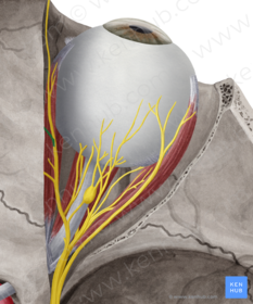
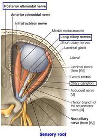
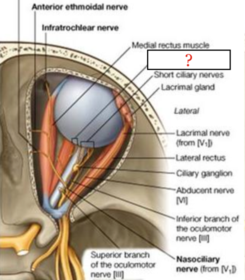
- 43Name Origin: Course: Supplies:Infratrochlear nerve
Origin: Nasociliary nerve which originates from the ophthalmic (V1)
Course: Runs along medial rectus and passes to medial commissure of eye
Supplies: Upper eyelid skin and conjunctiva, lacrimal sac and canaliculi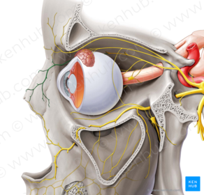
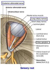
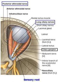
- 44Name Origin: Course: Supplies:Short ciliary nerves
Origin: Ciliary ganglion, situated behind the eyeball is where the preganglionic parasympathetic fibres from the edinger westphal nucleus of the midbrain at the superior colliculus level carried by the oculomotor nerve (cranial nerve 3) synapse.
Course: Fibres to muscles
Supplies: Ciliary muscle (accommodation) and pupillary constrictor muscle.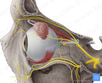
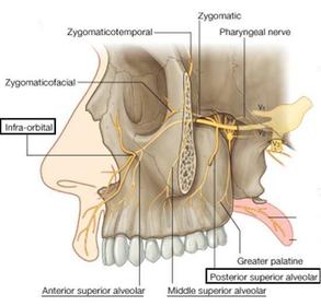
- 45Name Origin: Course: Branches that enter orbit:Maxillary division of trigeminal (V2)
Origin: Trigeminal nerve (V2)
Course: Exits cranium via the foramen rotundum to enter the pterygopalatine fossa. Within the pterygopalatine fossa gives off two branches that suspend the parasympathetic pterygopalatine ganglion. The sensory fibres of the maxillary nerve pass through the ganglion without synapsing to supply the nose, palate and the nasopharynx.
Branches that enter orbit: Infraorbital and zygomatic nerve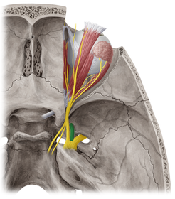
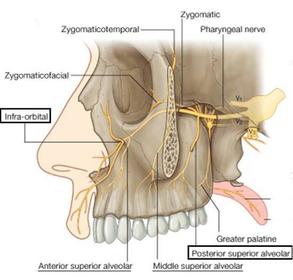
- 46Name Origin: Course: Supplies:Infratrochlear nerve
Origin: Maxillary division of trigeminal (V2)
Course: Enters the orbit through the infraorbital fissure and runs on the floor of the orbit emerging onto the face through the infraorbital foramen.
Supplies: The lower eyelid, cheek and upper lip. Also branches off to form the anterior superior alveolar nerve.
Anterior superior alveolar nerve: Arises from the infraorbital nerve to supply the anterior maxillary teeth.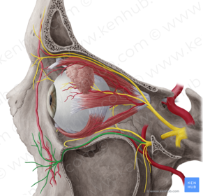
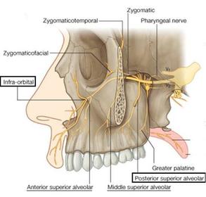
- 47Name Origin: Course: Supplies:Zygomatic nerve
Origin: Maxillary division of trigeminal (V2)
Course: Enters the orbit through the inferior orbital fissure, runs on the lateral wall of the orbit, divides into two branches, zygomaticofacial and zygomaticotemporal, which emerge onto the face through foramina in the zygomatic bone.
Supplies: The skin over the zygomatic bone.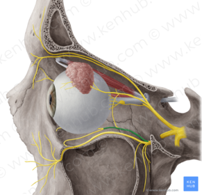
- 48Name Origin: Course: Supplies:Oculomotor nerve (cranial nerve 3)
Origin: From the anterior junction of the midbrain and pons of the brainstem.
Course: Enters the cranium via the tentorial notch and then enters the orbit via the superior orbital fissure.
Supplies: Levator palpebrae superioris, superior, inferior and medial recti, and inferior oblique muscles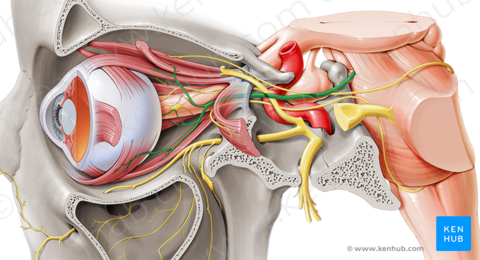
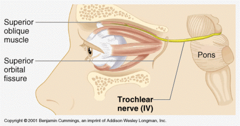
- 49Name Origin: Course: Supplies:Trochlear nerve (cranial nerve 4)
Origin: Posterior midbrain at the level of the inferior colliculus
Course: Enters the orbit via the superior orbital fissure
Supplies: Superior oblique muscle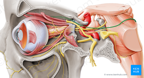
- 50Name Origin: Course: Supplies:Abducens nerve (cranial nerve 6)
Origin: Medial pontomedullary junction
Course: Enters orbit via superior orbital fissure
Supplies: Lateral rectus muscle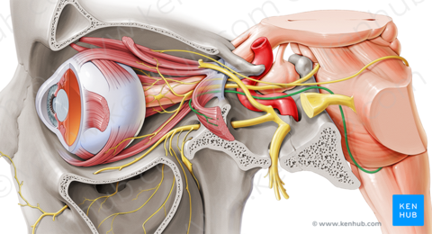
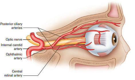
- 51Name Origin: Course: Supplies: Branches:Ophthalmic artery:
Origin: Arises from the internal carotid artery as it emerges from the cavernous sinus.
Course: It passes through the optic foramen together with the optic nerve and runs anteriorly close to the superomedial wall of the orbit. It gives off branches to structures in the orbit similar to the nasociliary nerve,
Branches: Central retinal artery and ends as the supraorbital artery.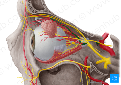
- 52Name Origin: Course: Supplies:Central retinal artery
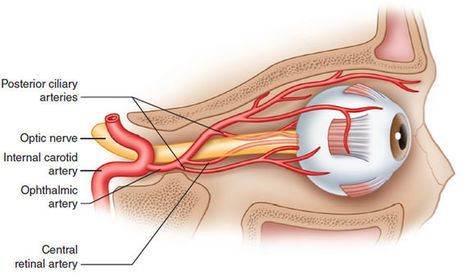
Origin: Ophthalmic artery
Course: Runs in the optic nerve
Supplies: The retina.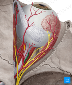
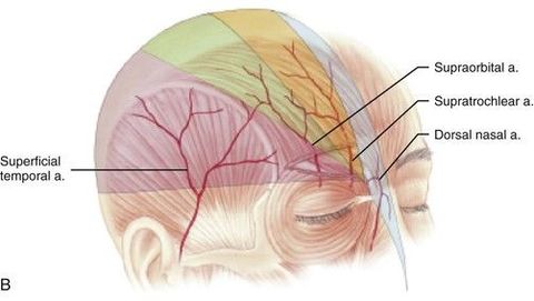
- 53Name Origin: Course: Supplies:Supraorbital artery
Origin: A terminal branch of the ophthalmic artery.
Course: It passes superiorly from the supraorbital foramen
Supplies: The muscles and skin of the forehead and scalp.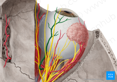
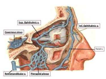
- 54Name Origin: Course:Superior ophthalmic vein
Origin: Drains into cavernous sinus and facial vein
Course: Travels through superior orbital fissure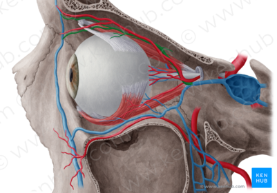
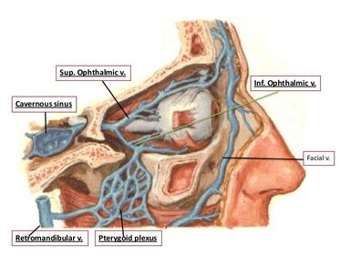
- 55Name Origin: Course:Inferior ophthalmic vein
Origin: Drains into cavernous sinus
Course: Travels through superior orbital fissure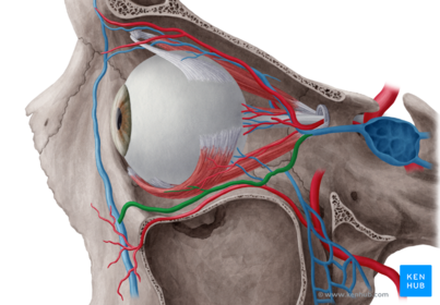
- 56Name A and describe in detail the origin and course of innervation:Lacrimal gland
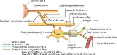
The lacrimal gland is innervated by parasympathetic fibres.
The preganglionic fibres leave the CNS with CN 7 and enter the greater petrosal nerve and continues until it becomes the nerve of the pterygoid canal.
It eventually joins the pterygopalatine ganglion and synapses onto the postganglionic neuron. This neuron joins V2 (maxillary nerve) and continues until the zygomatic nerve branches from the maxillary nerve.
Then, the parasympathetic fibres will travel with the zygomatic nerve until it eventually distributes postganglionic fibres in a small branch that joins the lacrimal nerve.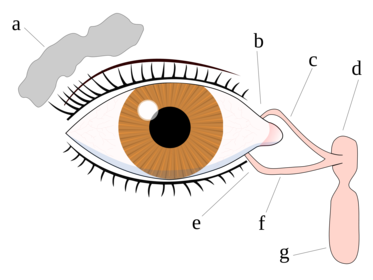
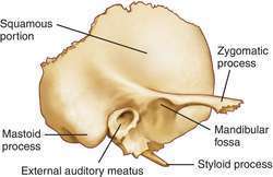
- 57Name allA; Squamous part of the temporal bone
B: Zygomatic process
C: Glenoid fossa
D: External auditory meatus (EAM)
E: Mastoid process
F: Styloid process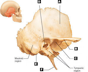
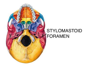
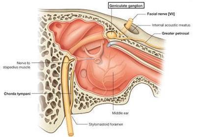
- 58Name Contents:Stylomastoid foramen
Contents: Facial nerve (Cranial nerve 7)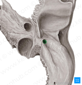
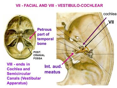
- 59Name Contents:Internal auditory meatus (IAM)
Contents: Facial nerve (cranial nerve 7) and vestibulocochlear nerve (cranial nerve 8)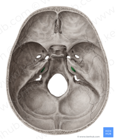
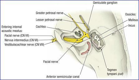
- 60Name Contents:Squamotympanic fissure
Contents: Chorda tympani nerve (branch of the facial nerve (cranial nerve 7)) running to join the lingual nerve which descends between the medial pterygoid muscle and medial surface of the mandible to supply the anterior 2/3 of the tongue; lingual-general, chorda tympani-taste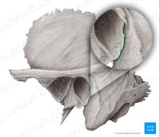
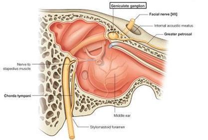
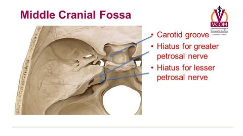
- 61Name Contents:Groove for greater petrosal nerve
Contents: Greater petrosal nerve (branch of facial, carrying preganglionic parasympathetic fibres from the lacrimal nucleus to the pterygopalatine ganglion in the pterygopalatine fossa)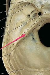
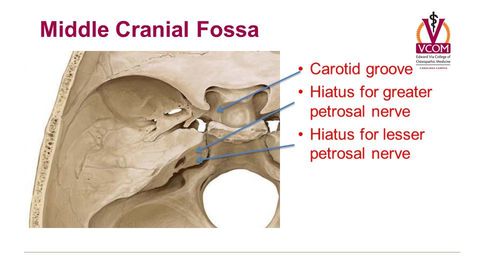
- 62Name Contents:Groove for lesser petrosal nerve
Contents: Lesser petrosal nerve (branch of facial nerve)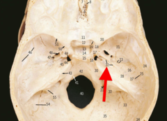
- 63NameGroove for superior petrosal sinus
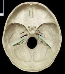
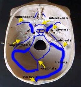
- 64NameGroove of lesser petrosal sinus
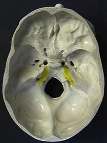
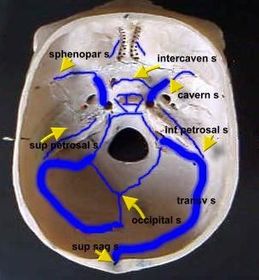
- 65Name Contents:Carotid canal
Contents: Internal carotid artery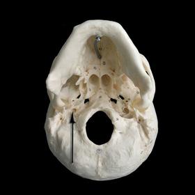
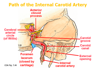
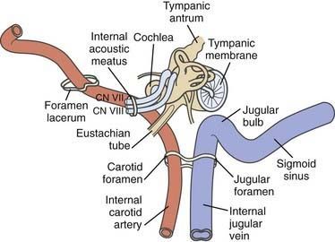
- 66Name JB and SSJB: Jugular bulb; the enlargement of the internal jugular vein
SS: Sigmoid sinus, which drains into the jugular bulb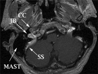
- 67Name Underlying structure:Arcuate eminence
Underlying structure: Superior semicircular canal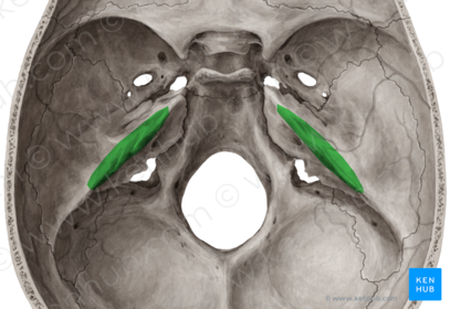
- 68Name 3 and 63: Oval window
6: Round window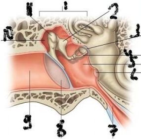
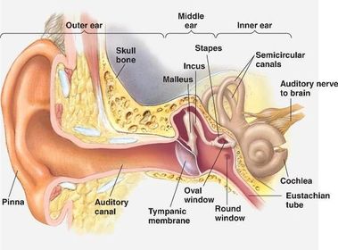
- 69—
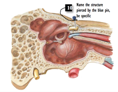
- 70Name 1, 6 and 8
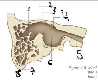
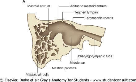
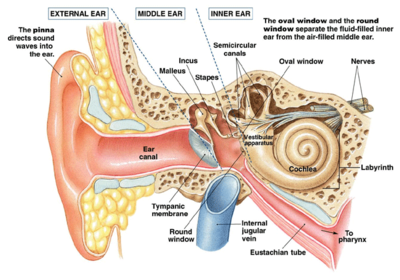
- 71Name Composition: Course:External auditory meatus (EAM)
Composition: Lateral third is cartilaginous whilst medial two thirds is petrous temporal bone
Course: From the auricle to the tympanic membrane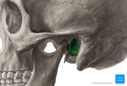
- 72Name Covering externally and internally:Tympanic membrane
Externally covered by skin and internally by mucous membrane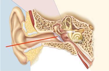
- 73Name all Type of joint connecting them:Ossicles
1. Foot plate of stapes
A. Stapes
B. Incus
C. Malleus
2. Handle of malleus
They are all linked via synovial joints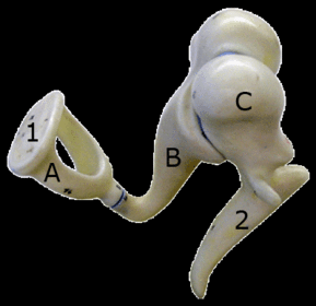
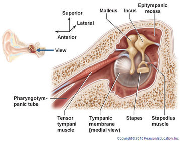
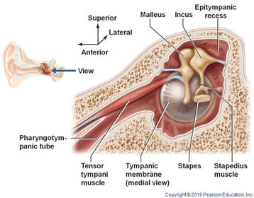
- 74Name Insertion: Function: Innervation:Tensor tympani
Insertion: Handle of the malleus
Function: Reduce excessive movement of the malleus helping dampen loud noises
Innervation: Mandibular division of trigeminal (V3) via medial pterygoid nerve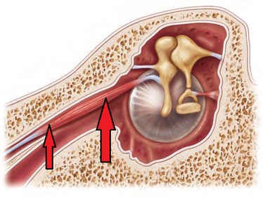
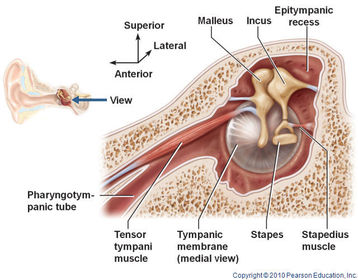
- 75Name Insertion: Function: Innervation:Stapedius
Insertion: Stapes
Function: Reduce excessive movement of the stapes helping dampen loud noises
Innervation: Motor branch of facial nerve (cranial nerve 7)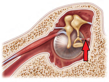
- 76Name Course:Auditory tube:
Course: Opens into the anterior wall of tympanic cavity at the lateral end and opens into the nasopharynx at the medial end.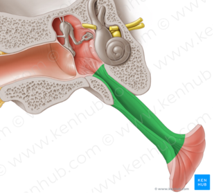
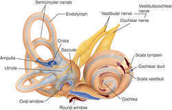
- 77Name Describe what is located anteriorly and posteriorly and the source of their innervationsThe vestibule/round window is the central part-windows opens to the tympanic cavity and is filled by the foot plate of the stapes.
Cochlea lies anterior to the vestibule.
Semicircular canals lie posterior to the vestibule, with one in each plane (superior, inferior and horizontal canals.
The cochlear and semicircular canals are supplied by the cochlear and vestibular nerves respectively; at the lateral end of the internal acoustic meatus the vestibulocochlear nerve (cranial nerve 8) divides into the cochlear and vestibular portions.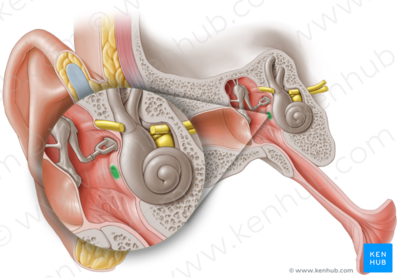
Informações
- Descrição
- RCA Practical 5-Oral and Nasal cavities, Paranasal Sinuses, Orbit, and the Ear
- Criado por
- Usuário eliminado · set 2016
- Atualizado
- 20 set 2016
- Visualizações
- 26
- Tags
- Histórico
-
- Criado por Ashutosh Kumar · 31 ago 2016
- Copiado por Binura Wijesinghe · 18 set 2016
- Copiado por Binura Wijesinghe · 19 set 2016
- Avaliação