
Quiz
Anatomy module 2
Este quiz tem tipos de pergunta que abrem no player de sempre.
Perguntas do quiz (61)
1From superficial to deep, list the layers of the thoracic wall:
1. [blank_start]Epidermis + dermis[blank_end]
2. [blank_start]Hypodermis[blank_end]
3. [blank_start]Deep fascia[blank_end]
4. [blank_start]Ribs (muscle, nerves, BV, LV)[blank_end]
5. [blank_start]Parietal pleura[blank_end]
6. [blank_start]Visceral pleura[blank_end]
7. [blank_start]Lung[blank_end]- AEpidermis + dermis
- BHypodermis
- CDeep fascia
- DRibs (muscle, nerves, BV, LV)
- EParietal pleura
- FVisceral pleura
- GPericardium
- HLung
- IPleural fluid
2Label features of the rib.
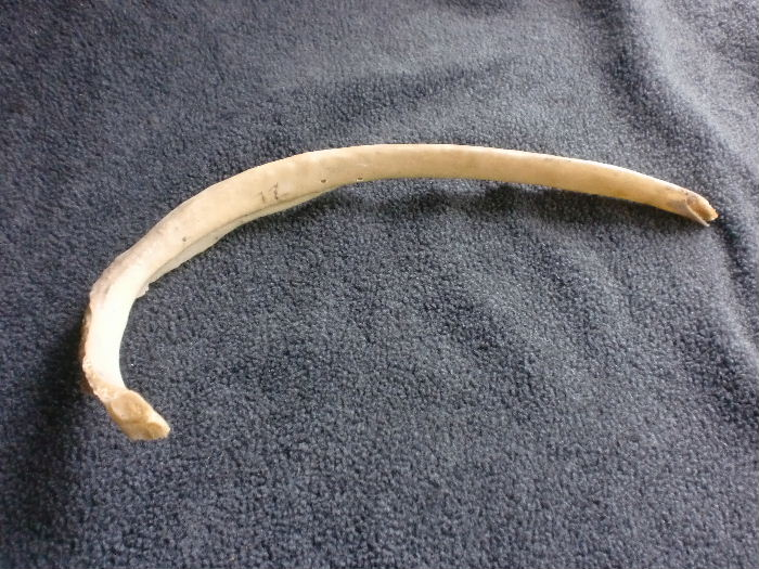
- Acostal groove
- Barticular facet
- Cneck
- Dangle
3Label the features of the rib.
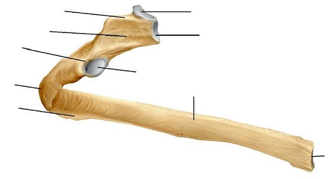
- Aarticular facet
- Btubercle
- Carticular facet
- Darticular facet
- Eneck
- Fcostal groove
- Gcrest
- Hcostal cartilage
4The [blank_start]tubercular facet[blank_end] of the 6th rib articulates with the [blank_start]costal facet[blank_end] of the transverse process of the [blank_start]6th[blank_end] vertebrae.
The articular facets on the [blank_start]head[blank_end] of the 6th rib articulates with the [blank_start]vertebral body[blank_end] of the [blank_start]5th and 6th[blank_end] vertebra.
The [blank_start]crest on the head[blank_end] of the rib articulates with the site where the [blank_start]intervertebral disc[blank_end] would be located.- Atubercular facet
- Bcostal facet
- Chead
- Dneck
- Ebody
- F5th and 6th
- G6th
- H5th
- Icrest on the head
- Jintervertebral disc
- Kintervertebral space
- Lvertebral body
- Mlamina
- Npedicle
- Ospinous process
5From superficial to deep, the costal joint with vertebral body contains:
[blank_start]joint capsule[blank_end], [blank_start]joint cavities[blank_end], [blank_start]intra-articular ligament[blank_end].
The transverse process of the vertebrae and the tubercular facet of the rib forms a [blank_start]costotransverse joint[blank_end].
The [blank_start]costotransverse ligament[blank_end] connects the transverse process of the previous vertebrae to the subsequent rib.- Acostotransverse joint
- Bcostotransverse ligament
- Cjoint capsule
- Djoint cavities
- Eintra-articular ligament
6Choose the CORRECT statement(s).
- ATrue ribs are I - VI
- BFloating ribs are XI - XII
- CFalse ribs have no anterior articulation, their cartilaginous tips end in the musculature of the thoracic body wall.
- DThe costal margin is formed by ribs VII - X and the xiphoid process.
- EThe false ribs are VIII - XII
7What is TRUE regarding the cervical rib?
- AIt is present in 5% of the population.
- BIt articulates with the 7th cervical vertebrae, always bilaterally.
- CA cervical rib can result in thoracic outlet syndrome.
- DNone of the above.
8Label parts of the sternum.
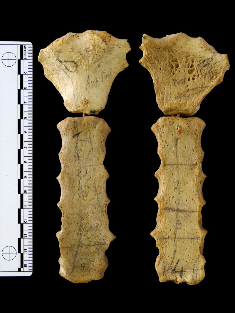
- Ajugular notch
- Bsternal angle
- Cmanubrium
- Dmanubriosternal joint
- Ebody
- Farticular site for clavicle
- Gattachment site for rib I
9Label the sternum diagram. For the dots on the right, write the number of the articulating ribs.
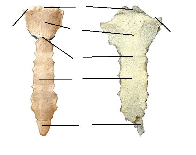
- Amanubrium
- Bxiphoid process
- Cbody
- Dsternal angle
- Etransverse ridges
- Fjugular notch
- Grib II
- Hrib VII
- Irib III - VI
- Jrib I
10The sternal angle is an important clinical landmark, because it is the reference point from which you measure the height of the [blank_start]jugular venous pressure[blank_end].
- Ajugular venous pressure
11Choose the CORRECT statement(s).
- AThe spaces between the ribs are called intercostal spaces.
- BThe space superior to the 8th rib is called the 8th intercostal space.
- CThe 5th intercostal space is important because that's where the apex of the heart is located.
- DThe space inferior to the 6th rib is the 6th intercostal space.
12Choose the CORRECT statement(s).
- AThe superior thoracic aperture is formed from the first thoracic vertebra and the first ribs.
- BThe thoracic outlet is flexible to accommodate all the viscera that needs to pass through.
- CThe thoracic outlet contains the apices of the right and left lungs, great vessels, lympathics.
- DThe thoracic outlet contains the trachea, the esophagus, and major nerves.
- EThoracic outlet syndrome occurs only when the there is a cervical rib.
- FVascular symptoms of thoracic outlet syndrome include a pale, swollen, cold, and/or aching upper limb.
- GNeurological signs of thoracic outlet syndrome shows exclusively with weakness and wasting of muscles on the hand.
13The inferior thoracic aperture is defined by the [blank_start]12th thoracic vertebra[blank_end], the [blank_start]11th ribs[blank_end], the [blank_start]12th ribs[blank_end], the [blank_start]costal margin[blank_end] and the [blank_start]ziphisternal joint[blank_end].
It is the origin of the thoracic [blank_start]diaphragm[blank_end].- A12th thoracic vertebra
- B11th ribs
- C12th ribs
- Dcostal margin
- Eziphisternal joint
- Fdiaphragm
14The [blank_start]ventral[blank_end] rami of thoracic spinal nerves continue between adjacent ribs as the [blank_start]intercostal nerves[blank_end].
The ventral ramus of the T12 spinal nerve is called the [blank_start]subcostal nerve[blank_end], it runs [blank_start]inferiorly[blank_end] to the 12th rib.
[blank_start]T7[blank_end] - T12 nerves enter the [blank_start]abdominal wall[blank_end] and contribute to its innervation.- Aventral
- Bdorsal
- Cintercostal nerves
- Dsubcostal nerves
- Eintervertebral nerves
- Fsubcostal nerve
- Gintercostal nerve
- Hinferiorly
- Isuperiorly
- Jabdominal wall
- Kchest wall
- LT7
- MT6
- NT5
- OT1
15Label the correct parts of the superior mediastinum.
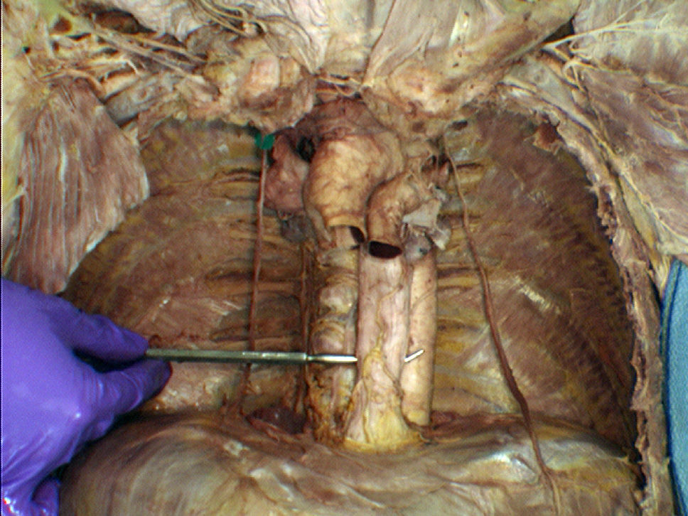
- Aaorta
- Besophagus
16Label parts of the superior mediastinum
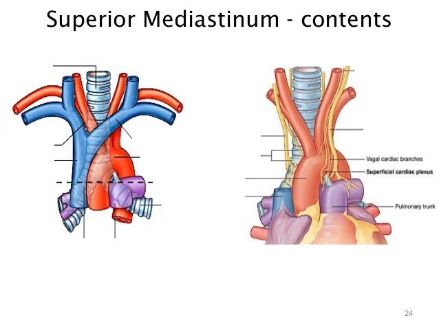
- Atrachea
- Baortic arch
- Caortic arch
- Dleft primary bronchus
- Eright primary bronchus
- Fleft subclavian artery
- Gleft common carotid artery
- Hbrachiocephalic trunk
- Isuperior vena cava
- Jleft brachiocephalic vein
- Kright common carotid artery
- Lright subclavian artery
- Mleft internal jugular vein
- Nleft subclavian vein
- Oright brachiocephalic vein
- Pright internal jugular vein
- Qright subclavian vein
- Rleft venous angle
- Sright venous angle
17The superior vena cava returns venous blood to the [blank_start]right atrium[blank_end] of the heart.
- Aright atrium
18Label branches of the aortic arch.
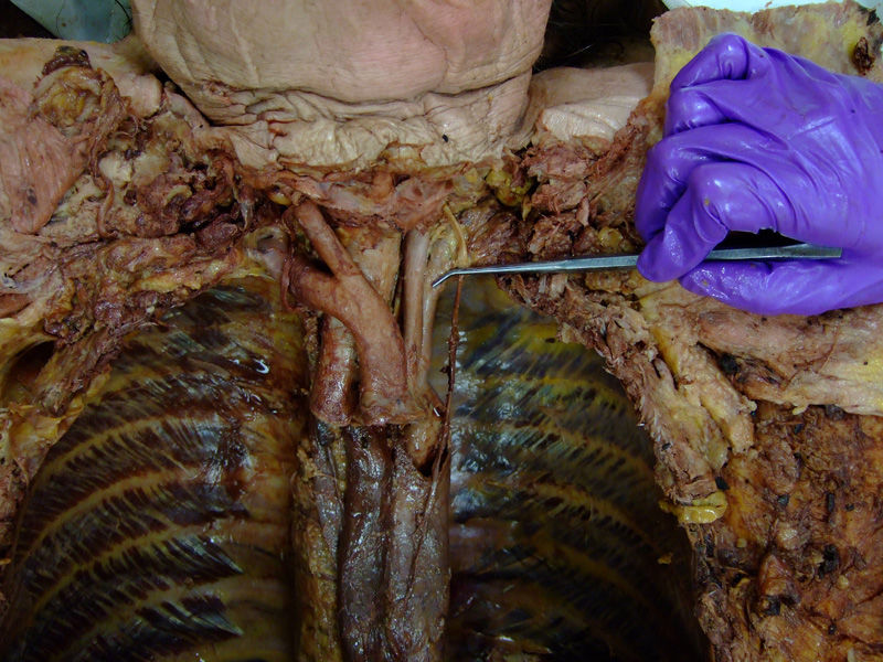
- Abrachiocephalic trunk
- Bleft common carotid artery
- Cleft subclavian artery
- Dright common carotid artery
- Eright subclavian artery
19Label the blood vessels.
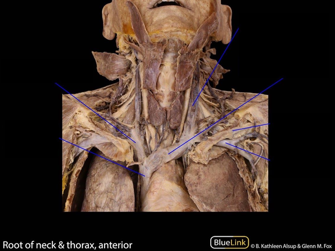
- Aright brachiocephalic vein
- Bsuperior vena cava
- Cleft internal jugular vein
- Dleft brachiocephalic vein
- Eleft subclavian vein
- Fleft common carotid artery
20Label the blood vessels.
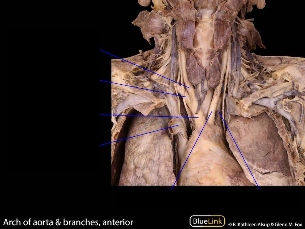
- Aright common carotid artery
- Bright subclavian artery
- Cbrachiocephalic trunk
- Dright brachiocephalic vein
- Eleft common carotid artery
- Fleft subclavian artery
21From superficial to deep, the three layers of intercostal muscles are: [blank_start]external[blank_end], [blank_start]internal[blank_end], and [blank_start]innermost[blank_end].
- Aexternal
- Binternal
- Cinnermost
22What is TRUE regarding the intercostal space?
- AIntercostal muscles do not move in order to keep the ribs in place.
- BThe intercostal neurovascular bundles are located between external and internal intercostal muscles.
- CThe intercostal neurovascular bundles disappear behind the innermost intercostal muscles as they pass anteriorly around the thoracic body wall.
- DNone of the above.
23Label the structure pinned.
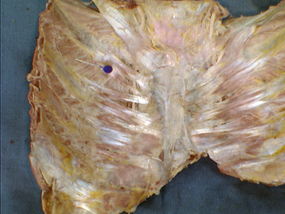
- Ainnermost intercostal muscle
24Label the structure (at the pin).
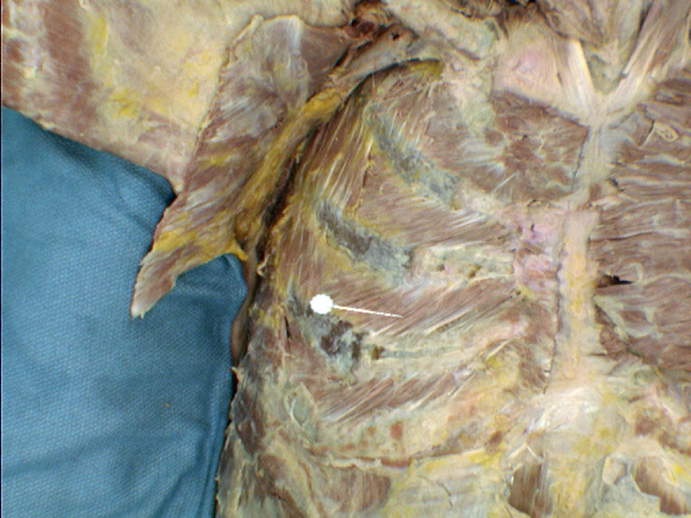
- Ainternal intercostal muscle
- Bexternal intercostal muscle
25Label the structure on the probe.
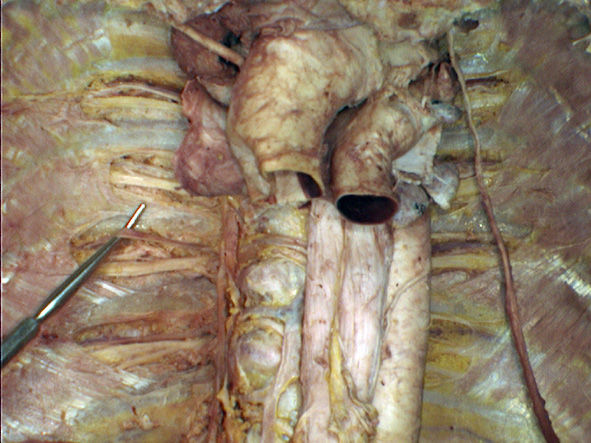
- Aintercostal vein
26Label.
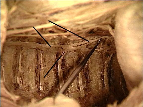
- Asympathetic chain
- Brami communicans
- Cgreater splanchnic nerve
- Dintercostal nerve
- Esympathetic ganglion
27The thoracic aorta has two types of branches:
[blank_start]parietal[blank_end] branches are the [blank_start]posterior intercostal arteries[blank_end], they supply the thickness of the chest wall;
[blank_start]visceral[blank_end] branches supply the [blank_start]esophagus[blank_end], [blank_start]bronchial tree[blank_end], and the [blank_start]pericardium[blank_end].- Aparietal
- Bposterior intercostal arteries
- Cvisceral
- Desophagus
- Ebronchial tree
- Fpericardium
28On the right side, the venous blood travel from the [blank_start]posterior intercostal vein[blank_end] to the [blank_start]azygous vein[blank_end].
On the left side, the venous blood travel from the posterior intercostal vein to the [blank_start]hemiazygous vein[blank_end] and [blank_start]accessory hemizaygous vein[blank_end], then to [blank_start]the azygous vein[blank_end].
From the [blank_start]azygous vein,[blank_end] the blood drains into the [blank_start]superior vena cava[blank_end], and eventually to the [blank_start]right atrium of the heart[blank_end].- Aposterior intercostal vein
- Banterior intercostal vein
- Cazygous vein
- Dhemiazygous vein
- Eaccessory hemizaygous vein
- Fthe azygous vein
- Gthe hemizygous vein
- Hthe accessory hemiazygous vein
- Iazygous vein,
- Jhemiazygous vein,
- Kleft brachiocephalic vein,
- Lleft internal jugular vein,
- Mright brachiocephalic vein
- Nright subclavian vein
- Oright internal jugular vein
- Pleft brachiocephalic vein
- Qleft internal jugular vein
- Rsuperior vena cava
- Sright atrium of the heart
- Tleft atrium of the heart
- Uright ventricle of the heat
- Vleft ventricle of the heart
29Label the structure on the probe.
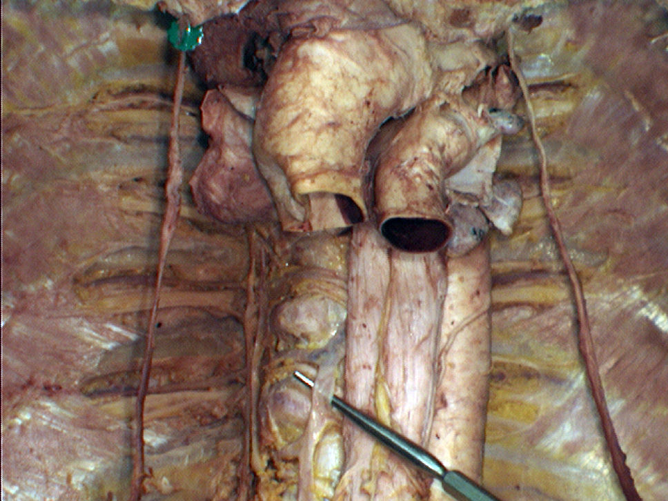
- Aazygous vein
30Label the structures.
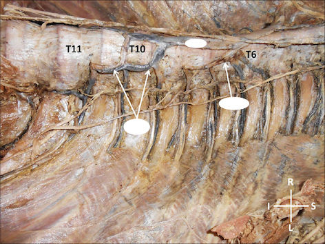
- Aazygous vein
- BHemiazygous vein
- Caccessory hemiazygous vein
31Label the structures.
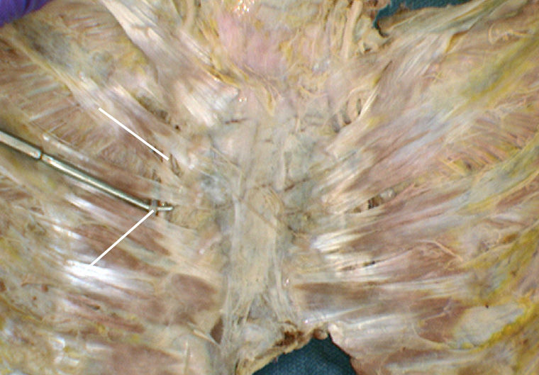
- Ainternal thoracic artery
- Binternal thoracic vein
32The anterior intercostal arteries arise from [blank_start]internal thoracic arteries[blank_end], which are from [blank_start]right and left subclavian arteries[blank_end].
The anterior intercostal veins drains into [blank_start]internal thoracic veins[blank_end], then [blank_start]brachiocephalic veins[blank_end], and then unite to form the [blank_start]superior vena cava[blank_end].
The anterior and posterior vasculature form [blank_start]anastomoses[blank_end] in the [blank_start]thoracic wall[blank_end] at the midclavicular line.- Ainternal thoracic arteries
- Bright and left subclavian arteries
- Cinternal thoracic veins
- Dbrachiocephalic veins
- Esuperior vena cava
- Fanastomoses
- Gthoracic wall
33Select the INCORRECT statement(s).
- AInterstitial fluid is formed from blood plasma.
- BAll of the interstitial fluid enters lymphatic capillaries to become lymph.
- CLymph vessels open into lymph nodes, which cleanses lymph of foreign elements and transformed cells.
- DCleansed lymph is excreted through the kidney.
- EAll statements are correct.
34Posteriorly, lymph from the chest wall drains into [blank_start]intercostal lymph nodes[blank_end], and then into the [blank_start]thoracic duct[blank_end].
Anteriorly, lymph from the chest wall drains into the [blank_start]parasternal lymph nodes[blank_end], and then into the [blank_start]bronchomediastinal trunk[blank_end].- Aintercostal lymph nodes
- Binternal thoracic lymph nodes
- Cthoracic duct
- Dbrachiocephalic duct
- Eparasternal lymph nodes
- Finternal lymph nodes
- Gthoracic lymph nodes
- Hbronchomediastinal trunk
- Isubclavian trunk
35Posteriorly, lymph from the chest wall drains into [blank_start]intercostal lymph nodes[blank_end], and then into the [blank_start]thoracic duct[blank_end].
Anteriorly, lymph from the chest wall drains into the [blank_start]parasternal lymph nodes[blank_end], and then into the [blank_start]bronchomediastinal trunk[blank_end].
In either case, lymph ultimately drains into venous blood at the right and left [blank_start]venous angles[blank_end].- Aintercostal lymph nodes
- Bthoracic duct
- Cparasternal lymph nodes
- Dbronchomediastinal trunk
- Evenous angles
36Select the CORRECT statements regarding lymphatic drainage.
- ARoughly 75% of the lymph draining from the breast passes superolaterally through axillary and subclavicular nodes to enter the brachiocephalic trunks.
- BThe remaining lymph drains medially into the parasternal nodes, then enters the bronchomediastinal drunks.
- CA smaller portion drains inferiorly into abdominal and superficial inguinal nodes.
- DBreast cancer can only be spread by lymph superolaterally.
- ETumour cells cannot travel from one breast to another.
37Lymph drainage from the breast can be classified into three directions:
superolaterally through [blank_start]axillary[blank_end] and [blank_start]supraclavicular[blank_end] nodes, enters the [blank_start]subclavian trunks[blank_end];
medially into the [blank_start]parasternal[blank_end] nodes, enters the [blank_start]bronchomediastinal trunk[blank_end];
inferiorly into [blank_start]abdominal[blank_end] and [blank_start]superficial inguinal[blank_end] nodes.- Aaxillary
- Bsupraclavicular
- Csubclavian trunks
- Dparasternal
- Ebronchomediastinal trunk
- Fabdominal
- Gsuperficial inguinal
38Sympathetic output for all body targets arises from the spinal cord segments T1 and [blank_start]L2[blank_end].
Sympathetic output for thoracic viscera is restricted to [blank_start]T1[blank_end] to [blank_start]T4[blank_end] spinal levels.
Preganglionic axons leave the spinal cord via the [blank_start]ventral root[blank_end] and [blank_start]spinal nerve[blank_end], then travel via a [blank_start]ramus communicans[blank_end] to a [blank_start]sympathetic chain ganglion[blank_end].- AL2
- BL1
- CS1
- DS2
- EL5
- FT1
- GT2
- HT3
- IT4
- JT5
- KT6
- LT7
- MT8
- NT9
- OT10
- PT11
- QT12
- Rventral root
- Sdorsal root
- Tspinal nerve
- Uramus communicans
- Vsympathetic chain ganglion
- Wventral rootlet
- Xdorsal rootlet
- Ysympathetic chain
39Sympathetic innervation of the thoracic viscera is the same as that of the body wall and limbs.
- AVerdadeiro
- BFalso
40Parasympathetic output arises from the [blank_start]brainstem[blank_end], and [blank_start]S2 - S4 spinal levels[blank_end].
Thoracic viscera receive parasympathetic pre-ganglionic output from the [blank_start]vagus nerve[blank_end].- Abrainstem
- BS2 - S4 spinal levels
- Cvagus nerve
41Sympathetic and parasympathetic inputs to viscera overlap in [blank_start]autonomic plexi[blank_end] as they approach their target organs. Examples include the [blank_start]cardia[blank_end], [blank_start]pulmonary[blank_end], and [blank_start]esophageal plexi[blank_end].
- Aautonomic plexi
- Bcardia
- Cpulmonary
- Desophageal plexi
42Label the structure on the probe.
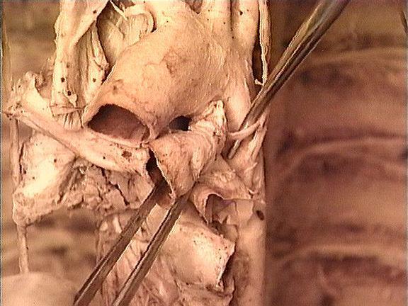
- Aleft recurrent laryngeal nerve
43Label the structures.
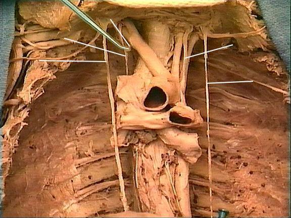
- Aright recurrent laryngeal nerve
- Bright vagus nerve
- Cright phrenic nerve
- Dleft vagus nerve
- Eleft phrenic nerve
44The right recurrent laryngeal nerve recurs around the [blank_start]right subclavian artery[blank_end].
The left recurrent laryngeal nerve recurs around the [blank_start]aortic arch[blank_end].- Aright subclavian artery
- Baortic arch
45Label the structure below.
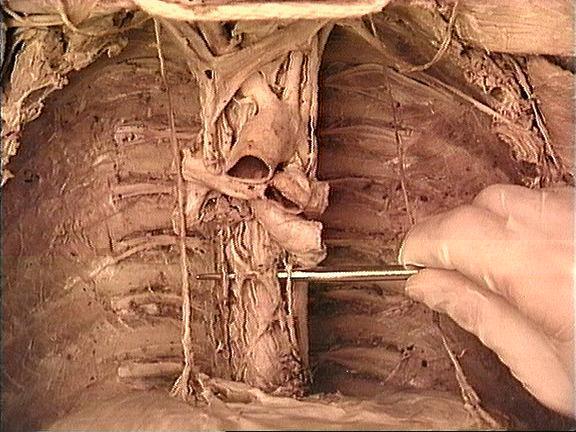
- Aesophagus plexus
46What is INCORRECT about the pleural cavity?
- AThe visceral pleura and the parietal pleura differ in that the visceral pleura is covered by a serous membrane, while the parietal pleura is fibrous.
- BThe two pleural cavities are separated by the mediastinum.
- CThe pleura produces pleural fluid that act as a lubricant for the lungs to move in a friction-free manner.
- DNone of the above.
47Label parts of the lung.
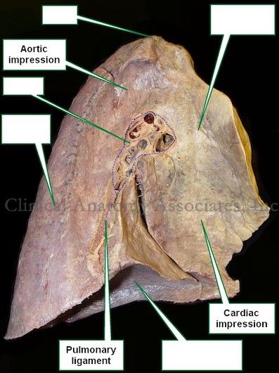
- AHilum
- Bapex
- Cdiaphragmatic surface
- Dmediastinal surface
- Ecostal surface
48Collectively, the structures in the hilum of the lung that are bound together by connective tissues is called the [blank_start]root of the lung[blank_end].
- Aroot of the lung
49Choose the INCORRECT statement(s) regarding pleural effusion.
- AIn pleural effusion, excess fluid accumulates in the pleural space.
- BThe lung volume increases due to build up of fluid.
- CTo analyze the underlying cause of the pleural effusion, an X-ray can be taken.
- DFluid accumulates in the lowest part of the chest in response to gravity.
50The diaphragm originates from the [blank_start]inferior thoracic aperture[blank_end].
It is defined by five structures: [blank_start]T12[blank_end] vertebra, the [blank_start]12th and 11th[blank_end] ribs, the [blank_start]costal margin[blank_end], and the [blank_start]xiphisternal joint[blank_end].- Ainferior thoracic aperture
- BT12
- C12th and 11th
- Dcostal margin
- Exiphisternal joint
51What is TRUE regarding the diaphragm?
- AThe posterior attachment of the diaphragm to the T12 vertebra and ribs is significantly superior to its anterior attachment to the xiphisternal joint.
- BThe fibers of the diaphragm are organized radially, and insert into the contractile central tendon.
- CWhen the muscle fibres contract, the volume of the thoracic cavity decreases.
- DNone of the above.
52There are three main openings in the diaphragm:
[blank_start]caval opening[blank_end] for the interior vena cava, at vertebral level [blank_start]T8[blank_end];
[blank_start]esophageal hiatus[blank_end] for the esophagus, at vertebral level [blank_start]T10[blank_end];
[blank_start]aortic hiatus[blank_end] for the thoracic/abdominal aorta, at vertebral level [blank_start]T12[blank_end].- Acaval opening
- BT8
- Cesophageal hiatus
- DT10
- Eaortic hiatus
- FT12
53The diaphragm is innervated primarily by the [blank_start]phrenic nerves[blank_end], formed from the [blank_start]ventral[blank_end] primary rami of the [blank_start]C3[blank_end], [blank_start]C4[blank_end], and [blank_start]C5[blank_end] spinal nerves.
Both the vegus and phrenic nerve descend in the neck and pass into the thorax via the [blank_start]thoracic inlet[blank_end].
The phrenic nerves run along the lateral aspect of the [blank_start]mediastinum[blank_end], deep to the [blank_start]mediastinal parietal pleura[blank_end], [blank_start]anterior[blank_end] to the root of the lung.
The vagus nerves pass [blank_start]posterior[blank_end] to the root of the lung.- Aphrenic nerves
- Bvagus nerves
- Cventral
- Ddorsal
- EC3
- FC1
- GC2
- HC4
- IC5
- JC6
- KC7
- LT1
- MT2
- NT3
- OT4
- PT5
- QT6
- Rthoracic inlet
- Sinferior thoracic aperture
- Tmediastinal parietal pleura
- Umediastinum
- Vanterior
- Wposterior
54Choose the CORRECT statement(s) regarding innervation of the diaphragm.
- AThe phrenic nerve contains parasympathetic but not sympathetic postganglionic fibres.
- BThe cell bodies of the somatic motor neurons from the phrenic nerve are located in the C3-C5 segments of the spinal cord.
- CThe sensory fibres are responsible for the diaphragm, the serous coverings, mediastinal parietal pleura, and the pericardial sac.
- DPain on the shoulder may be pain originating from the diaphragm because C3 - C5 spinal levels also receive sensory afferents from the corresponding dermatome.
55Label the subdivisions of the mediastinum.
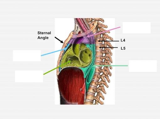
- Asuperior mediastinum
- Bposterior mediastinum
- Canterior mediastinum
- Dmiddle mediastinum
56The superior mediastinum extends from the [blank_start]superior thoracic aperture[blank_end] to a horizontal plane passing through the [blank_start]sternal angle[blank_end] and the intervertebral disc between the [blank_start]T4[blank_end] and [blank_start]T5[blank_end] vertebrae.
The middle mediastinum consists of the [blank_start]pericardial sac and its contents[blank_end].
The anterior mediastinum is the region between the [blank_start]sternum[blank_end] and the [blank_start]pericardial sac[blank_end].
The posterior mediastinum is the region between the pericardial sac and the [blank_start]T5 - T12[blank_end] vertebrae.- Asuperior thoracic aperture
- Binferior thoracic aperture
- Csternal angle
- Djugular notch
- ET4
- FT3
- GT2
- HT1
- IT5
- JT6
- KT7
- LT8
- Mpericardial sac and its contents
- Npleural cavities
- Osternum
- Pmanubrium
- Qpericardial sac
- Rvisceral pleura
- ST5 - T12
- TS1 - S5
- UL1 - L5
- VT1 - 5
57The bulk of the thymus gland is located in the [blank_start]superior mediastinum[blank_end], [blank_start]posterior[blank_end] to the [blank_start]manubrium[blank_end] of the sternum, but it can extend [blank_start]inferiorly[blank_end] into the [blank_start]anterior mediastinum[blank_end].
It is involved in the development of [blank_start]the immune system[blank_end], largest during [blank_start]children and adolescents[blank_end].- Asuperior mediastinum
- Binferior mediastinum
- Cposterior mediastinum
- Dmiddle mediastinum
- Eposterior
- Fanterior
- Gmanubrium
- Hbody
- Ixiphoid process
- Jinferiorly
- Ksuperiorly
- Lanterior mediastinum
- Mthe immune system
- Nsenescence
- Opuberty
- Pchildren and adolescents
- Qthe elderly
58Label the layers of pericardial sac.
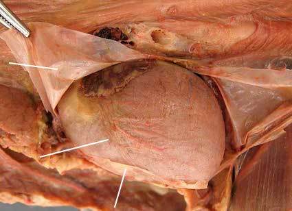
- Aparietal serous pericardium
- Bvisceral serous pericardium
- Cfibrous pericardium
59What is TRUE regarding the pericardium?
- AThe visceral pericardium is completely separated from the parietal pericardium.
- BThe fibrous pericardium becomes continuous with the outermost connective tissue layer of the great vessels (tunica adventitia).
- CThe fibrous layer and the serous layer of the pericardium are two distinctive separated layers.
- DNone of the above.
60The apex of the heart is pointed [blank_start]downward[blank_end], [blank_start]forward[blank_end], and to the [blank_start]left[blank_end]. The base is [blank_start]posterior[blank_end].
The pyramid rests on its [blank_start]diaphragmatic[blank_end] surface, its [blank_start]anterior[blank_end] surface faces the sternum, and has a right and left [blank_start]pulmonary[blank_end] surface.- Adownward
- Bforward
- Cleft
- Dposterior
- Ediaphragmatic
- Fanterior
- Gpulmonary
61Label parts of the heart (for dots around the heart, write the corresponding surface).
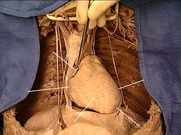
- Aright auricle
- Bright atrium
- Cright ventricle
- Dapex
- Eleft ventricle
- Fdiaphragmatic surface
- Gleft pulmonary surface
- Hright pulmonary surface
- Ianterior surface
Informações
- Descrição
- Thorax
- Autor
- M Z
- Idioma
- English
- Visualizações
- 23
- Etiquetas
- Avaliação
- Ainda sem avaliações Avalie este quiz