5511353

Questão 1
Questão
The four basic types of tissue are...? (check all that apply)
Responda
-
Cardiac
-
Skeletal
-
Muscle
-
Smooth
-
Epithelial
-
Nervous
-
Connective
-
Ependymal
Questão 2
Questão
The cells of [blank_start]connective[blank_end] tissue tend to be widely separated from each other. The large amounts of [blank_start]extra cellular matrix[blank_end] are found in these [blank_start]spaces[blank_end]. Loose connective tissue keeps [blank_start]organs[blank_end] and epithelial in place while [blank_start]dense[blank_end] connective tissue forms both [blank_start]ligaments[blank_end] and tendons. [blank_start]Adipose[blank_end] tissue (fat) is also connective tissue. [blank_start]Blood[blank_end], bone and [blank_start]cartilage[blank_end] are also kinds of connective tissue.
Responda
-
connective
-
extracellular matrix
-
spaces
-
organs
-
dense
-
ligaments
-
Adipose
-
Blood
-
cartilage
Questão 3
Questão
The main role of connective tissue in the body is to transport substances, and provide structure and support.
Responda
- True
- False
Questão 4
Questão
[blank_start]Epithelial[blank_end] tissue is arranged next to one another and there is little [blank_start]extracellular matrix[blank_end]. The tissue protects outer surfaces (our skin) and is lines [blank_start]internal cavities[blank_end].
Responda
-
Epithelial
-
extracellular matrix
-
internal cavities
Questão 5
Questão
What are the two types of bone tissue?
Responda
-
long and short
-
compact and cancellous/spongy
-
flat and short bones
-
compact and soft
-
diaphysis and epiphysis
Questão 6
Questão
Long bones are wider than they are long
Responda
- True
- False
Questão 7
Questão
Carpal bones are what type of bone?
Responda
-
long bones
-
short bones
-
flat bones
-
irregular bones
Questão 8
Questão
Metacarpals are in which type of bone class?
Responda
-
long
-
short
-
flat
-
irregular
Questão 9
Questão
The scapula bone is a __________.
Responda
-
long bone
-
short bone
-
flat bone
-
irregular bone
Questão 10
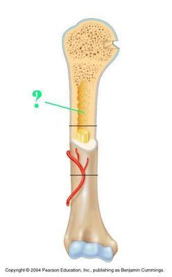
Questão
What type of bone is in this picture?
Responda
-
long
-
short
-
flat
-
irregular
Questão 11
Questão
What are the two divisions of the skeleton?
Responda
-
Axial and appendicular
-
Axial and biaxial
-
Central and anterior
-
anterior and interior
Questão 12
Questão
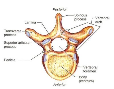
How many bones are there for the cervical division in the vertebrae?
Responda
-
5
-
7
-
8
-
9
-
12
Questão 13
Questão
How many bones are there for the Thoracic division in the vertebrae?
Responda
-
5
-
7
-
8
-
9
-
12
Questão 14
Questão
How many bones are there for the lumbar division in the vertebrae?
Responda
-
5
-
7
-
8
-
9
-
12
Questão 15
Questão
Flexion is in the ___________ plane.
Responda
-
sagittal plane
-
transverse plane
-
median plane
-
coronal plane
-
lateral plane
Questão 16
Questão
Extension is in the ________ plane.
Responda
-
sagittal
-
transverse
-
medial
-
coronal
-
lateral
Questão 17
Questão
How many ribs make up the ribcage?
Responda
-
10 paired ribs; 20 in total
-
12 paired ribs; 24 in total
-
15 paired ribs; 30 in total
-
16 paired ribs; 32 in total
Questão 18
Questão
Which bone does the clavicle articulate with?
Responda
-
sternum
-
scapula
-
ribs
-
humerus
Questão 19
Questão
It is possible to palpate the whole length of the clavicle because it is more [blank_start]superficial[blank_end] and no [blank_start]muscle[blank_end] is covering it.
Responda
-
superficial
-
muscle
Questão 20
Questão
Why bone does the scapula articulate with?
Responda
-
clavicle
-
pectoral girdle
-
pelvic girdle
-
humerus
Questão 21
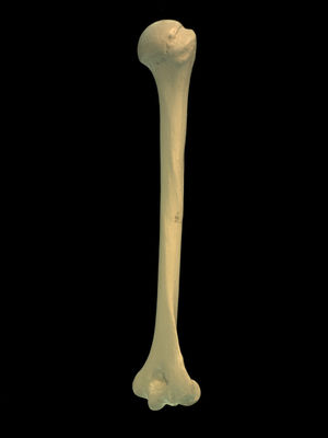
Responda
-
humerus
-
femur
-
tibia
-
fibula
Questão 22
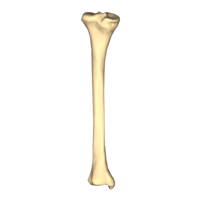
Responda
-
humerus
-
tibia
-
fibula
-
femur
Questão 23
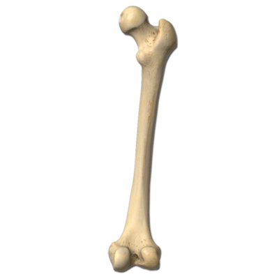
Responda
-
humerus
-
tibia
-
fibula
-
femur
Questão 24
Questão
How many carpal bones are on the hand?
Responda
-
2
-
3
-
5
-
7
-
8
Questão 25
Questão
How many metacarpals are in the hand?
Responda
-
3
-
5
-
6
-
8
Questão 26
Questão
How many phalanges are in the hand?
Responda
-
3
-
5
-
6
-
8
Questão 27
Questão
How many tarsals are in a foot?
Responda
-
3
-
5
-
6
-
7
Questão 28
Questão
How many metatarsals are in the foot?
Responda
-
3
-
5
-
6
-
7
Questão 29
Questão
How many phalanges are in a toe of a foot (other than the big toe)?
Responda
-
3
-
5
-
6
-
7
Questão 30
Questão
An epicondyle is a [blank_start]prominence[blank_end] on the [blank_start]distal[blank_end] part of a [blank_start]long[blank_end] bone serving for the attachment of muscles and [blank_start]ligaments[blank_end].
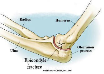
Responda
-
prominence
-
distal
-
long
-
ligaments
Questão 31
Questão
The forearm is defined as the region from [blank_start]elbow[blank_end] to [blank_start]wrist[blank_end]. The forearm has two [blank_start]long[blank_end] bones, the [blank_start]radius[blank_end] and [blank_start]ulna[blank_end].
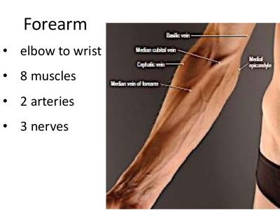
Responda
-
elbow
-
wrist
-
long
-
radius
-
ulna
Questão 32
Questão
Bones are made of [blank_start]connective[blank_end] tissue; they consist of cells embedded in an [blank_start]extracellular matrix[blank_end]. Bones begin as [blank_start]cartilaginous[blank_end] preforms in infancy and [blank_start]ossify[blank_end] as we age. Because bones are [blank_start]alive[blank_end] and contain cells, they have the ability to adapt and [blank_start]repair[blank_end].
Responda
-
connective
-
extracellular matrix
-
cartilaginous
-
ossify
-
alive
-
repair
Questão 33
Questão
The [blank_start]ECM[blank_end] of a bone is different from other types of [blank_start]connective[blank_end] tissue as it only consists of [blank_start]1/3[blank_end] of organic material and [blank_start]2/3[blank_end] of [blank_start]inorganic[blank_end] material
Responda
-
ECM
-
connective
-
1/3
-
2/3
-
inorganic
Questão 34
Questão
What type of cell in the cellular component of the bone build the extracellular matrix?
Responda
-
osteoblasts
-
osteocytes
-
osteoclasts
-
osteocyotes
Questão 35
Questão
What type of cell in the cellular component of the bone break down the extracellular matrix?
Responda
-
osteoblasts
-
osteoclasts
-
osteocytes
-
osteocyotes
Questão 36
Questão
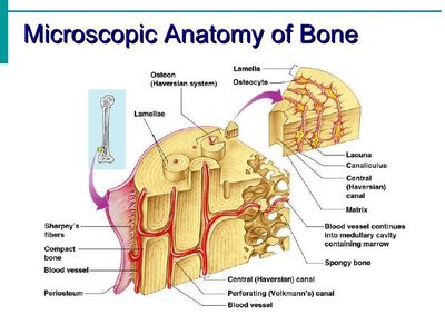
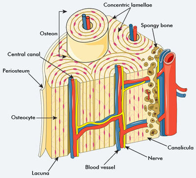
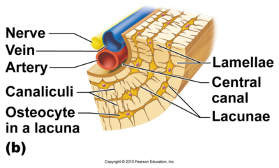
The outer surface of bone is covered and protected by the [blank_start]periosteum[blank_end], a fine sheet of [blank_start]connective[blank_end] tissue. There are small holes in the periosteum called [blank_start]foraminae[blank_end]. The foraminae allow blood vessels and [blank_start]nerves[blank_end] to pass into the bone and [blank_start]supply[blank_end] the cells. Osteocytes are found in living spaces called [blank_start]lacunae[blank_end], trapped in sheets of [blank_start]bone[blank_end] known as [blank_start]lamellae[blank_end]. Each lamellae is made up of [blank_start]collagen[blank_end] fibres that are [blank_start]arranged[blank_end] in a specific direction. Bone has many [blank_start]layers[blank_end] of lamellae and their [blank_start]collagen[blank_end] fibres run in many different directions, so bone is resistant to [blank_start]tension[blank_end] in many directions. Lamellae arrange themselves into [blank_start]cylinder[blank_end]-shaped structures known as [blank_start]osteons[blank_end].
Responda
-
periosteum
-
connective
-
foraminae
-
nerves
-
supply
-
lacunae
-
bone
-
lamellae
-
collagen
-
arranged
-
layers
-
collagen
-
tension
-
cylinder
-
osteons
Questão 37
Questão
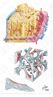
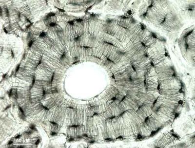
A [blank_start]central canal[blank_end], through which blood vessels and [blank_start]nerves[blank_end] pass, runs down the middle of an [blank_start]osteon[blank_end]. Blood and nerves penetrate into the [blank_start]lacunae[blank_end] to supply the [blank_start]osteocytes[blank_end] through channels called [blank_start]canaliculi[blank_end] that connects to the lacunae to the central canal and to each other. These allow delivery of nutrients and [blank_start]removal[blank_end] of wastes.
Responda
-
central canal
-
nerves
-
osteon
-
lacunae
-
osteocytes
-
canaliculi
-
removal
Questão 38
Questão
In the middle of shafts of long bones is a cavity in which the bone marrow is found: the [blank_start]medullary cavity[blank_end]. Lining this is a fine sheet of connective tissue called [blank_start]endosteum[blank_end].
Responda
-
endosteum
-
medullary cavity
Questão 39
Questão
Cancellous bone is comprised of the same tissue as compact bone but it is arranged quit differently. [blank_start]Lamaellae[blank_end] form struts of bone known as [blank_start]trabeculae[blank_end], which interconnect to form a [blank_start]honeycomb[blank_end] or sponge-like structure. [blank_start]Bone marrow[blank_end] is found between the trabeculae. Trabeculae are thinner than [blank_start]osteons[blank_end], so osteocytes can communicate with each other and with nerves and blood vessels directly.
Responda
-
Lamaellae
-
trabeculae
-
honeycomb
-
Bone marrow
-
osteons
Questão 40
Questão
Bone growth first occurs in the [blank_start]diaphysis[blank_end]; as the bone develops, more [blank_start]bone[blank_end] is formed from the center [blank_start]outwards[blank_end] towards the [blank_start]end[blank_end] of bones. Then, blood vessels grow into the bone and are reshaped by [blank_start]osteoclast[blank_end]s so that the central cavity contains bone marrow.
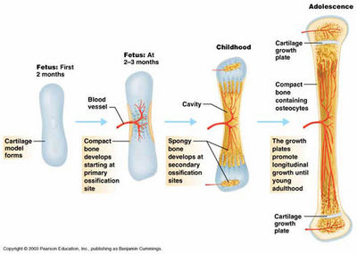
Responda
-
diaphysis
-
bone
-
outwards
-
end
-
osteoclast
Questão 41
Questão
Osteocytes reside in [blank_start]lacunae[blank_end].
Responda
-
lacunae
Questão 42
Questão
Which fracture occurs when the bone breaks completely into two but there is no penetration of the skin, and very little damage to the surrounding soft tissue?
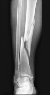
Responda
-
closed
-
open
-
greenstick
-
anterior
Questão 43
Questão
What type of fracture occurs when the bone breaks and pierces the skin, and results in serious soft tissue damage (the bone rips through muscles)? *greatly increases the possibility of infection as the bone is exposed.
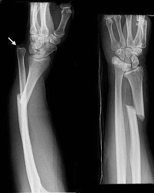
Responda
-
closed
-
open
-
greenstick
-
anterior
Questão 44
Questão
Thickening, or addition of the bone, occurs at the [blank_start]subperiosteal[blank_end] surface, and bone is removed or resorbed (by [blank_start]osteoclasts[blank_end]) at the [blank_start]endosteal[blank_end] surface.
Responda
-
subperiosteal
-
endosteal
-
osteoclasts
Questão 45
Questão
Osteoclasts remove bone by releasing [blank_start]lysosomes[blank_end] and [blank_start]acid[blank_end]. Enzymes break down the [blank_start]organic part[blank_end] of the bone tissue, and acid breaks down the [blank_start]inorganic part[blank_end].
Responda
-
lysosomes
-
acid
-
organic part
-
inorganic part
Questão 46
Questão
In the first stage of fracture repair, broken blood vessels lead to a formation of a [blank_start]haematoma[blank_end] (a soft [blank_start]clot[blank_end]), which takes 0 to [blank_start]3[blank_end] days to form. [blank_start]Capillaries[blank_end] grow into the haematoma. [blank_start]Phagocytes[blank_end] (white blood cells) engulf the [blank_start]dead[blank_end] [blank_start]tissues[blank_end].
Responda
-
haematoma
-
clot
-
3
-
Capillaries
-
Phagocytes
-
dead
-
tissues
Questão 47
Questão
In stage 2 of a fracture repair, the next [blank_start]3[blank_end] days to [blank_start]2[blank_end] weeks, [blank_start]fibroblasts[blank_end] arrive through the [blank_start]capillaries[blank_end] and begin producing [blank_start]collagen[blank_end] fibres to form a matrix of collagen. Some of these fibroblasts differentiate into [blank_start]chondroblasts[blank_end]. They then form a [blank_start]soft[blank_end] [blank_start]cartilaginous[blank_end] [blank_start]callus[blank_end] that splints the bone ends together and [blank_start]reduces[blank_end] movement.
Responda
-
3
-
2
-
fibroblasts
-
capillaries
-
collagen
-
chondroblasts
-
soft
-
cartilaginous
-
callus
-
reduces
Questão 48
Questão
In stage 3 of bone fracture repair, which is during the next [blank_start]3[blank_end] to [blank_start]4[blank_end] weeks, [blank_start]osteoblasts[blank_end] transform the fibrocartilage callus ([blank_start]soft callus[blank_end]) into a [blank_start]bony callus[blank_end].
Responda
-
3
-
4
-
osteoblasts
-
soft callus
-
bony callus
Questão 49
Questão
In stage 4 of fracture repair, the bone is completely [blank_start]united[blank_end] and converts [blank_start]bony[blank_end] callus into more organized [blank_start]osteon[blank_end] structure of bone tissue, and refashioning the bone into its original shape. This starts to happen [blank_start]2[blank_end] to [blank_start]3[blank_end] months after initial fracture has occurred, but it may take [blank_start]2[blank_end] years to completely [blank_start]remodel[blank_end].
Responda
-
united
-
bony
-
osteon
-
2
-
3
-
2
-
remodel
Questão 50
Questão
The lower the [blank_start]bony[blank_end] congruence, the more [blank_start]soft[blank_end] [blank_start]tissue[blank_end] [blank_start]support[blank_end].
Responda
-
bony
-
soft
-
tissue
-
support
Questão 51
Questão
The overall function of a tendon is to restrict movement. and keep structures stable.
Responda
- True
- False
Questão 52
Questão
Chondrocytes are a [blank_start]cartilage[blank_end] cell; a former [blank_start]chondroblast[blank_end] that has become enclosed in a [blank_start]lacuna[blank_end] in the cartilage matrix.
Responda
-
cartilage
-
chondroblast
-
lacuna
Questão 53
Questão
Cartilage is [blank_start]avascular[blank_end] (contains no blood vessels), so [blank_start]nutrients[blank_end] and oxygen must diffuse through the [blank_start]ECM[blank_end] to nourish the [blank_start]chondrocytes[blank_end], so it takes a long time for cartilage to heal after an [blank_start]injury[blank_end] and it can never grow very thick.
Responda
-
avascular
-
nutrients
-
ECM
-
chondrocytes
-
injury
Questão 54
Questão
Hyaline cartilage is [blank_start]thin[blank_end] and contains [blank_start]collagen[blank_end] fibres arranged randomly throughout the [blank_start]matrix[blank_end]. This arrangement traps [blank_start]water[blank_end] and makes hyaline cartilage appear smooth. It has less [blank_start]collagen[blank_end] and more [blank_start]water[blank_end] than fibrocartilage. It covers the [blank_start]articular[blank_end] surfaces of bones, and also allows bone surfaces involved with a joint to move against one another [blank_start]fluidly[blank_end] and with little friction.
Responda
-
thin
-
collagen
-
matrix
-
water
-
collagen
-
water
-
articular
-
fluidly
Questão 55
Questão
The extracellular matrix of [blank_start]dense[blank_end] [blank_start]fibrous[blank_end] connective tissue consists of [blank_start]collagen[blank_end] fibres embedded in ground substances. [blank_start]Fibroblasts[blank_end] are spread throughout the [blank_start]ECM[blank_end]. The collagen is densely packed in [blank_start]one[blank_end] [blank_start]direction[blank_end], which makes the DFCT extremely good at resisting [blank_start]tension[blank_end]. DFCT is partially [blank_start]avascular[blank_end], it takes a long time for it to repair, but not as long as cartilage.
Responda
-
dense
-
fibrous
-
collagen
-
Fibroblasts
-
ECM
-
one
-
direction
-
tension
-
avascular
Questão 56
Questão
The shape of ends of [blank_start]bones[blank_end] involved in the [blank_start]joint[blank_end] determines the amount and type of [blank_start]movement[blank_end] possible at the joint. For example, the [blank_start]round[blank_end] head of the [blank_start]femur[blank_end] allows a wide range of [blank_start]movement[blank_end] at the [blank_start]hip[blank_end], and its deep [blank_start]articulation[blank_end] with the hip gives the joint stability.
Responda
-
bones
-
joint
-
movement
-
round
-
femur
-
movement
-
hip
-
articulation
Questão 57
Questão
Articular cartilage covers the bones where they [blank_start]meet[blank_end] and promotes a [blank_start]frictionless[blank_end] surface.
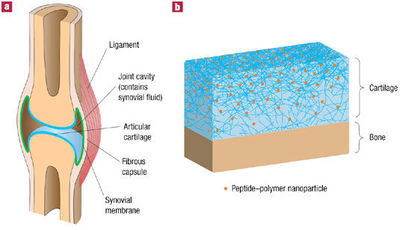
Responda
-
frictionless
-
meet
Questão 58
Questão
In a synovial joint, the thickenings of capsule form the [blank_start]capsular[blank_end] ligaments. These reinforce joints and [blank_start]restrict[blank_end] movement where it is not [blank_start]required[blank_end]. For example, the capsular ligaments at the knee joint are called [blank_start]collateral[blank_end] ligaments and prevent [blank_start]adduction[blank_end] and abduction.
Responda
-
capsular
-
restrict
-
required
-
collateral
-
adduction
Questão 59
Questão
The ligaments of a synovial joint may be divided into [blank_start]extra-[blank_end]capsular ligaments (those that lie outside the capsule), [blank_start]capsular[blank_end] ligaments (those that are thickened and parts of the capsule) and [blank_start]intra[blank_end]-capsular ligaments (those that lie within the capsule).
Responda
-
extra-
-
capsular
-
intra
Questão 60
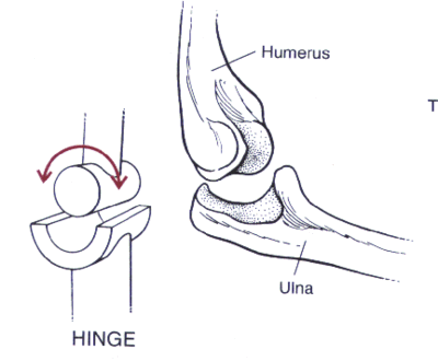
Questão
The hinge joint is a bone joint in which the [blank_start]articular[blank_end] surfaces are [blank_start]molded[blank_end] to each other in such a manner as to permit motion only in [blank_start]one[blank_end] plane. It permits motion back and forth like a [blank_start]door[blank_end]. This joint creates movement of [blank_start]flexion[blank_end] and extension. An example of a hinge joint are the [blank_start]interphalangeal[blank_end] joints in the [blank_start]finger[blank_end].
Responda
-
articular
-
molded
-
one
-
door
-
flexion
-
interphalangeal
-
finger
Questão 61
Questão
One type of a synovial joint, is a [blank_start]pivot[blank_end] joint, which is [blank_start]uniaxial[blank_end]. This joint allows for [blank_start]supination[blank_end] and pronation. An example includes the [blank_start]radioulnar[blank_end] joints (both proximal and [blank_start]distal[blank_end]).
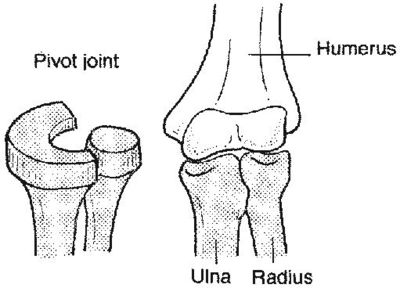
Responda
-
pivot
-
uniaxial
-
supination
-
radioulnar
-
distal
Questão 62
Questão
The saddle joint is [blank_start]biaxial[blank_end] and creates movements such as flexion & [blank_start]extension[blank_end] and also abduction & [blank_start]adduction[blank_end], which ultimately causes [blank_start]circumduction[blank_end]. An example of this joint is [blank_start]carpometacarpal[blank_end] joint at the base of the [blank_start]thumb[blank_end].
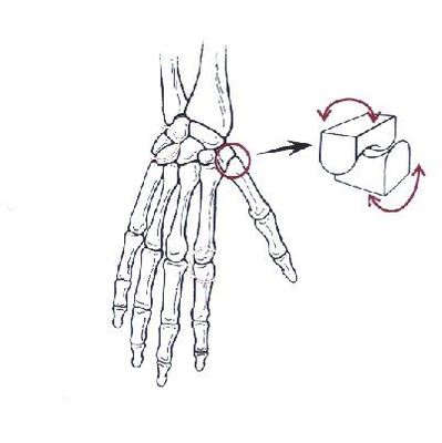
Responda
-
biaxial
-
extension
-
adduction
-
circumduction
-
carpometacarpal
-
thumb
Questão 63
Questão
The ellipsoid joint is [blank_start]biaxial[blank_end] and causes flexion, [blank_start]extension[blank_end], adduction, [blank_start]abduction[blank_end] and [blank_start]circumduction[blank_end]. An example is the [blank_start]radiocarpal[blank_end] joint which is located in the [blank_start]wrist[blank_end].
Responda
-
biaxial
-
extension
-
abduction
-
circumduction
-
radiocarpal
-
wrist
Questão 64
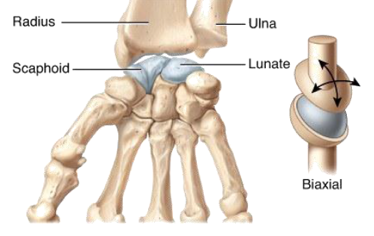
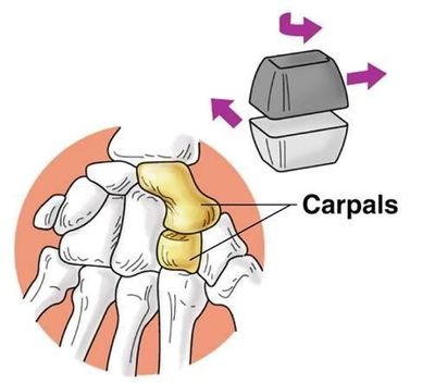
Questão
The plane joint is [blank_start]multi-axial[blank_end] and causes sliding/[blank_start]gliding[blank_end]. The [blank_start]intercarpal[blank_end] and intertarsal joints, which are between carpal and [blank_start]tarsal[blank_end] bones, are an example.
Responda
-
multi-axial
-
gliding
-
intercarpal
-
tarsal
Questão 65
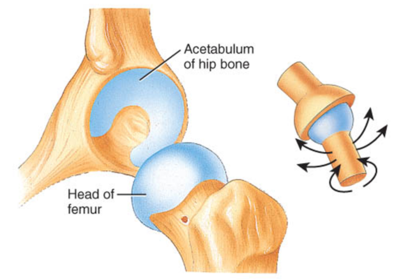
Questão
Ball and socket joints are [blank_start]multiaxial[blank_end], and therefore can cause [blank_start]adduction[blank_end], abduction, [blank_start]flexion[blank_end], extension, circumduction and [blank_start]rotation[blank_end]. An examples include the [blank_start]hip[blank_end] joint and the shoulder joint.
Responda
-
multiaxial
-
adduction
-
flexion
-
rotation
-
hip
Questão 66
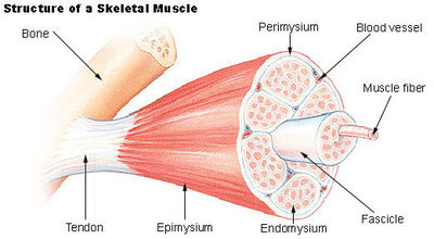
Questão
The three types of tissue involved in the structure of a muscle are the [blank_start]epimysium[blank_end], perimysium and endomysium. The epimysium is a layer of connective tissue that wraps around the whole muscle. The [blank_start]perimysium[blank_end] wraps around the fascicles inside the muscle. The [blank_start]endomysium[blank_end] wraps around each [blank_start]myocyte[blank_end]. All of these tissues will eventually connect to one another and form a [blank_start]tendon[blank_end] that connects the skeletal muscle to the [blank_start]skeleton[blank_end]. Therefore, the layers of connective tissue support the muscle and transmit the [blank_start]force[blank_end] of contraction to the bones.
Responda
-
epimysium
-
perimysium
-
endomysium
-
myocyte
-
tendon
-
skeleton
-
force
Questão 67
Questão
Each skeletal muscle is comprised of [blank_start]fascicles[blank_end] and each fascicle is made up of muscle cells called [blank_start]myocytes[blank_end]. Myocytes are long , skinny and [blank_start]cylindrical[blank_end] cells and are multi-[blank_start]nucleated[blank_end]. They are made of [blank_start]myofibrils[blank_end], which are made up of [blank_start]myofilaments[blank_end]. Myofilaments come in two different varieties: [blank_start]thick[blank_end] filament of myosin and [blank_start]thin[blank_end] filament of [blank_start]actin[blank_end]. The myosin and actin proteins are arranged in a contractile units called [blank_start]sarcomeres[blank_end] and work together to produce muscle [blank_start]contraction[blank_end].
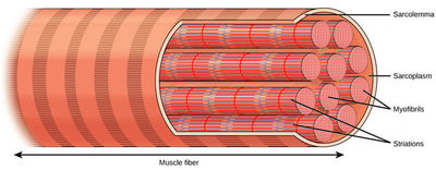
Responda
-
fascicles
-
myocytes
-
cylindrical
-
nucleated
-
myofibrils
-
myofilaments
-
thick
-
thin
-
actin
-
sarcomeres
-
contraction
Questão 68
Questão
Muscle is striated because....
Responda
-
the thin and thick filaments are contractile units that cause muscle contraction
-
sarcomeres are arranged end on end along the myofibril
-
the Z lines are placed at regular intervals
-
the thick myosin filaments pull on the thin actin filaments during contraction.
Questão 69
Questão
The boundaries of sarcomeres are marked by [blank_start]Z[blank_end]-lines. These are placed at [blank_start]regular[blank_end] intervals along the [blank_start]myofibril[blank_end], and anchor the thin [blank_start]actin[blank_end] filaments together. The [blank_start]thick[blank_end] myosin filament lies in the [blank_start]middle[blank_end] of the sarcomere. During muscle contraction, the thick myosin filaments [blank_start]pull[blank_end] on the thin actin filaments, causing the [blank_start]actin[blank_end] to slide over the [blank_start]myosin[blank_end]. This draws Z lines together and shortens the [blank_start]sarcomeres[blank_end].
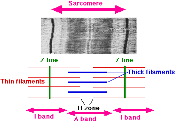
Responda
-
Z
-
regular
-
myofibril
-
actin
-
thick
-
middle
-
pull
-
actin
-
myosin
-
sarcomeres
Questão 70
Questão
The size of a motor unit can vary. A small motor unit enables [blank_start]fine[blank_end] control as each [blank_start]action[blank_end] [blank_start]potential[blank_end] only causes the contraction of a small number of [blank_start]myocytes[blank_end]. Small motor units are therefore found in muscles that move eyes and fingers. Large motor units are found in areas where fine control is not found, such as the [blank_start]gluteus[blank_end] maximus.
Responda
-
fine
-
action
-
potential
-
myocytes
-
gluteus
Questão 71
Questão
Activation of motor units is an 'all' or 'nothing' response: either all the [blank_start]myocytes[blank_end] innervated will contract, or none of them will. However, whole muscles can contract with different amounts of [blank_start]force[blank_end]. The force of contraction depends on the number of [blank_start]active[blank_end] motor units, the [blank_start]frequency[blank_end] of [blank_start]action[blank_end] potentials arriving at the motor unit and the [blank_start]size[blank_end] of the motor unit.
Responda
-
myocytes
-
force
-
active
-
frequency
-
action
-
size
Questão 72
Questão
The muscle is active and develops tension, and shortening of sarcomeres follows. This leads to the whole muscle shortening and therefore a change in joint position. What type of muscle action is this?
Responda
-
isometric
-
eccentric
-
concentric
Questão 73
Questão
The muscle is active and develops tension, but there is no change in sarcomere length. The muscle itself does not change in length, and there is no change in joint positions. What muscle action is this?
Responda
-
isometric
-
concentric
-
eccentric
Questão 74
Questão
The muscle is active and develops tension, and the lengthening of the sarcomeres follows. This leads to the whole muscle lengthening, and therefore a change in joint position. Which type of muscle action is this?
Responda
-
isometric
-
concentric
-
eccentric
Questão 75
Questão
A muscle that is creating a movement by contracting concentrically. Which muscle role is this?
Responda
-
agonist
-
antagonist
-
stabilizer
-
neutralizer
Questão 76
Questão
A muscle that is opposing a movement by contracting eccentrically. Which muscle role is this?
Responda
-
agonist
-
antagonist
-
stabilizer
-
neutralizer
Questão 77
Questão
A muscle that is holding a joint still by contracting isometrically. What type of muscle role is this?
Responda
-
agonist
-
antagonist
-
stabilizer
-
neutralizer
Questão 78
Questão
A muscle that eliminates the unwanted movement, so that another movement may occur. What type of muscle role is this?
Responda
-
agonist
-
antagonist
-
neutralizer
-
stabilizer
Questão 79
Questão
If the muscle is [blank_start]anterior[blank_end] to the joint, concentric action will produce flexion.
Responda
-
anterior
Questão 80
Questão
If a muscle is [blank_start]posterior[blank_end] to the joint, concentric action will cause extension.
Responda
-
posterior
Questão 81
Questão
If a muscle is lateral to the joint, concentric action will produce [blank_start]adduction[blank_end].
Responda
-
abduction
Questão 82
Questão
If the muscle is [blank_start]medial[blank_end] to the joint, concentric action will produce adduction.
Responda
-
medial
Questão 83
Questão
The [blank_start]controlled variable[blank_end] is the variable that the system is trying to keep the same. The [blank_start]set point[blank_end] is the optimal target for the variable to be at. The [blank_start]reference range[blank_end] is the range of acceptable limits of that variable.
Responda
-
controlled variable
-
set point
-
reference range
Questão 84
Questão
[blank_start]Feedback loops[blank_end] can either be positive or negative. A [blank_start]positive[blank_end] loop causes an increase in response (stretch in giving birth), where as a negative loop causes a decrease in response (change in temperature).
Responda
-
Feedback loops
-
positive
Questão 85
Questão
What is the receptor that detects a change in a controlled variable?
Responda
-
sensor
-
control/integration center
-
effector
-
feedforward
Questão 86
Questão
The [blank_start]control center[blank_end] compares the change in the [blank_start]controlled variable[blank_end] to the set point and elicits an appropriate response.
Responda
-
control center
-
controlled variable
Questão 87
Questão
The [blank_start]effector[blank_end] uses mechanisms to bring about change in the controlled variable, according to information received from the [blank_start]control center[blank_end].
Responda
-
effector
-
control center
Questão 88
Questão
[blank_start]Feedforward[blank_end] is when a sensor senses a [blank_start]change[blank_end] in the controlled variable and immediately effects a change to resist it, bypassing the integration center (anticipation). Usually is complimentary to [blank_start]negative[blank_end] [blank_start]feedback[blank_end], so any mistakes can be fixed, or response can be exaggerated. An example of this is gag reflex.
Responda
-
Feedforward
-
change
-
negative
-
feedback
Questão 89
Questão
Flexion and extension occur on the [blank_start]sagittal[blank_end] [blank_start]plane[blank_end]. Abduction and adduction occur on the [blank_start]coronal[blank_end] [blank_start]plane[blank_end].
Responda
-
sagittal
-
plane
-
coronal
-
plane
Questão 90
Questão
[blank_start]Cancellous[blank_end] bone has a high surface area to mass ratio, as it is less [blank_start]dense[blank_end] than compact bone. The main functional unit is the [blank_start]trabeculae[blank_end], which are aligned towards the distribution of mechanical force on the bone, helping to absorb the [blank_start]impact[blank_end]. Often found in the [blank_start]ends[blank_end] of the bone.
Responda
-
Cancellous
-
dense
-
trabeculae
-
impact
-
ends
Questão 91
Questão
[blank_start]Osteocytes[blank_end] originate from osteoblasts once they become trapped in the [blank_start]ECM[blank_end]. Their function is to maintain [blank_start]bone[blank_end] tissue. Osteoblasts originate from [blank_start]osteoprogenitor[blank_end] cells and are found in areas of growth. Their function is to secrete bone [blank_start]matrix[blank_end]. Osteoclasts originate from [blank_start]macrophages[blank_end] and their function is to [blank_start]break[blank_end] [blank_start]down[blank_end] bone tissue during the remodeling and healing of bone.
Responda
-
Osteocytes
-
ECM
-
bone
-
osteoprogenitor
-
matrix
-
macrophages
-
break
-
down
Questão 92
Questão
Each lacunae is connected to one another via [blank_start]canaliculi[blank_end].
Responda
-
canaliculi
Questão 93
Questão
A fracture occurs when bone tissue is broken as a result of external force. Bone tissue is highly vascularized and so there is often a lot of [blank_start]bleeding[blank_end] associated with the injury. Bone tissue also has an extensive [blank_start]nerve[blank_end] supply which is why there is a lot of pain involved with a fracture. The first stage is [blank_start]haematoma[blank_end] [blank_start]formation[blank_end], which is when a blood clot forms around the site of injury to stop excessive bleeding. This is important because excessive blood [blank_start]loss[blank_end] leads to death and reparative cells arrive to the site of injury through [blank_start]blood[blank_end] [blank_start]supply[blank_end]. A process known as [blank_start]angiogenesis[blank_end] then occurs to increase [blank_start]vascular[blank_end] supply to the fracture. [blank_start]Phagocytes[blank_end] arrive from the blood capillaries and clean up the dead tissue and debris. After that, stage 2 occurs when the [blank_start]soft[blank_end] callus forms. [blank_start]Fibroblasts[blank_end] arrive at the site of injury where they differentiate into [blank_start]chondroblasts[blank_end]. The chondrocytes secrete a [blank_start]cartilaginous[blank_end] matrix forming a fibrocartilaginous callus (this callus is soft because it is made up of cartilage, not bone). Stage three is when the [blank_start]hard[blank_end] callus forms; the [blank_start]osteoblasts[blank_end] turn fibrocartilaginous callus into a hard boney callus is a process known as ossification. The last stage is when [blank_start]osteoclasts[blank_end] and osteoblasts work together to remodel the bone into its normal [blank_start]arrangement[blank_end].
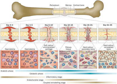
Responda
-
bleeding
-
nerve
-
haematoma
-
formation
-
loss
-
blood
-
supply
-
angiogenesis
-
vascular
-
Phagocytes
-
soft
-
Fibroblasts
-
chondroblasts
-
cartilaginous
-
hard
-
osteoblasts
-
osteoclasts
-
arrangement
Questão 94
Questão
Fibrocartilage are high quantities of [blank_start]collagen[blank_end] bundles aligned towards the [blank_start]direction[blank_end] or pressure. Main function is to resist [blank_start]tension[blank_end]. Found where some movement is required but [blank_start]stability[blank_end] is also needed, for example the [blank_start]menisci[blank_end] of the knee joint.
Responda
-
collagen
-
direction
-
tension
-
stability
-
menisci
Questão 95
Questão
This structure in a synovial joint is found at points of articulation within the joint. This acts to facilitate frictionless movement between the two bones.
Responda
-
articular cartilage
-
capsular ligament
-
synovial membrane
-
synovial fluid
-
intra-capsular
-
extra-capsular
Questão 96
Questão
A ___________ surrounds the joint, which is thicker where support is required and thinner where movement is required.
Responda
-
articular cartilage
-
synovial membrane
-
synovial fluid
-
capsular ligament
-
intra-capsular
Questão 97
Questão
What provides the articular cartilage with nutrients and lubricates the synovial joint?
Responda
-
synovial membrane
-
synovial fluid
-
capsular ligament
Questão 98
Questão
what does the synovial membrane do?
Responda
-
secretes synovial fluid
-
surrounds joint where support is required
-
resists compression
Questão 99
Questão
Which of the following is the correct order?
Responda
-
fascia, epimysium, perimysium, endomysium, myofibrils, myofilaments
-
fascia, epimysium, perimysium, endomysium, myofilaments, myofibrils
-
fascia, endomysium, perimysium, epipmysium, myofibrils, myofilaments
-
fascia, perimysium, epimysium, perimysium, myofibrils, myofilaments
Questão 100
Questão
Muscle form depends on three factors:
1) Length of muscle fibres: [blank_start]Long[blank_end] muscle fibres = larger [blank_start]ROM[blank_end].
2) Number of muscle fibres: higher number of fibres means more [blank_start]tension[blank_end] can be generated, thus more strength
3) Arrangement of muscle fibres: Fibres can be parallel or [blank_start]pennate[blank_end].
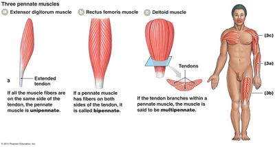
Responda
-
ROM
-
Long
-
tension
-
pennate
Quer criar seus próprios Quizzes gratuitos com a GoConqr? Saiba mais.