
Quiz
E2. Anatomy, Lecture 1. Thorax
Este quiz tem tipos de pergunta que abrem no player de sempre.
Perguntas do quiz (28)
1Thorax extends from the [blank_start]inferior[blank_end] thoracic aperture, superiorly to the superior thoracic [blank_start]aperture[blank_end].
The diaphragm divides the [blank_start]thoracic[blank_end] cavity from the [blank_start]abdominal[blank_end] cavity- Athoracic
- Babdominal
- Caperture
- Dinferior
2Superficial to the pectoralis [blank_start]major[blank_end] is the mammary gland. It consists of [blank_start]glandular[blank_end] tissue, [blank_start]ducts[blank_end] and when inactive, mostly [blank_start]adipose[blank_end] tissue
- Aglandular
- Bducts
- Cadipose
- Dmajor
3Label
PICA=Posterior Intercostal A
Br= branch(es)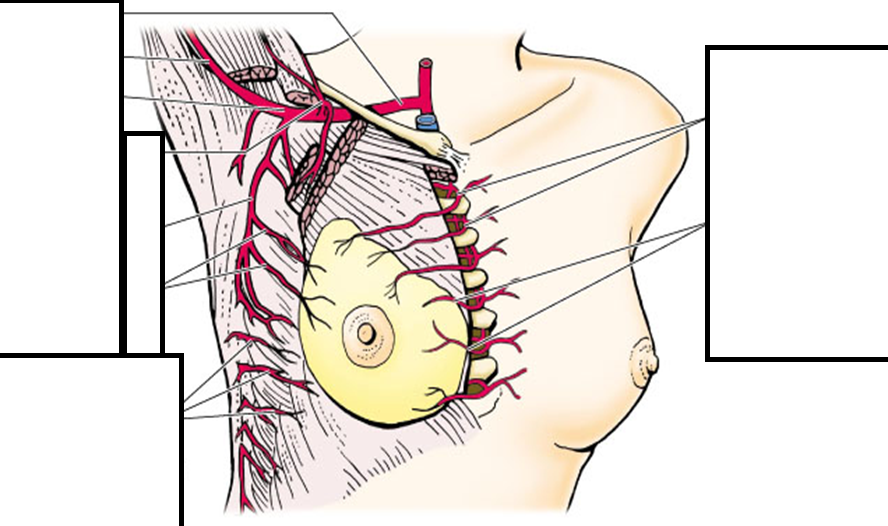
- ASubclavian
- BBrachial
- CAxillary
- DThoracoacromial trunk
- ELateral Thoracic
- FLat Mammary Brs
- GLat Mammary Br of Lat Cut Br of PICA
- HInternal Thoracic and its perforating Br
- IMedial Mammary Br
4Lymphatic drainage is to the _______ nodes?
[blank_start]Axillary[blank_end] (lateral), [blank_start]parasternal[blank_end] (medial), [blank_start]inferior phrenic[blank_end] (inferior).- Ainferior phrenic
- Bparasternal
- CAxillary
5Label
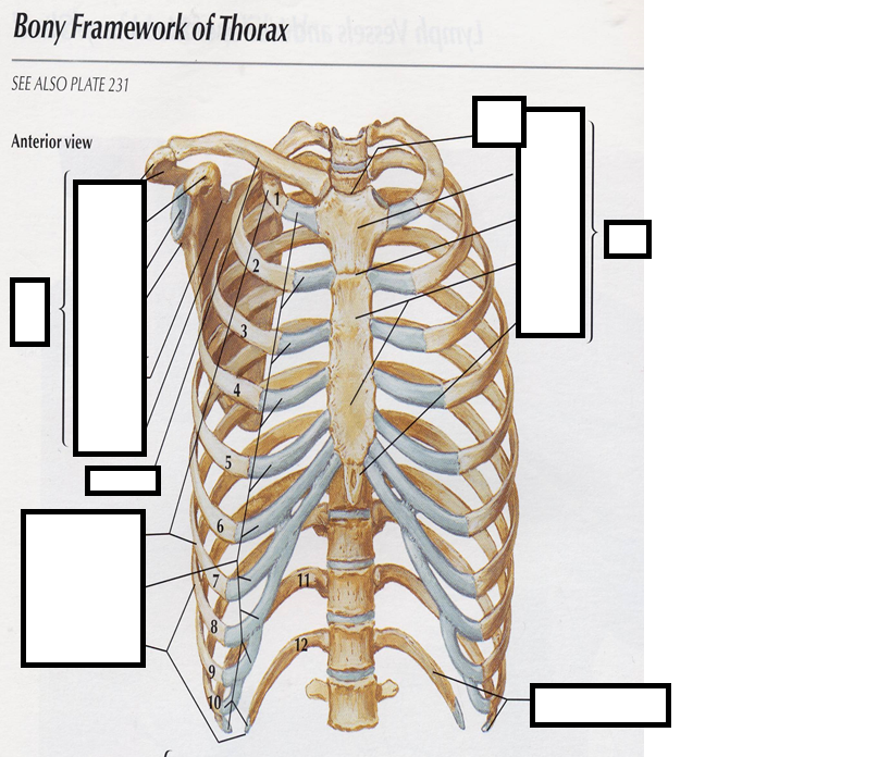
- AAcromion
- BCorcacoid Process
- CGlenoid Cavity
- DScapula
- EClavicle
- FTrue ribs
- GCostal Cartliage
- HFalse ribs (includes floating ribs)
- IFloating Ribs
- JSternum
- KXiphoid Process
- LBody
- MAngle
- NManubrium
- OJugular Notch
6The thoracic cavity is bounded by the [blank_start]sternum[blank_end], [blank_start]ribs[blank_end] and bodies of the [blank_start]vertebrae[blank_end].
There are [blank_start]12[blank_end] pairs of ribs, [blank_start]1-7[blank_end] true,[blank_start]8-10[blank_end] false and [blank_start]11-12[blank_end] floating.- Asternum
- Bribs
- Cvertebrae
- D11-12
- E8-10
- F1-7
- G12
7Posterior Thorax:
Other bones associated with the thoracic wall are the [blank_start]clavicle[blank_end] and [blank_start]scapula[blank_end].- Ascapula
- Bclavicle
8The first rib articulates through a [blank_start]cartilagenous[blank_end] joint (immovable)
Second through seventh are [blank_start]synovial[blank_end] joints (some movement)- Acartilagenous
- Bsynovial
9The [blank_start]head[blank_end] of the rib articulates with the body of [blank_start]2[blank_end] vertebrae
The [blank_start]tubercle[blank_end] articulates with the [blank_start]transverse[blank_end] process
The [blank_start]costal[blank_end] cartilage of the true ribs articulate directly with the [blank_start]sternum[blank_end]
The [blank_start]false[blank_end] ribs articulate with the with the costal cartilage of the [blank_start]seventh[blank_end]
The [blank_start]floating[blank_end] ribs have no anterior articulation- Ahead
- B2
- Ctubercle
- Dtransverse
- Ecostal
- Fsternum
- Gfalse
- Hseventh
- Ifloating
10Label
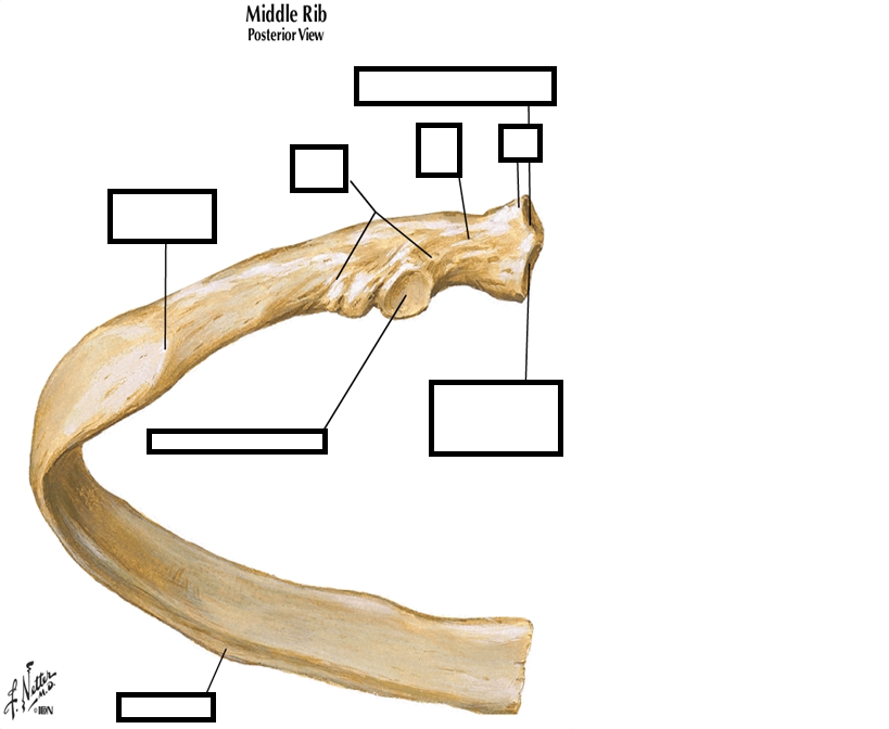
- ASup Articular Facet for the Vert Body
- BHead
- CInferior Articular Facet for Vert Body
- DNeck
- ETubercle
- FAngle
- GArticular Facet for Transverse process
- HCostal Groove
11Label
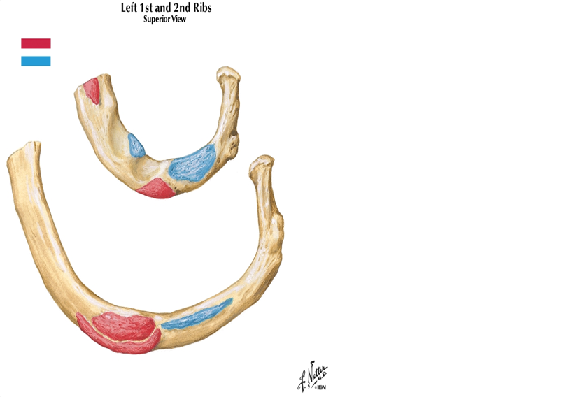
- ASubclavian A
- BSubclavian V
- CAnterior Scalene
- DMiddle Scalene
- EPost Scalene
- FSubclavius
- GSerratus Ant 1st rib
- HSerratus Anterior 2nd rib,1stdigitation
- ISerratus Anterior 2nd rib,2nddigitation
- JHead of Rib 1
- KNeck of Rib 1
- LTubercle of rib 1
- Mtubercle of rib 2
- NAngle
- ONeck of Rib 2
- PHead of Rib 2
12The first rib is flattened [blank_start]superior-inferior[blank_end], instead of [blank_start]anterior-posterior[blank_end]
- Asuperior-inferior
- Banterior-posterior
131st rib knowledge!
the [blank_start]groove[blank_end] for the subclavian vein is [blank_start]anterior[blank_end], the groove for the s-c artery is [blank_start]posterior[blank_end] to the [blank_start]scalene[blank_end] tubercle. This is important knowledge, since you will frequently insert catheters into the s-c [blank_start]vein[blank_end] for pacemakers, ICU monitoring, IV medication.The inserted needle may inadvertently cause damage to the [blank_start]artery[blank_end], [blank_start]brachial[blank_end] plexus, or pleura and [blank_start]lung[blank_end]- Agroove
- Banterior
- Cposterior
- Dscalene
- Evein
- Fartery
- Gbrachial
- Hlung
14Label
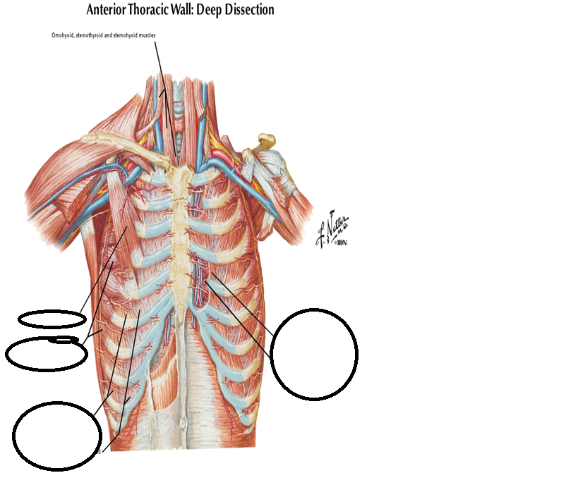
- APec Minor
- BSerratus Ant
- CExternal intercostal Muscles
- DExternal Intercostal Membranes
- EInternal Intercostals (cut)
- FTransverse Thoracis
15The external intercostal muscle becomes [blank_start]membranous[blank_end] at around the junction of the [blank_start]ribs and costal cartilage[blank_end]. It continues as a [blank_start]membrane[blank_end] anteriorly
The course of the fibers is [blank_start]lateral[blank_end] to [blank_start]medial[blank_end] as you travel [blank_start]superior[blank_end] to [blank_start]inferior[blank_end]- Amembranous
- Bribs and costal cartilage
- Cmembrane
- Dlateral
- Emedial
- Fsuperior
- Ginferior
16The internal intercostals are muscular [blank_start]anteriorly[blank_end] and membranous [blank_start]posterior[blank_end].
The fiber direction is [blank_start]medial[blank_end] to [blank_start]lateral[blank_end] as you go [blank_start]inferiorly[blank_end]- Aanteriorly
- Bposterior
- Cmedial
- Dlateral
- Einferiorly
17Label
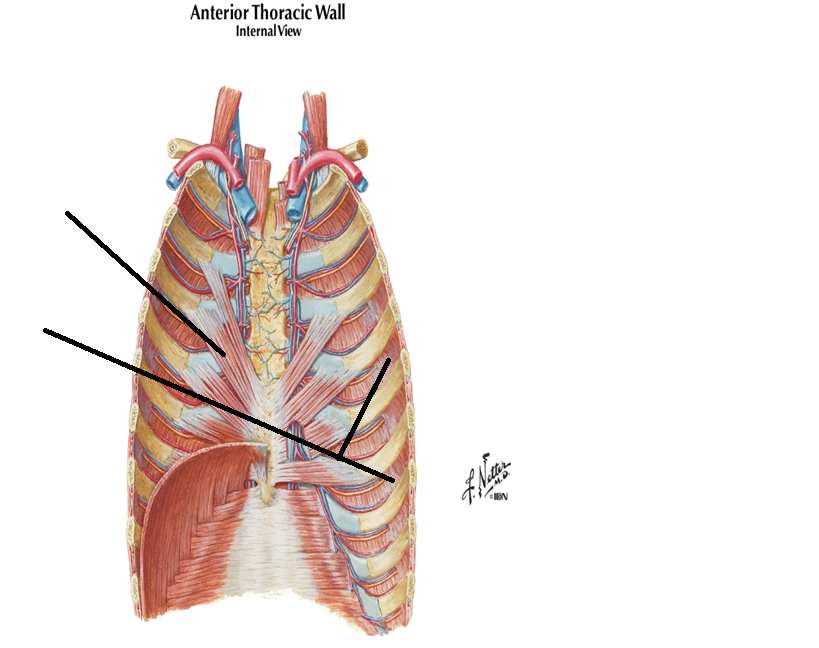
- ATransversus Thoracis
- BInnermost Intercostals
18Transversus [blank_start]thoracis[blank_end] and innermost [blank_start]intercostals[blank_end]:
Both incomplete layers of [blank_start]muscle[blank_end] which lie [blank_start]internal[blank_end] to the intercostal [blank_start]neurovascular[blank_end] bundle
They both traverse across more than one [blank_start]intercostal[blank_end] space- Athoracis
- Bintercostals
- Cmuscle
- Dinternal
- Eneurovascular
- Fintercostal
19Label
ICoIIC =Interchondral part of int intercostals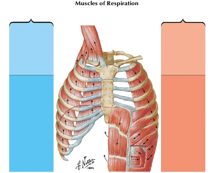
- AAccessory Muscles: SCM and Scalenes
- BExternal Intercostals (EIC)
- CInterchondral part of int intercostals
- DDiaphragm
- ERectus Abdominis
- FTransversus abdominis
- GInternal Oblique
- HExternal Oblique
- IInternal Intercostals
- JSternocleidomastoid (SCM)
- KScalene ant
- LScalene Middle
- MScalene Post
- NAction: to elevate and fix the upper rib
- OInspiration
- PExpiration
- QPrinciple: EIC, ICoIIC, Diaphragm
- RElevation or ribs, expand Thor cavity
- Sincrease inf thor cavity, raises InfRibs
- TQuiet breathing: passive recoil of lung
- UActive breathing: IIC, Abdominals
- VDepress ribs,
- WCompress abdominal, push up diaphragm
20Label
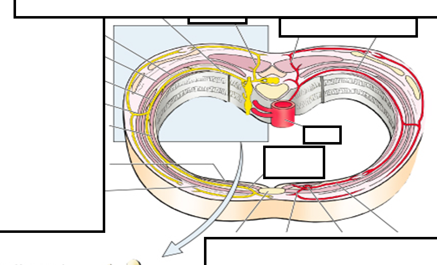
- ADorsal Primary Ramus
- BVentral Primary Nerve (intercostalNerve)
- CInternal Intercostal membrane
- DExternal intercostal muscle
- EInternal Intercostal Muscle
- FLateral Cutaneous Branch
- GInnermost Intercostal Muscle
- HTransverse Thoracic Muscle
- IExternal Intercostal Membrane
- JSternum
- KPerforating (cutaneous) Branch
- LInternal Thoracic Artery
- MAnterior Intercostal Artery
- NPosterior Intercostal Artery
- ODorsal Br of Post Intercostal A
- PAorta
- QAnterior (cutaneous) branch
21Label what you can
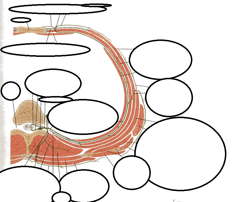
- ASternum
- BExternal Intercostal Membrane
- CTransverse Thoracis
- DMedial Branch
- EAnterior Cutaneous Branch
- FLateral Branch
- GExternal Intercostal Muscle
- HInternal Intercostal Muscle
- IInnermost Intercostal Muscle
- JAnterior Branch
- KLateral Cutaneous Branch
- LPosterior Branch
- MSerratus Anterior
- NLatissimus Dorsi
- OSubscapularis
- PTeres Major
- QScapula
- RInfraspinatus
- STrapezius
- TRhomboid Major
- UErector Spinae
- VInternal Intercostal Membrane
- WPost Cutaneous Branch
- XLateral Branch (posterior)
- YMedial branch (posterior)
- ZDorsal Ramus
- AAVentral Ramus
- ABSmypatetic Ganglion
- ACSpinal Nerve
- ADSpinal Sensory Ganglion
22The lungs are covered by [blank_start]parietal[blank_end] and [blank_start]visceral[blank_end] pleura. The [blank_start]visceral[blank_end] is the layer against the lung itself.
- Aparietal
- Bvisceral
- Cvisceral
23Label!
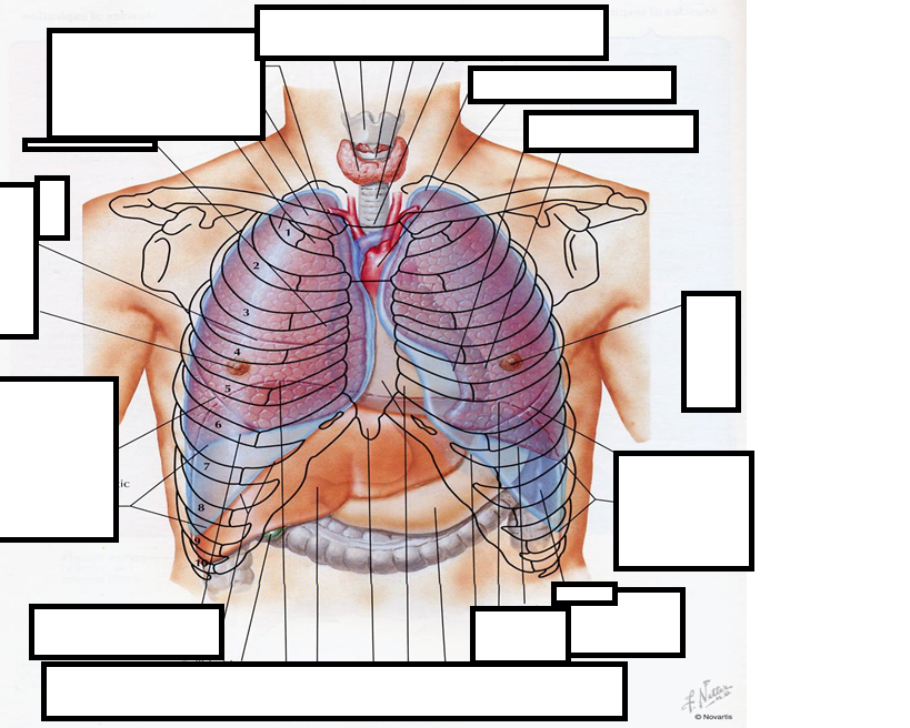
- AHorizontal Fissure
- Boblique fissure (R)
- CCostaldiaphragmatic recess of pleural ca
- DXiphoid Process
- EOblique fissure (L)
- FHeart Border (Greater)
- GCardiac Notch
- HArch of Aorta
- IApex of lung
- JJugular Notch
- KCervical (of pleura)
- LSternoclavicular joint
- MCardiac Border (lesser)
24placeholder
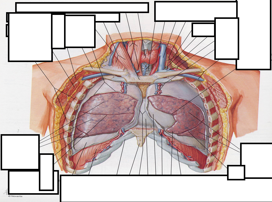
- Athis one
25Label
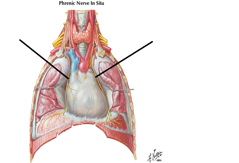
- APhrenic Nerve Right
- BPhrenic Nerve Left
26Running between the [blank_start]pleura[blank_end] and the [blank_start]paricardium[blank_end] is the [blank_start]phrenic[blank_end] nerve(C[blank_start]3-5[blank_end]). The [blank_start]phrenic[blank_end] nerve supplies innervation to the [blank_start]Diaphragm[blank_end]
- Aparicardium
- Bpleura
- Cphrenic
- D3-5
- Ephrenic
- FDiaphragm
27Label
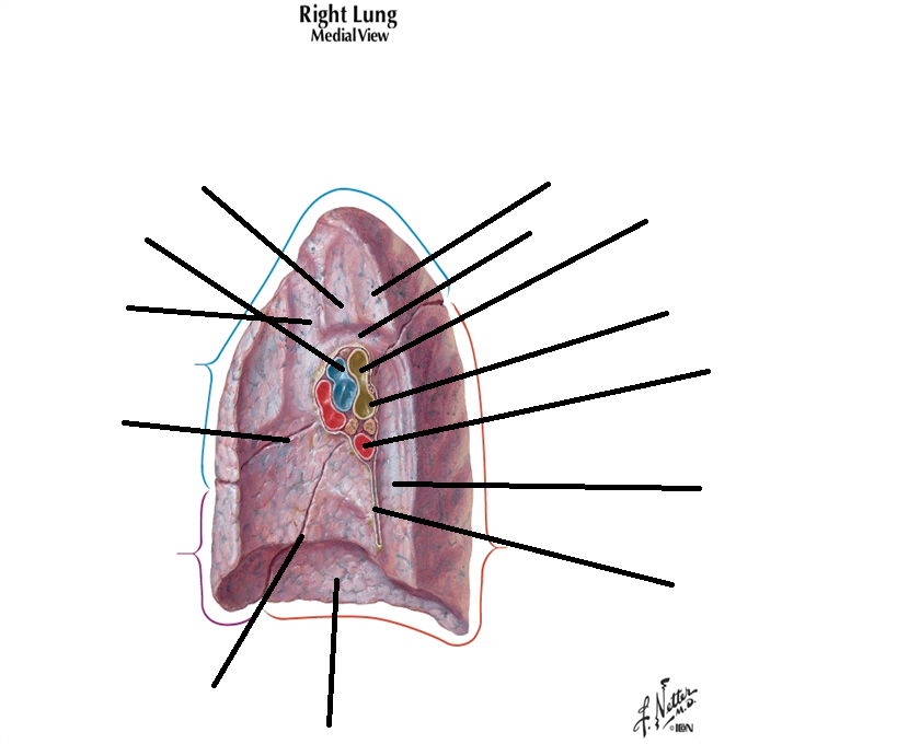
- AArterties
- BHorizontal fissure
- Coblique fissuren (inf)
- DPulmonary Ligament
- EPulmonary Vein
- FInf Bronchus
- GSuperior Bronchus
- HEsophagus
- IArch of Azygos
- JTrachea
- KBrachiocephalic Vein
- LDiaphragm
- MGroove for Esophages
- NSuperior Lobe
- OMiddle Lobe
- PInferior Lobe
- QOblique Fissure (sup)
28label
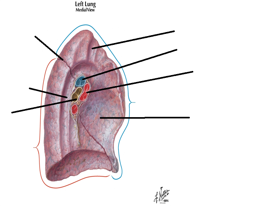
- AOblique fissure (superior)
- BAorta
- CEsophagus
- DCardiac
- ESubclavian artery
- FPulmonary Vein
- GPulmonary Artery
- HBronchus
- ISuperior Lobe
- JInferior Lobe
- KOblique Fissure (inferior)
- LArea for Trachea and Esophagus
- MPulmonary Ligament
- NGroove for Left Braciocephalic V
- ODiaphragmic Surface
Informações
- Descrição
- Gotta look up a couple things when I get home but good to go for the msot part
- Autor
- Sahil Bharwani
- Idioma
- English
- Visualizações
- 45
- Avaliação
- Ainda sem avaliações Avalie este quiz