
Quiz
IHS Test 2 - Intraoperative Monitoring Parts 1 and 2
Este quiz tem tipos de pergunta que abrem no player de sempre.
Perguntas do quiz (51)
1A [blank_start]Standard of Care[blank_end] is the highest mandate for clinical behavior.
- AStandard of Care
2Select the standard of nursing practice that most applies to CRNAs.
- AStandard III
- BStandard IV
- CStandard V
- DStandard VI
3Fill in the blanks for the AANA Standards for Nurse Anesthesia Practice:
Standard V
A. Monitor [blank_start]ventilation[blank_end] continuously.
B. Monitor [blank_start]oxygenation[blank_end] continuously.
C. Monitor [blank_start]cardiovascular[blank_end] status continuously.
D. Monitor [blank_start]body temperature[blank_end] continuously.
E. Monitor [blank_start]neuromuscular function[blank_end] and status.
F. Monitor and assess the [blank_start]patient positioning[blank_end].- Aventilation
- Boxygenation
- Ccardiovascular
- Dbody temperature
- Eneuromuscular function
- Fpatient positioning
4One of the key components to patient safety is to: [blank_start]Avoid turning your back on the patient[blank_end] and surgical procedure for long periods of time.
- AAvoid turning your back on the patient
5[blank_start]Monitors[blank_end] are placed first and removed last.
- AMonitors
6Name two ways (in alpha order) that CRNAs monitor the respiratory system:
1. [blank_start]Capnography[blank_end]
2. [blank_start]Pulse oximetry[blank_end]- ACapnography
- BPulse oximetry
7What is the standard of care for monitoring of oxygenation?
- AArterial blood gas
- BCapnography
- CPulse oximetry
8The Beer-Lambert Law is a method for measuring what?
- ACapnography
- BArterial blood gas
- CPulse oximetry
9Drag and drop the spectrum of light to the appropriate type.
Red: [blank_start]660[blank_end] nm
Infrared: [blank_start]940[blank_end] nm- A660
- B940
10The Beer-Lambert Law and pulse oximetry measures the difference between absorbed light by [blank_start]oxyhemoglobin[blank_end] relative to [blank_start]deoxyhemoglobin[blank_end] in a pulsatile ([blank_start]arterial[blank_end]) bed.
- Aoxyhemoglobin
- Bdeoxyhemoglobin
- Carterial
11[blank_start]Oxyhemoglobin Dissociation Curve[blank_end]: The percentage of hemoglobin saturation with oxygen at different partial pressures of oxygen in blood is described by this S-shaped curve.
- AOxyhemoglobin Dissociation Curve
12Insert the appropriate corresponding numbers for the oxygen-hemoglobin dissociation curve.
At an 02 reading of 97 percent, the Pa02 is likely [blank_start]100[blank_end].
At an 02 reading of [blank_start]90[blank_end] percent, the Pa02 is usually 60.
At an 02 reading of 83 percent, the Pa02 is usually [blank_start]50[blank_end].- A100
- B50
- C90
13Your patient's pulse oximeter isn't reading well. What are some potential causes?
Low flow conditions
• [blank_start]Hypotension[blank_end]→vasoconstrictionoftheperiphery
– Motion artifact
– Nail polish
– Ambient light interference – [blank_start]Dysfunctional hemoglobin[blank_end]
• Fetal hemoglobin
• Hemoglobin S
– Carboxyhemoglobinemia
• 240timestheaffinityforhgbvs.O2. – Methemoglobinemia
– Methylene blue, indigo carmine- AHypotension
- BDysfunctional hemoglobin
14Name the three ways we verify intubation:
1. [blank_start]Auscultation[blank_end]
2. [blank_start]Chest excursion[blank_end]
3. Confirmation of [blank_start]CO2 in expired gases[blank_end].- AAuscultation
- BChest excursion
- CCO2 in expired gases
15What is an early indicator of esophageal intubation and airway disconnect?
- AArterial blood gas
- BCapnography
- CPulse oximetry
16[blank_start]Capnography[blank_end] continuously monitors all of the following:
– Alveolar ventilation
– Pulmonary perfusion
– Respiratory patterns
– Correct placement of endotracheal tube- ACapnography
17Match the type of capnography with its description:
[blank_start]Main-Stream Capnographs[blank_end] (non-diverting or flow-through):
CO2 sensor located between endotracheal tube and breathing circuit
[blank_start]Side-Stream Capnographs[blank_end]: (Diverting or aspiration):
Sensor is located in the main unit and CO2 is aspirated via a sampling tube connected to a T-piece adapter located between endotracheal tube and breathing circuit.- AMain-Stream Capnographs
- BSide-Stream Capnographs
18Name two potentially fatal conditions that may first be indicated by capnographic changes:
[blank_start]Pulmonary embolism[blank_end]
[blank_start]Malignant hyperthermia[blank_end]- APulmonary embolism
- BMalignant hyperthermia
19Use of [blank_start]capnography[blank_end] has markedly decreased incidence of unrecognized esophageal intubation and their associated brain injuries and deaths
- Acapnography
20Label the following image with the appropriate portions of the expiratory segment.
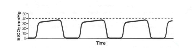
- ABeginning of exhalation
- BGas exhalation alveolar capillary bed
- CAlveolar plateau, C02-rich gas
- DInspiration
21Label the appropriate angle segments of the expiration of capnography.
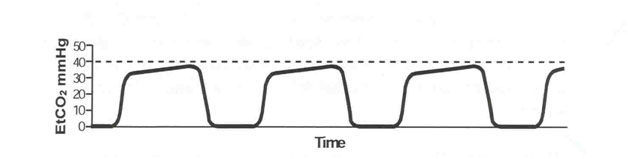
- AAlpha angle
- BBeta angle
22Which of the following options describes Phase I of the expiratory segment?
- AExhalation of gas from the alveolar capillary bed mixing with dead space gas
- BPositive slope due to continuous excretion of CO2 into alveoli
- CGas exchange that is free of CO2
- DUsed to assess extent of breathing
23In capnography, what is part of the expiratory segment?
- APhase 0
- BPhase I
- CPhase II
- DPhase III
24In capnography, the [blank_start]alpha[blank_end] angle is between Phases II and III.
The [blank_start]alpha[blank_end] angle is an indirect indication of the V/Q status of the lung.
The [blank_start]beta[blank_end] angle is between Phase III and descending limb of inspiratory segment.
The [blank_start]beta[blank_end] angle is used to assess the extent of rebreathing.- Aalpha
- Bbeta
- Calpha
- Dbeta
- Ealpha
- Fbeta
- Galpha
- Hbeta
25Fill in the blanks for the five characteristics of capnogram that should be evaluated:
-Frequency
-Rhythm
-[blank_start]Height[blank_end]
-Baseline
-[blank_start]Shape[blank_end]- AHeight
- BShape
26Your patient has low or no ETCO2 -- what are two of the major causes?
[blank_start]Decreased CO2 production/delivery[blank_end].
Causes: Hypothermia
[blank_start]Decreased pulmonary perfusion[blank_end].
Causes: Hypovolemia
Hypotension
Pulmonary embolism
Decreased cardiac output (arrest)
- ADecreased CO2 production/delivery
- BDecreased pulmonary perfusion
27Select some causes of low ETCO2.
- AAltered alveolar ventilation
- BIncrease CO2 production/delivery
- CIncreased pulmonary perfusion
- DTechnical errors/machine faults
28Match the causes of low ETCO2 to the examples (in alpha order)
Altered Alveolar Ventilation
[blank_start]Apnea (accidental extubation)[blank_end]
[blank_start]Hyperventilation[blank_end]
[blank_start]Increase in dead space ventilation[blank_end]
[blank_start]Partial/total airways obstruction[blank_end]
Technical Errors/Machine Faults
[blank_start]Esophageal intubation[blank_end][blank_start]Sampling line leak[blank_end]
[blank_start]Ventilator malfunction[blank_end]- AApnea (accidental extubation)
- BHyperventilation
- CIncrease in dead space ventilation
- DPartial/total airways obstruction
- EEsophageal intubation
- FSampling line leak
- GVentilator malfunction
29Name the two chief causes of elevated CO2.
[blank_start]Increased CO2 production/delivery[blank_end].
[blank_start]Increased pulmonary perfusion[blank_end]- AIncreased CO2 production/delivery
- BIncreased pulmonary perfusion
30Drag and drop the cause of the elevated ETCO2 with its cause:
Increased CO2 production/delivery.
[blank_start]Fever[blank_end]
[blank_start]Hyperthyroid storm[blank_end]
[blank_start]Laparoscopic surgeries[blank_end]
[blank_start]Malignant hyperthermia[blank_end]
[blank_start]Right-to-Left Shunts[blank_end]
[blank_start]Sepsis[blank_end]
[blank_start]Shivering[blank_end]
[blank_start]Sodium bicarbonate[blank_end]
[blank_start]Tourniquet[blank_end]
Increased pulmonary perfusion
[blank_start]Increased blood pressure[blank_end]
[blank_start]Increased cardiac output[blank_end]- AFever
- BHyperthyroid storm
- CLaparoscopic surgeries
- DMalignant hyperthermia
- ERight-to-Left Shunts
- FSepsis
- GShivering
- HSodium bicarbonate
- ITourniquet
- JIncreased blood pressure
- KIncreased cardiac output
31Select some examples of elevated CO2 causes.
- AHyperventilation
- BHypoventilation
- CBronchial intubation
- DExhausted CO2 absorber
- EApnea/circuit disconnect
- FMalignant hyperthermia
- GSepsis
32You notice your patient is experiencing small breaths at various points in the ETCO2 waveform. What is NOT a potential cause of this?
- AInadequate muscle paralysis
- BImproper ventilator settings
- CExcessive oxygenation
- DPressure on the patient's chest
33Cardiac oscillations are most prominent in what patient population?
- AAdult
- BElderly
- CPediatric
- DPregnant
34[blank_start]Cardiac oscillations[blank_end] are small gas movements produced by pulsations of the aorta and heart.
- ACardiac oscillations
35This image is an example of what on a capnograph?
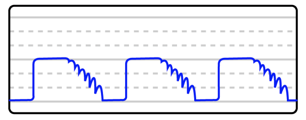
- ACardiac oscillations
36Other considerations/things that may mimic [blank_start]cardiac oscillations[blank_end]
– negative intrathoracic pressure
– low respiratory rate
– low I:E ratio
– Waning muscle relaxation- Acardiac oscillations
37Phase II and III are prolonged or slanted when a patient experiences a [blank_start]prolonged expiratory upstroke[blank_end].
- Aprolonged expiratory upstroke
38You are administering anesthesia when you suddenly notice your patient's baseline is elevated. You immediately:
- ACheck for disconnections
- BLook at your CO2 absorbent
- CChange the I:E ratio
- DIncrease your patient's sedative
39You see the capnograph below during your case. You know this is a sign of:
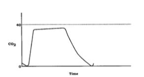
- AExhausted CO2 absorbent
- BAirway obstruction
- CCardiac oscillations
- DInspiratory valve malfunction
40An [blank_start]expiratory valve[blank_end] defect allows exhaled CO2-rich gases to be rebreathed with each inspiration.
- Aexpiratory valve
41ETC02 is indicative of [blank_start]alveolar[blank_end] CO2.
- Aalveolar
- Barterial
42You have two CO2 readings: 38 and 43. Which one is the arterial concentration or PACO2?
- A38
- B43
43The [blank_start]facial[blank_end] nerve is the better site to monitor for onset of block because the larynx also mimics the response of the diaphragm.
- Afacial
- Bulnar
44The [blank_start]ulnar[blank_end] nerve is the best nerve to monitor for recovery.
- Afacial
- Bulnar
45Match the pattern of stimulation to its stimulus.
[blank_start]Single stimulus:[blank_end] The simplest mode of stimulation; consists of a single supramaximal electrical stimulus that is delivered from every 1 to 10 seconds (1‐0.1 Hz)
[blank_start]Train of Four:[blank_end] Four successive 200 μs stimuli at 2Hz delivered every 0.5 seconds for 2 seconds
[blank_start]Double Burst Stimulation:[blank_end] delivers two bursts (at 50Hz) of three electrical stimulations separated by 750 sec followed later by two such impulses
[blank_start]Tetanus:[blank_end] Repetitive, high-frequency stimulation at frequencies of 50 Hz or greater for five seconds- ASingle stimulus:
- BTrain of Four:
- CDouble Burst Stimulation:
- DTetanus:
46Which form of peripheral nerve monitoring is based on the concept that acetylcholine is depleted by successive stimulation?
- ASingle stimulus
- BTrain of four
- CDouble-burst stimulation
- DTetanus
47In Train of Four stimulation, no twitches equals what kind of blockage?
- A75
- B80
- C90
- D100
48Clinical relaxation usually require what percentage of neuromuscular blockage?
- A50 to 60 percent
- B60 to 70 percent
- C75 to 95 percent
- D100 percent
49What PNM improves accuracy over the T4/T1 ratio by delivering a stronger stimulation and eliminating the second and third twitches?
- ADouble burst stimulation
- BSingle burst stimulation
- CTetanus
50Which PNM option may provide an indication of the time until return of the first response in the TOF and subsequent readiness for reversal of neuromuscular blockage?
- ATetanus
- BPost-Tetanic Potentiation
- CTrain of Four
- DDouble-Burst Stimulation
51Which of the following muscle groups are most sensitive to neuromuscular blocking agents?
- ADiaphragm and Masseter
- BExtra ocular and adductor pollicis
- CDiaphragm and Orbicularis oculi
- DAdductor pollicis and Orbicularis oculi
Informações
- Descrição
- Quiz sobre IHS Test 2 - Intraoperative Monitoring Parts 1 and 2, criado por Rachel Nall em 29-06-2016.
- Autor
- Rachel Nall
- Idioma
- English
- Visualizações
- 55
- Avaliação
- Ainda sem avaliações Avalie este quiz