
Flashcards
Week 4
Flashcards do conjunto (52)
- 1How thick is the synovial membrane in a synovial joint?2-3 cells thick
- 2What gives synovial joints their stability?Fibrous capsule
Ligaments
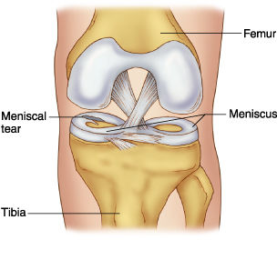
Muscles - 3What does the meniscus in the knee do?Meniscus: extra area of cartilage in the knee only
-Crescentic plates of fibrocartilage
-Increases congruity of articular surface - 4What are the clinical presentations of knee pain?-Pain + stiffness
-Overlying skin is hot/red
-Swelling in suprapatellar pouch
-limited knee movement
-limp on walking
-history of leg giving way
-trauma - 5What are signs of inflammation?Redness, swelling, warmth
- 6What are the common causes of knee pain?-Infection (septic arthritis-treatable)
-Crystal arthritis (ie. gout-caused by increase in uric acid)
-Osteoarthritis
-Chronic inflammatory arthritis (ie. RI)
-Trauma (to menisci, ligaments or tendon)
-Bursitis - 7Define infective (septic) arthritis In which populations is it more common in?Inflammation of a joint caused by bacterial infection
Children/elderly
People with diabetes
Debilitated - 8How do bacteria gain access?-Local trauma
-Spread from adjacent sites of infection
-Bloodstream - 9What are the pathological changes due to septic arthritis?-Ioint destruction (osteomyelitis)
-Can drain to the surface via a sinus
-Bone formation stimulated
-Ankylosis
-Dislocation - 10What are the signs on a X-ray of septic arthritis?-Loss of joint space
-Lytic areas
-Erosion on bone edge
-New bone formation (looks denser)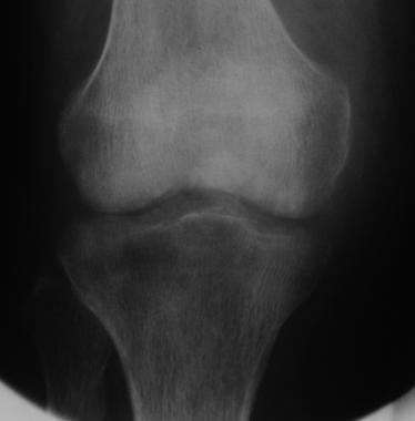
- 11What is gout? In which populations is it more common in?A crystal induced arthritis due to the deposition of uric acid crystals in joints which leads to inflammation
-Men (middle age onwards)
-Post-menopause
-Hypertensive patients - 12Describe the aetiology of goutPrimary: idiopathic
-impaired excretion of uric acid by kidney (?genetic)
Secondary:
-Increased production of uric acid eg. in high alcohol consumption
-Impaired excretion by kidneys due to disease/drugs (diuretics) - 13What are the pathological changes of goat?-Cartilage degeneration
-Synovial hyperplasia
-Erosion of bone
-Secondary degenerative change (eg. osteoarthritis)
-Tophaceous depositis of uric acid in skin - 14What is osteoarthritis/crepitus? In which populations is it more common in?Degenerative disease leading to joint failure. There is a disregulation of normal tissue turnover + repair
60+ years - 15Describe the aetiology of osteoarthritisPrimary
Secondary:
-complications of other joint disorders - 16Pathological changes with osteoarthritis?-Involve cartilage, bone, synovial and joint capsule with secondary effects on muscle
-Fibrillation/erosion of articular cartilage
-Pseudocysts - 17What are the radiological changes of osteoarthritis?-Joint space narrowing
-Subchondral bone sclerosis and cysts
-Marginal osteophyte formation - 18What is rheumatoid arthritis? In which populations is it more common in?Chronic inflammatory joint disease
-Females
-Onset occurs between 35-45 years old - 19What is the aetiology of rheumatoid arthritis? Clinical manifestations?Autoimmune
-Symmetrical deforming polyarthritis
-Diffuse small joint involvement (hands/feet. Larger joints can be affected)
-Can be associated with non-articular disease - 20What pathological changes does rheumatoid arthritis result in?-Rheumatoid synovial hyperplasia and inflammatory infiltration which can result in pannus formation (abnormal layer of fibrovascular/granulation tissue)
-Invasion + destruction of articular cartilage
-Erosions - 21What radiological changes would be seen in rheumatoid arthritis?-Soft tissue swelling
-Joint space narrowing
-Osteopenia (reduce bone density)
-Erosions - 22What is the temporomandibular joint (TMJ)? Where is it found?The joint formed by the articulation of the mandible with the cranium. Modified synovial joint
Anterior to and vertically level with the tragus of the ear - 23What are the superior and inferior articulatory surfaces of the TMJ?Superior:
-Under surface of the squamous part of the temporal bone
-2 articular sites: mandibular fossa (posterior and concave) and articular tubercle (anterior and convex)
Inferior:
-Condyle of the mandible has a rounded superior edge and ellipsoid circumference with its major axis postero-mandial (oblique) - 24Describe the movement of the TMJMovement in the mediolateral plane and opening/closing
- 25Describe the features of the TMJ-Fibrous capsule
-Capsule is very strong
-Capsule is thin and loose to permit movement (has laxity)
-Has a meniscus to prevent bony surfaces making direct contact
-Upper and lower cavity present on either side of the meniscus - 26What does the presence of the upper/lower cavities of the TMJ allow?Upper cavity: translational movements due to a gliding joint
Lower joint: rotational movements due to a modified hinge joint - 27What are the articulating surfaces of the upper/lower joint cavity? What is the purpose of the meniscus?Upper: under surface of the temporal bone and upper surface of the articular disc
Lower: inferior surface of the articular disc and the mandibular condyle
Meniscus improves congruity - 28What lines the articulatory surfaces of the TMJ?-Fibrocartilage (can withstand big forces eg. bites)
-At birth, the fibrocartilage is lined by synovial membrane - 29Describe the articular disc/meniscus of the TMJ-Composed of dense fibrous connective tissue
-Upper surface is concavo-convex (antero-posteriorly)
-Under surface is concave to fit condyle of mandible
-Thicker at periphery where it attaches to the articular capsule (thinner centrally)
-Incompressible
-Can recoil/stretch a bit due to its attachments - 30What are the capsular attachments of the TMJ?Superior: circumference of the mandibular fossa and articular tubercle
Inferior: neck of condyle of the mandible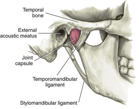
- 31—Green- TMJ ligament
Orange- Joint capsule
Red- Stylomandibular ligament
Purple- Sphenomandibular ligament
- 32Describe the TMJ ligament-Strongest ligament of TMJ
-Deep fibres blend with the capsule
-Attaches to lower border of zygoma to posterior border of neck and ramus of mandible
-Tightens the head in retrusion (closing + pulling jaw backwards) - 33Describe the sphenomandibular ligament-Accessory ligament
-Remains constant in length and tension
-Prevents inferior dislocation of the joint
-Originates from the sphenoid spine to attach to the mandible - 34Describe the stylomandibular ligament-Accessory ligament
-Extends from apex of the styloid process to the posterior border of the ramus of the mandible near its angle
-Tickening of parotid gland fascia
-Separates parotid gland from submandibular gland - 35Describe the stabilisation of the TMJ-More stable when jaw is closed since mandibular condyle are in contact with mandibular fossa
-Teetch are in occlussal contact when jaw is closed (ie. zig-zag)
-Least stable when jaw is open
-Liable to spontaneous dislocation in people who lack teeth - 36What happen during opening movements of the TMJ?-Condyles are pulled forwards (protrusion)- a gliding movement. The lateral pterygoid muscles are involved
-Chin is pulled down and back, a hinge movement. This involves the digastric muscles - 37What happen during closing movements of the TMJ?-Retraction of the mandible by the posterior fibres of the temporalis muscles (pull mandible backwards)
-Elevation of the mandible by the rest of the temporalis muscles, masseter muscles and medial pterygoid muscles - 38What anatomical factors are present to add stability to the TMJ?Post glenoid tubercle: limits posterior displacement
Articular tubercle: limits passive anterior displacement
Sphenomanibular/stylomandibular ligament: inferior dislocation - 39What are some disorders of the TMJ?-Knacking
-Bruxism
Temporomandibular pain dysfunction disorders
-Mal-occlusal syndromes (teeth do not align properly) - 40What are the general features of fibrous joints? Examples?-Don't allow movement/locomotion
-Any movement is not normal/pathological
Syndesmosis (sheet of fibrous tissue uniting bones eg. interosseus membrane between ulna/radius) and gomphosis (joint between tooth and socket) - 41Diagram of a fibrous joint?
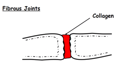
- 42Describe a cartilaginous jointPrimary cartilaginous joint: united by hyaline cartilage. Usually a temporary joint during growth
Secondary cartilaginous joint: articular surface covered in hyaline cartilage. Bones are united by strong fibrocartilage and strong joints allow slight movement - 43—Blue- fibrous capsule
Green- fibrocartilage (movement between bone ends)
Red- hyaline cartilage
- 44What are synovial joints? What specialised features do they contain?-Provides free movements
Features:
-articular discs (fibrocartilage pads) which act as shock absorbers
-fibrocartilage ring (labrum) which deepens joints esp ball and socket joints
-tendons - 456 common features of synovial joints-Bone ends covered in articular (hyaline) cartilage
-Connective tissue capsule
-Joint cavity
-Inner surface is lined with synovial membrane
-Reinforced by ligaments
-Wide range of movements - 46—

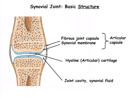
- 47Describe the specialisation of the hip joint as a synovial joint-Head of femur is deep into the socket
-Ligament is inside the joint capsule
-The hip is rarely dislocated (compared to the shoulder) but movement is limited - 48What covers the surface of the tibia in the knee joint?2 Menisci (form a C shape on either side) with 2 articular cartilages in the centre
This prevents wear/tear as there is no bone/bone grinding - 49Describe the glenoid labrum Purpose of it?A fibrocartilaginous 'lip' around the glenoid cavity of the scapula
Acts to deepen the cavity for better articulation - 50What is the rotator cuff? Contents?A specialised muscle group which fixes the head of the humerus into the glenoid cavity. It increases stability to a joint that is prone is dislocation.
Supraspinatus, infraspinatus, teres minor, subscapularis - 51Describe the dislocation of the shoulder joint Consequences?-Humeral head rarely moves upwards during dislocation due to presence of coracoid process
-Dislocation occurs inferiorly and the head lies in a subcoracoid position (under coracoid process)
-Tears joint capsule and can damage to axillary nerve (since it wraps around the head of the humerus)
-Damage to the axillary nerve can result in loss of sensation to skin of regimental badge area (deltoid muscle) - 52What happens in a pulled elbow?-Radial head is subluxed
-Radial head is held in place by a annular ligament
-Ligament is softer in children so is more prone to dislocation (eg. when child is lifted up by the arm)
Informações
- Descrição
- Common joint pathologies Temporomandibular joint Joints- Stability, support and movement
- Criado por
- Dolu Falowo
- Visualizações
- 155
- Avaliação