14885166
Description
Quiz by Matthew Coulson, updated more than 1 year ago
|
|
Created by Matthew Coulson
over 7 years ago
|
|
Question 1
Question
How many turns are there in the cochlea?
Answer
-
1
-
1.5
-
2
-
2.5
Question 2
Question
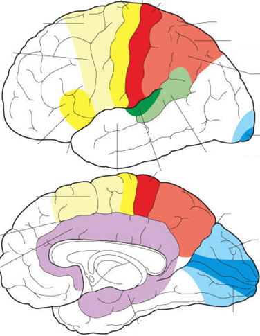
Label the visual and auditory areas on the below diagram.
Answer
-
Primary Visual Cortex
-
Primary Auditory Cortex
-
Auditory Association Cortex (Wernicke's)
-
Broca's Area
-
Primary Motor Cortex
-
Primary Somatosensory Cortex
Question 3
Question
[blank_start]Expressive Dysphasia/Aphasia[blank_end]: Patient's have difficulty in producing language, often only using few words or the most important words in a sentence.
[blank_start]Receptive Dysphasia/Aphasia[blank_end]: Patients have difficulty comprehending language.
Answer
-
Expressive Dysphasia/Aphasia
-
Receptive Dysphasia/Aphasia
Question 4
Question
[blank_start]Wernicke's Area[blank_end]: Processes incoming speech signals; this area is responsible for comprehending language
[blank_start]Broca's Area[blank_end]: Processes the motor aspect of speech; expressing language through means of speech
Answer
-
Broca's Area
-
Wernicke's Area
-
Primary Auditory Cortex
Question 5
Question
Therefore, damage to Wernicke's area will cause [blank_start]receptive[blank_end] dysphasia/aphasia whereas damage to Broca's area will cause [blank_start]expressive[blank_end] dysphasia/aphasia
Answer
-
receptive
-
expressive
-
expressive
-
receptive
Question 6
Question
Strangely, Wernicke's & Broca's areas tend to only exist unilaterally (on one side of the brain). In most people, they tend to both be found on the right side of the brain.
Answer
- True
- False
Question 7
Question
In terms of the visual pathway, after the optic nerves cross at the optic [blank_start]chiasm[blank_end] they become optic tracts. These optic tracts move up into the Thalamus where they synapse with optic radiations at the [blank_start]lateral geniculate[blank_end] nuclei, which then radiate towards and into the primary visual cortex.
Answer
-
chiasm
-
nucleus
-
radiation
-
lateral geniculate
-
medial geniculate
-
lentiform
-
solitary
Question 8
Question
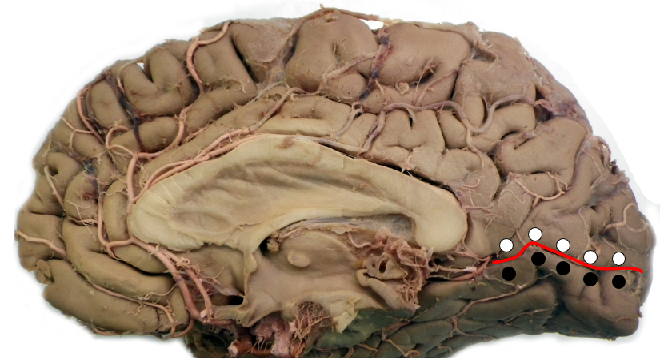
Which part of the visual cortex sees upper retinal projections, and which sees lower retinal projections?
Answer
-
Superior Visual field
-
Inferior Visual Field
-
Inferior Visual Field
-
Superior Visual Field
Question 9
Question
What does 'saccadic' eye movements tend to refer to?
Answer
-
Smooth, tracking movements of the eye
-
Jumpy movements of command (not tracking anything)
Question 10
Question
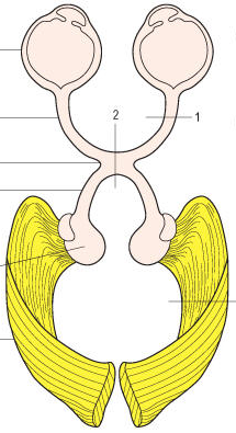
Name the visual field defect that would be produced at each area if it were to be damaged.
Answer
-
Monocular Blindness
-
Bitemporal Hemianopia
-
Homonymous Hemianopia
Question 11
Question
The primary [blank_start]gustatory[blank_end] cortex is a brain structure responsible for the perception of taste.
Answer
-
gustatory
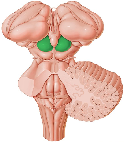
Question 12
Answer
-
Superior colliculus
-
Inferior colliculus
Question 13
Question
The [blank_start]inferior[blank_end] colliculus is the principal midbrain nucleus of the auditory pathway.
Answer
-
inferior
-
superior
Question 14
Question
The superior colliculus plays a large role in which of the following?
Answer
-
It helps to provide motor innervation to the neck muscles, facilitating rotation of the neck
-
As a part of the visual pathway it co-ordinates the movement of the eyes
-
Forms the fine touch part of the dorsal column/medial lemniscus pathway
Question 15
Question
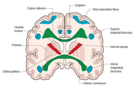
Types of White Matter Fibres:
[blank_start]Association fibres[blank_end]: Connect cortical sites lying in the same hemisphere.
[blank_start]Commissural fibres[blank_end]: Connect one hemisphere to the other, usually connecting areas with similar function.
[blank_start]Projection fibres[blank_end]: Connect hemispheres to deeper structures including thalamus, corpus striatum, brain stem and spinal cord.
Answer
-
Association fibres
-
Commissural fibres
-
Projection fibres
Question 16
Answer
-
Commissural Fibres
-
Projection Fibres
-
Association Fibres
Question 17
Question
Medial Geniculate Nucleus --> [blank_start]Auditory Cortex[blank_end]
Lateral Geniculate Nucleus --> [blank_start]Visual Cortex[blank_end]
Answer
-
Visual Cortex
-
Auditory Cortex
-
Auditory Cortex
-
Visual Cortex
Question 18
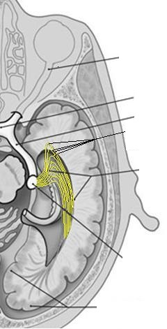
Answer
-
Meyer's Loop
Question 19
Question
When testing one eye for the pupillary light reflex, it is expected that the pupil of the other eye will remain dilated as there is no bright light entering it.
Answer
- True
- False
Want to create your own Quizzes for free with GoConqr? Learn more.