15284173
Description
Quiz by Tamara Podnosova, updated more than 1 year ago
|
|
Created by Tamara Podnosova
about 7 years ago
|
|
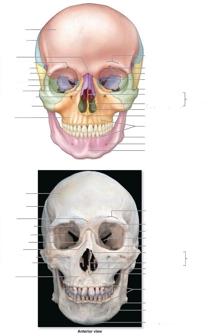
Question 1
Answer
-
frontal bone
-
parietal bone
-
supraorbital foramen
-
temporal bone
-
sphenoid bone
-
nasal bone
-
zygomatic bone
-
maxilla
-
mandible
-
infraorbital foramen
-
mental foramen
-
mental protuberance
-
alveolar process
-
inferior nasal concha
-
vomer
-
perpendicular plate
-
superior orbital fissure
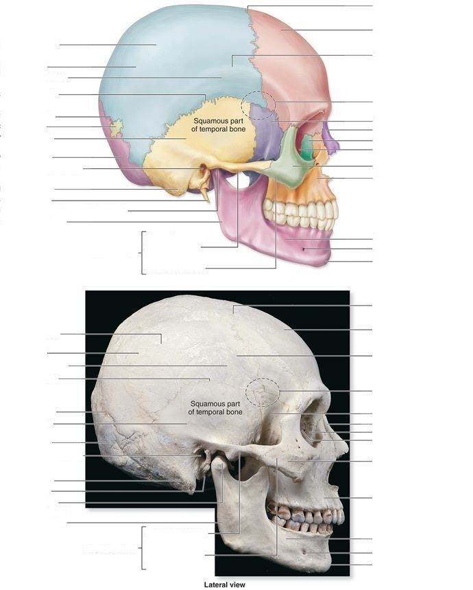
Question 2
Answer
-
coronal suture
-
frontal bone
-
parietal bone
-
squamous suture
-
lambdoid suture
-
temporal bone
-
occipital bone
-
external acoustic meatus
-
mastoid process
-
styloid process
-
mandibular ramus
-
temporal process of zygomatic bone
-
mental protuberance
-
mental foramen
-
maxilla
-
zygomatic bone
-
ethmoid bone
-
lacrimal bone
-
nasal bone
-
sphenoid bone
-
mandibular condyle
-
coronoid process
-
mandibular notch
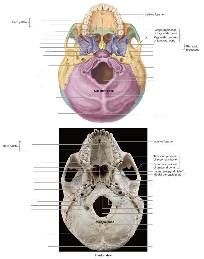
Question 3
Answer
-
palatine process
-
horizontal plate
-
vomer
-
foramen ovale
-
sphenoid bone
-
jugular foramen
-
carotid canal
-
lambdoid suture
-
foramen magnum
-
occipital condyle
-
mastoid process
-
mandibular fossa
-
styloid process
-
lateral pterygoid plate
-
medial pterygoid plate
-
external occipital protuberence
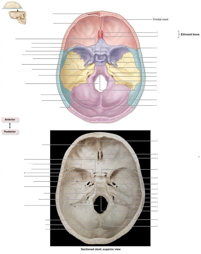
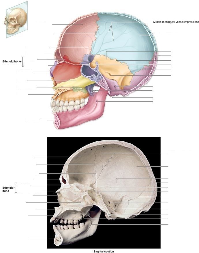
Question 4
Answer
-
frontal sinus
-
crista galli
-
cribriform plate
-
optic canal
-
lesser wing of sphenoid
-
sella turcica
-
foramen rotundum
-
greater wing of sphenoid
-
foramen ovale
-
internal acoustic meatus
-
jugular foramen
Question 5
Answer
-
sphenoid sinus
-
frontal sinus
-
crista galli
-
perpendicular plate
-
mandibular foramen
Question 6
Answer
-
coronal suture
-
sagittal suture
-
lambdoid suture
Question 7
Question
[blank_start]Long bones[blank_end] are longer tan they are wide and include the bones of the upper and lower limbs, excluding the ankle and wrist bones.
Answer
-
Long bones
Question 8
Question
[blank_start]Flat bones[blank_end] are shaped exactly as they're named. These bones include the ribs, sternum, certain skull bones, and hip bones.
Answer
-
Flat bones
Question 9
Question
[blank_start]Irregular bones[blank_end] are those whose shape doesn't fit into any of the other classes. These bones include the vertebrae, the sacrum, and certain bones of the skull, such as the sphenoid bone.
Answer
-
Irregular bones
Question 10
Question
[blank_start]Sesamoid bones[blank_end] are usually small, round, and flat, and shaped somewhat like a sesame seed.
Answer
-
Sesamoid bones
Question 11
Question
[blank_start]Sutural bones[blank_end], or Wormian bones, are small, flat, irregularly shaped bones between the flat bones of the skull. There are individual variations in the number, shape, and position of the sutural bones.
Answer
-
Sutural bones
Question 12
Question
The axial skeleton consists of
Answer
-
skull
-
upper and lower limbs
-
thoracic cage
-
pelvic girdle
-
bones associated with the skull
-
vertebral column
Question 13
Question
The appendicular skeleton consists of
Answer
-
Upper and lower limbs
-
Skull
-
Pectoral girdle
-
Vertebrae
-
Pelvic girdle
Question 14
Question
The vertebral column consists of 24 unfused vertebrae and 5 fused vertabrae of the sacrum and coccyx. Of the 24 unfused vertebrae, [blank_start]7[blank_end] are cervical, [blank_start]12[blank_end] are thoracic, and [blank_start]5[blank_end] are lumbar.
Answer
-
7
-
12
-
5
Question 15
Question
[blank_start]C1[blank_end], or [blank_start]atlas[blank_end], articulates with the occipital condyles, and holds up the head. It also allows us to nod our head yes.
Answer
-
C1
-
atlas
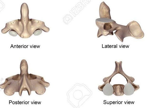
Question 16
Question
[blank_start]C2[blank_end], or [blank_start]axis[blank_end], fuses with C1 and allows us to rotate our head left and right.
Answer
-
C2
-
axis
Question 17
Answer
-
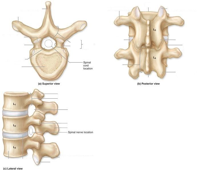
cervical vertebrae
-
thoracic vertebrae
-
lumbar vertebrae
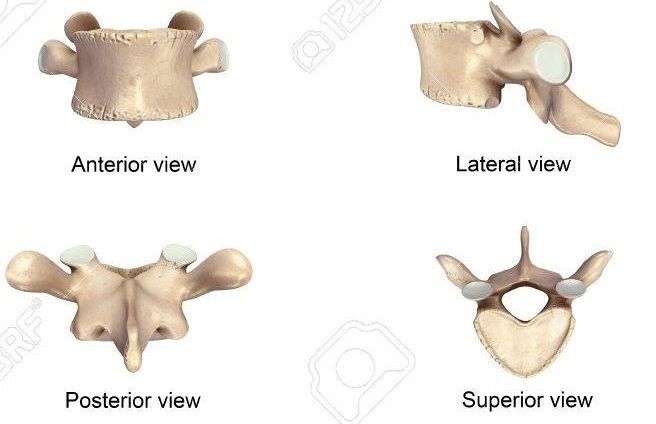
Question 18
Answer
-
cervical vertebrae
-
thoracic vertebrae
-
lumbar vertebrae
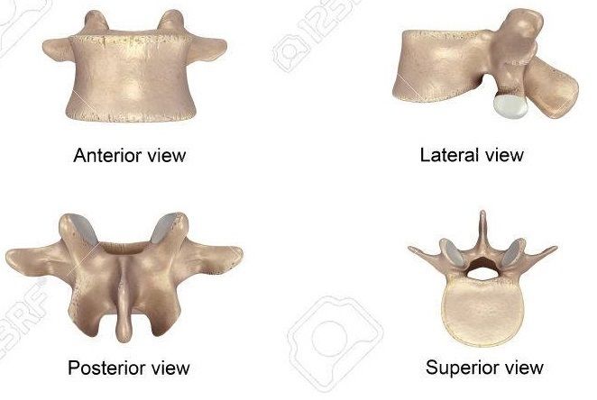
Question 19
Answer
-
cervical vertebrae
-
thoracic vertebrae
-
lumbar vertebrae
Question 20
Answer
-
spinous process
-
transverse process
-
lamina
-
pedicle
-
vertebral arch
-
superior articular facet
-
superior articular process
-
vertebral foramen
-
body
-
superior articular process
-
pedicle
-
intervertebral foramen
-
transverse process
-
spinous process
-
inferior articular process
-
inferior articular facet
-
body
-
transverse process
-
superior articular facet
-
inferior articular process
-
superior articular process
-
inferior articular process
-
spinous process
-
lamina
-
intervertebral disc
-
vertebral arch
Question 21
Answer
-
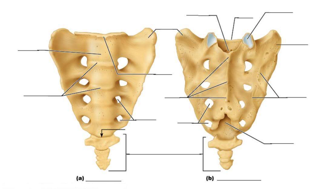
auricular surface
-
sacral foramina
Question 22
Answer
-
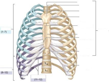
true ribs
-
false ribs
-
floating ribs
-
manubrium
-
body
-
xiphoid process
-
sternum
-
jugular notch
Question 23
Answer
-
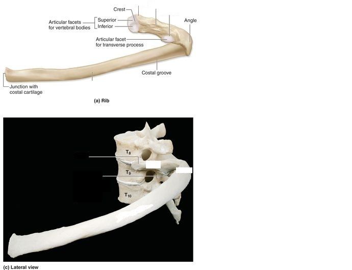
head
-
neck
-
tubercle
-
body
-
sternal end
Question 24
Question
Ribs 1-7 are considered [blank_start]true ribs[blank_end], or vertebrosternal ribs, because they attach directly to the sternum by their own costal cartilage. Ribs 8-12, on the other hand, are classified as [blank_start]false ribs[blank_end] because they lack this direct attachment to the sternum. Ribs 8-10 are [blank_start]vertebrochondral ribs[blank_end] as they have an indirect attacment to the sternum, as their cartilage attaches to the costal cartilage of the true ribs. Ribs 11-12 have no attachment to the sternum at all, so they are often referred to as [blank_start]floating ribs[blank_end] or vertebral ribs.
Answer
-
false ribs
-
vertebrochondral ribs
-
true ribs
-
floating ribs
-
floating ribs
-
vertebrochondral ribs
-
true ribs
-
false ribs
-
vertebrochondral ribs
-
vertebrosternal ribs
-
floating ribs
-
true ribs
-
false ribs
-
floating ribs
-
vertebrochondral ribs
-
vertebrosternal ribs
Question 25
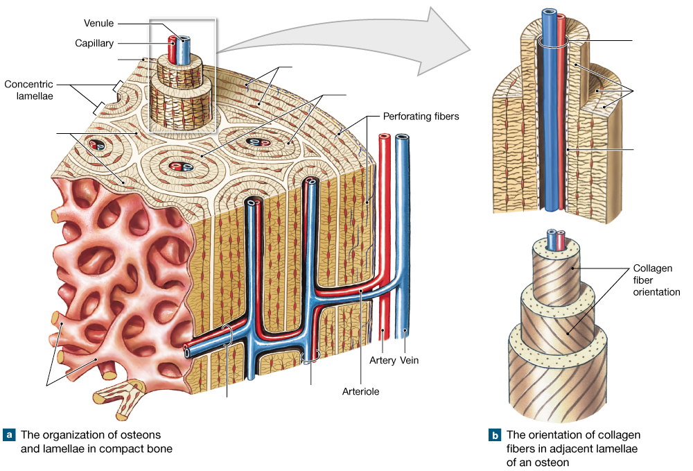
Answer
-
circumferential lamellae
-
periosteum
-
interstitial lamellae
-
trabeculae of spongy gone
-
osteons
-
central canal
-
perforating canal
-
central canal
-
concentric lamellae
-
endosteum
Question 26
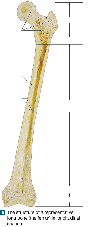
Answer
-
epiphysis
-
epiphysis
-
metaphysis
-
metaphysis
-
diaphysis
-
compact bone
-
medullary cavity
-
spongy bone
Question 27
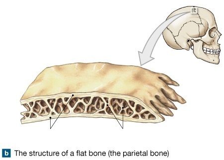
Answer
-
compact bone
-
diploe
Question 28
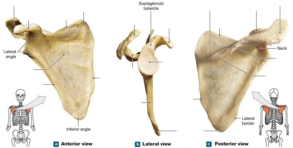
Answer
-
acromion process
-
acromion process
-
acromion process
-
coracoid process
-
coracoid process
-
coracoid process
-
superior border
-
superior border
-
superior angle
-
supraspinous fossa
-
subscapular fossa
-
spine
-
spine
-
glenoid cavity
-
infraspinous fossa
-
medial border
-
lateral border
-
inferior angle
-
lateral border
-
medial border
Question 29
Answer
-
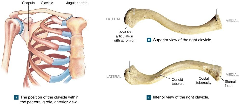
sternal end
-
acromial end
Question 30
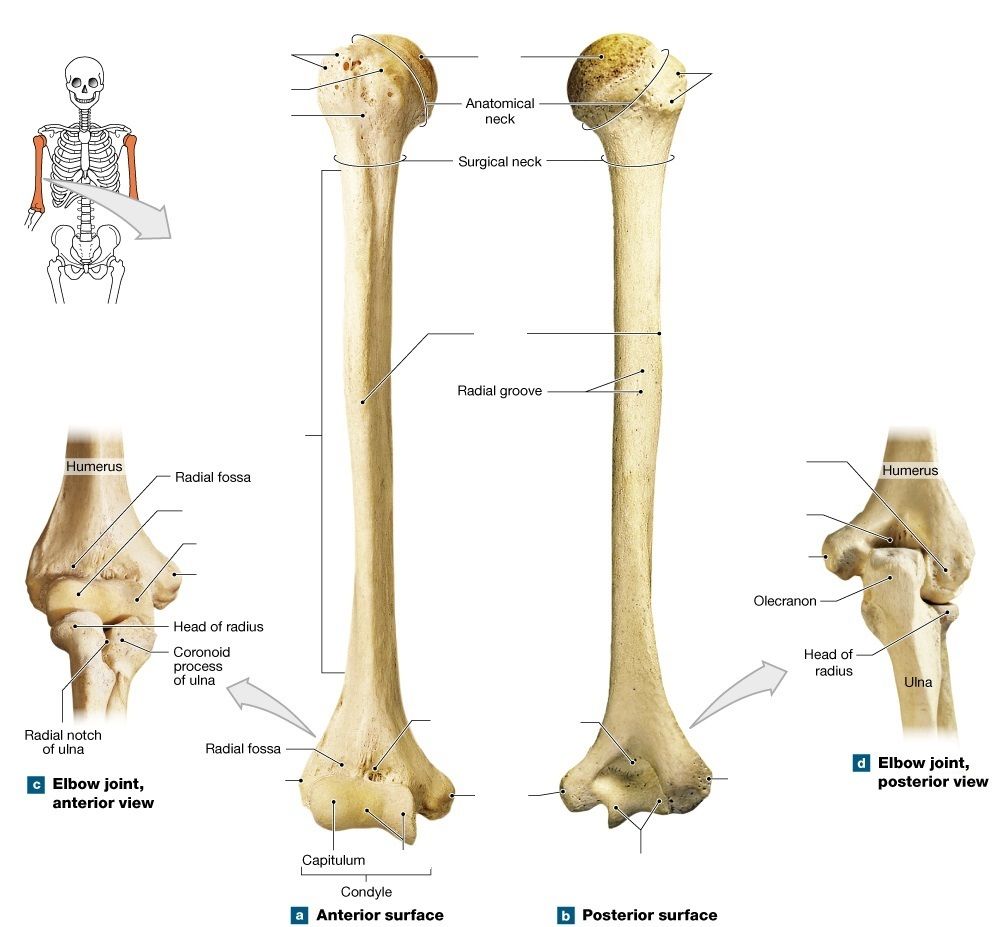
Answer
-
head
-
greater tubercle
-
greater tubercle
-
lesser tubercle
-
intertubercular groove
-
shaft
-
deltoid tuberosity
-
capitulum
-
trochlea
-
medial epicondyle
-
trochlea
-
medial epicondyle
-
coronoid fossa
-
olecranon fossa
-
trochlea
-
lateral epicondyle
-
medial epicondyle
-
olecranon fossa
-
trochlea
Question 31
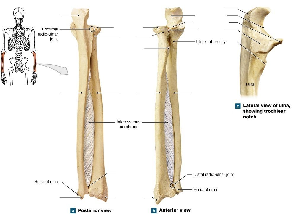
Answer
-
olecranon process
-
head of radius
-
neck of radius
-
radial tuberosity
-
radius
-
ulna
-
ulna
-
ulnar styloid process
-
radial styloid process
-
ulnar notch
-
olecranon process
-
trochlear notch
-
coronoid process
-
radial notch
Question 32
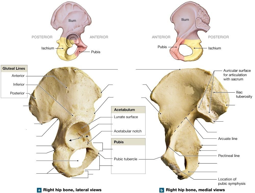
Answer
-
iliac crest
-
anterior superior iliac spine
-
anterior inferior iliac spine
-
acetabulum
-
superior pubic ramus
-
inferior pubic ramus
-
ischial ramus
-
ischial tuberosity
-
obturator foramen
-
lesser sciatic notch
-
ischial spine
-
greater sciatic notch
-
posterior inferior iliac spine
-
posterior superior iliac spine
-
posterior superior iliac spine
-
ischial ramus
-
ischial tuberosity
-
lesser sciatic notch
-
ischial spine
-
greater sciatic notch
-
posterior inferior iliac spine
Question 33
Answer
-
iliac crest
-
posterior superior iliac spine
-
posterior inferior iliac spine
-
sacro-iliac joint
-
pubic symphysis
-
greater sciatic notch
-
ischial spine
-
ischial tuberosity
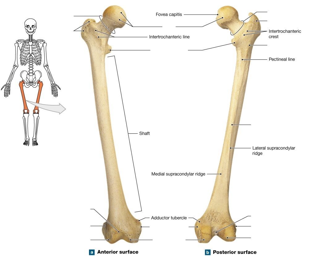
Question 34
Answer
-
head
-
neck
-
neck
-
greater trochanter
-
greater trochanter
-
lesser trochanter
-
gluteal tuberosity
-
linea aspera
-
patellar surface
-
lateral epicondyle
-
lateral condyle
-
medial epicondyle
-
medial condyle
-
intercondylar fossa
-
lateral epicondyle
-
lateral condyle
Question 35
Answer
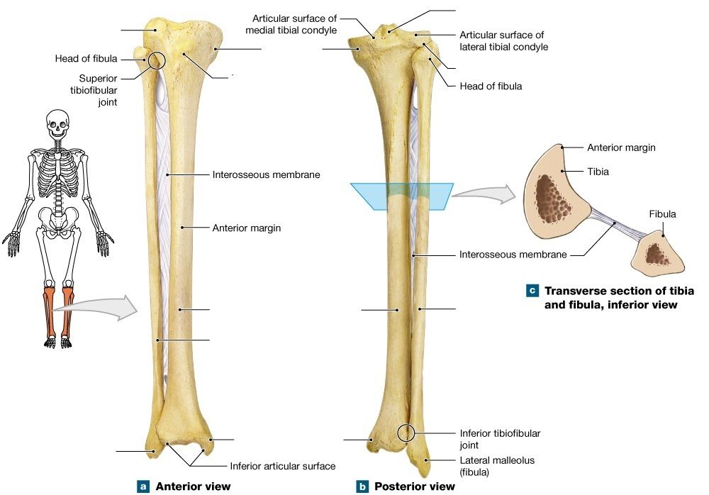
-
medial tibial condyle
-
lateral tibial condyle
-
tibial tuberosity
-
tibia
-
fibula
-
tibia
-
fibula
-
lateral malleolus
-
medial malleolus
-
intercondylar eminence
-
lateral tibial condyle
Question 36
Question
[blank_start]Fibrous joints[blank_end] consists of two bones joined by short collagen fibers. Most [blank_start]fibrous joints[blank_end] allow no motion. [blank_start]Cartilaginous joints[blank_end] consist of bones united by cartilage rather than fibrous connective tissue. Most [blank_start]cartilagenous joints[blank_end] allow some motion. [blank_start]Synovial joints[blank_end] are freely movable joints.
Answer
-
Fibrous joints
-
Cartilaginous joints
-
Synovial joints
-
fibrous joints
-
cartilaginous joints
-
synovial joints
-
Cartilaginous joints
-
Fibrous joints
-
Synovial joints
-
cartilagenous joints
-
fibrous joints
-
synovial joints
-
Synovial joints
-
Cartilaginous joints
-
Fibrous joints
Question 37
Question
Fibrous joints include
Answer
-
suture
-
synchondrosis
-
syndesmosis
-
pivot
-
gomphosis
Question 38
Question
Cartilaginous joints include
Answer
-
Suture
-
Synchondrosis
-
Syndesmosis
-
Symphysis
Question 39
Question
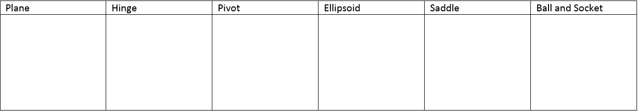
Synovial joints include
Answer
-
plane
-
suture
-
hinge
-
symphysis
-
pivot
-
synchondrosis
-
ellipsoid
-
syndesmosis
-
saddle
-
ball and socket
Question 40
Question
The cartilagenous connection between the ends of ribs 1 and 2 to the manubrium and sternum is an example of syndesmosis.
Answer
- True
- False
Question 41
Question
Gomphosis is a type of joint that binds what to what?
Answer
-
teeth to bony sockets
-
ribs to sternum
-
skull bones to each other
-
long bones to each oher
Question 42
Question
Bones connected by a ligament, such as the distal joint between the tibia and fibula is an example of syndesmosis.
Answer
- True
- False
Question 43
Question
Drag and Drop the joint into the correct category
Answer
-
intercarpal joints
-
vertebrocostal joints
-
sacroiliac joint
-
acromioclavicular joint
-
claviculosternal joint
-
elbow joint
-
knee joint
-
ankle joint
-
interpharangeal joint
-
atlanto-axial joint
-
proximal radio-ulnar joint
-
radiocarpal joint
-
metacarpophalangeal joint
-
metatarsophalangeal joint
-
first carpometacarpal joint
-
shoulder joint
-
hip joint
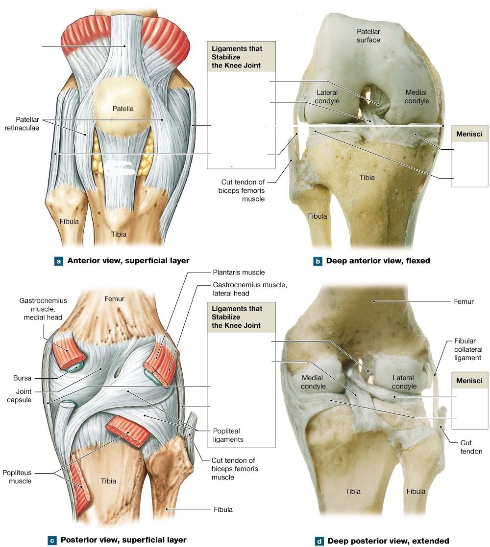
Question 44
Answer
-
quadriceps tendon
-
patellar ligament
-
posterior cruciate ligament
-
anterior cruciate ligament
-
tibial collateral ligament
-
fibular collateral ligament
-
medial meniscus
-
lateral meniscus
-
lateral meniscus
-
medial meniscus
-
anterior cruciate ligament
-
posterior cruciate ligament
-
tibial collateral ligament
-
fibular collateral ligament
Question 45
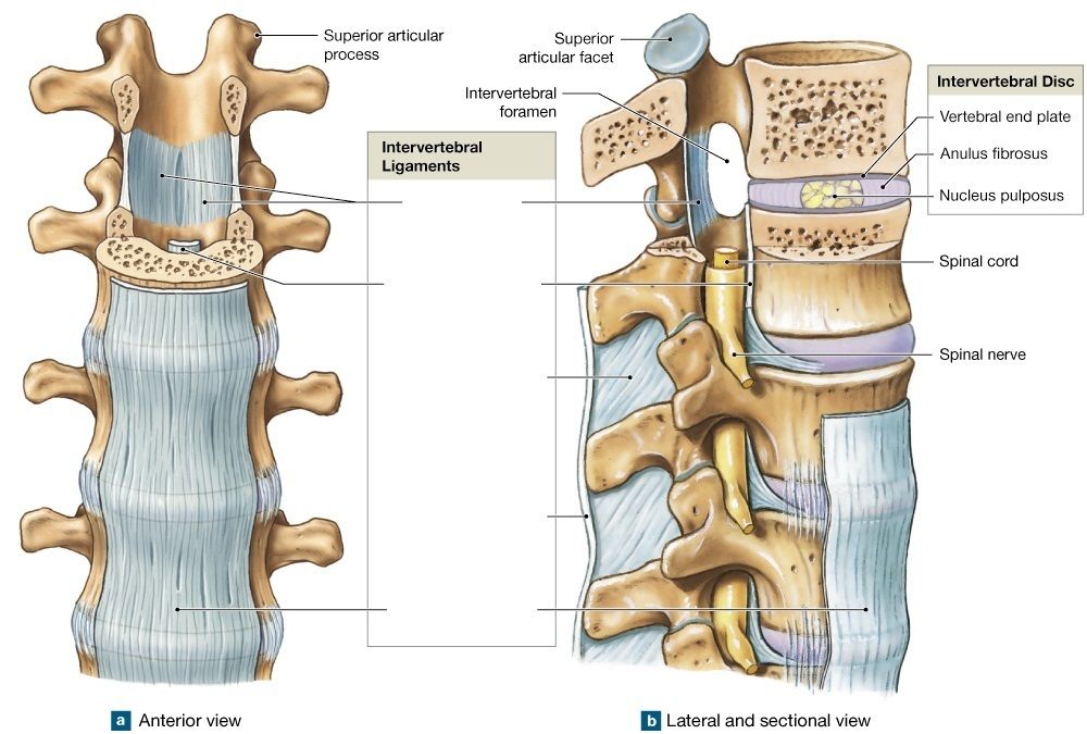
Answer
-
ligamentum flavum
-
posterior longitudinal ligament
-
interspinous ligaments
-
supraspinous ligament
-
anterior longitudinal ligament
Question 46
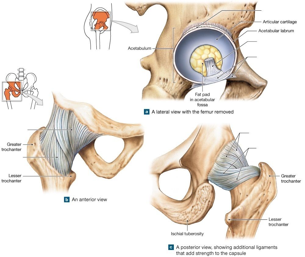
Answer
-
ligamentum teres
-
transverse acetabular ligament
-
pubofemoral ligament
-
iliofemoral ligament
-
ischiofemoral ligament
Question 47
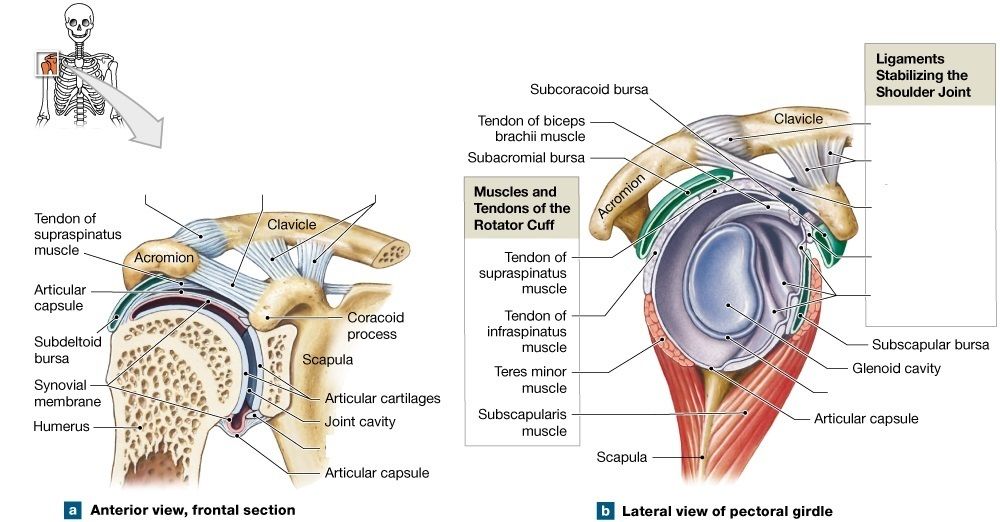
Answer
-
acromioclavicular ligament
-
coraco-acromial ligament
-
coracoclavicular ligaments
-
acromioclavicular ligament
-
coracoclavicular ligaments
-
coraco-acromial ligaments
-
coracohumeral ligament
-
glenohumeral ligaments
Question 48
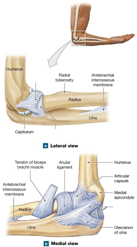
Answer
-
anular ligament
-
ulnar collateral ligament
-
radial collateral ligament
Want to create your own Quizzes for free with GoConqr? Learn more.