4316788
Description
Flashcards by t.whittingham, updated more than 1 year ago
|
|
Created by t.whittingham
over 8 years ago
|
|
| Question | Answer |
| The Auditory Process | Converts sound waves (air vibrations) into vibrations within the inner ear (fluid vibrations). -> These fluid vibrations then excite receptor cells in the inner ear and cause impulses in the auditory nerve. -> The nerve impulses travel to the auditory cortex and are perceived by the brain as sound. |
| Function of the external ear | Collection & transmission of sound. |
| Function of the middle ear | Amplification of sound. Transfers sound from air into fluid-membrane waves in the inner ear. |
| Function of the inner ear. | Conversion of sound to nerve impulses (cochlea). Balance function (Vestibular labyrinth) |
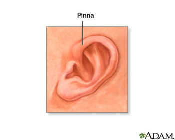
| Components of the Outer Ear (2) | 1. Pinna 2. External Auditory Canal (External Auditory Meatus) |
| Pinna | Visible part of ear. Acts as a funnel to collect sound -> directs it to the auditory canal. Skin closely adherent to cartilage. |
| External Auditory Meatus (External Auditory Canal) | Resonating tube -> transmits sound to eardrum. Extends from the pinna to the eardrum. Approximately 24mm long. Outer 1/3 = cartilage Inner 2/3 = bone |
| Function of wax in the Ear Canal | Protects ear -> traps dust & particles. Self-cleaning -> moves laterally (outwards) - migrates skin from surface of eardrum and deep external meatus. |
| Physiology of the external ear | 1. Collection + Localisation of sound -> detects sound source e.g. left/right - differential volume & sound shadowing effect of head - ear trumpet effect. 2. Resonance -> ear canal & pinna act like wind instrument. -> speech frequencies resonate in ear canal & amplify. |
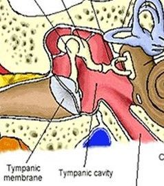
| Components of the Middle Ear (4) | 1. Tympanic Membrane 2. Tympanic Cavity 3. Ossicles 4. Eustachian Tube Middle ear cavity = inside temporal bone of the skull. |
| Tympanic Membrane (Eardrum) | Separates ear canal from middle ear cavity -> functionally part of middle ear. Approximately 1cm diameter. Function = transmits sound from air to ossicles in middle ear. |
| 3 layers of the Tympanic Membrane | 1. Outer layer = squamous epithelium (normal skin - continuous with ear canal). 2. Middle layer = fibrous, with radial & circular fibers to stiffen ear drum. 3. Inner layer = mucous membrane. |
| Tympanic Cavity | Small cavity surrounding the bones of the middle ear. Bound by the: - Oval window - Round window - Promontory |
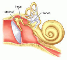
| Ossicular Chain | Whole system acts as a vibrating membrane with attached bony levers working a small piston to pump sound into fluid of inner ear. 1. Malleus 2. Incus 3. Stapes |
| Malleus | Also called the hammer. Largest of the ossicles. Vibration of tympanic membrane moves whole malleus. |
| Incus | Also called Anvil. Acts as a lever transferring vibration from malleus to the stapes. |
| Stapes | Stirrup shaped bone. Attaches to long process of incus. Smallest bone in the human body. Footplate lies in oval window of cochlea - transmits vibrations into inner ear. |
| Stilleto-Heel Effect | Size ratio of tympanic membrane to oval window = approximately 20:1. = Middle ear amplifier. |
| Eustachian Tube | Ventilates & drains middle ear cavity. Opens into back of nasal cavity (nasopharynx). - 1/3 bone, 2/3 cartilage. - Closed at rest, opens with yawning & swallowing. |
| Muscles of the Middle Ear | 1. Tensor Tympani Muscle 2. Stapedius |
| Tensor Tympani Muscle | Attaches to upper end of handle of malleus. Protects ear against sudden loud sound -> dampens sounds. |
| Stapedius muscle | The smallest skeletal muscle in the human body. Stiffens stapes as reflex against loud noise. |
| Nerves of the Middle Ear | 1. Chorda Tympani Nerve = crosses middle ear - transfers taste fibers from tongue & sends fibers to salivary glands in mouth. 2. Facial Nerve (C.N. VII) |
| Glue Ear | Build up of mucus in middle ear cavity & eustachian tube -> prevents ear drum from vibrating properly -> causes conductive hearing loss. |
| Treatment of Glue Ear | 1. Grommets = small tube inserted into ear - helps drain fluid and maintain ear pressure. 2. T-Tubes = long stay ventilation tube - bigger version of grommet. |
| Tympanometry | Objective test of middle-ear function. Tests mobility of ear drum & conduction bones by creating variations of ear pressure in ear canal. |
| Acute Otitis Media | Inflammatory disease of the middle ear -> dysfunction of the eustachian tube. Difference to Glue Ear = also includes other symptoms. |
| Perforated Ear Drum | Hole in ear drum -> pressure leaks through hole -> eardrum doesn't vibrate properly. |
| Attic Perforation | Perforation in the superior part of the eardrum. |
| Cholesteatoma | An uncommon abnormal collection of skin cells inside the ear. Associated with chronic infection. Can lead to erosion of ossicles, inner ear damage, facial nerve damage & in extreme cases, meningitis or brain abscess. |
| Causes of conductive hearing loss (6) | 1. Wax 2. Perforations 3. Glue Ear 4. Chronic Infection 5. Scarring 6. Damaged Ossicles |
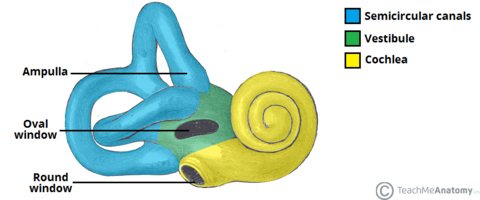
| Inner Ear - 2 main functional parts | 1. Cochlear system = dedicated to hearing 2. Vestibular system = dedicated to balance |
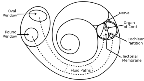
| Bony Labyrinth | One of the hardest bones in the body - protects nervous system. - Oval window = way in - Round window = way out Composed of 3 parts: 1.Cochlea 2. Vestibule 3. Semicircular Canals |
| Membranous Labyrinth. | Collection of fluid-filled tubes inside the bony labyrinth. Separated from bony labyrinth by fluid called Perilymph. |
| Cochlea | Spinal-shaped cavity. Receives vibrations from Stapes. Ascending passage = scala vestibuli Descending passage = scala tympani |
| Cochlear Duct (Scala Media) | Lies between the Scala Vestibuli & Scala Tympani. Filled with Endolymph. |
| Basiliar Membrane & Reissner's Membrane | Flexible - respond to vibrations travelling up the Scala Vestibuli. Movement of membranes send vibrations back down to the Scala Tempani. |
| Organ of Corti | Situated on the basiliar membrane. Stimulated as the basiliar membrane vibrates -> sends nerve impulses to the brain via the cochlear nerve. Nerve impulses are generated by hair cells within the Organ of Corti. |
| Tonotopic Organization | Low frequency sound are detected at the apex (top of cochlea) High frequency sound detected at basal turn (base of cochlea) |
| Pure Tone Audiometry | Key hearing test used to identify hearing threshold levels of individual -> can determine degree, type & configuration of hearing loss. Normal hearing threshold = less than 20dB HL in young healthy adults with no ear disease & no family history of deafness or noise trauma. |
| Sensorineural Hearing Loss | Type of hearing loss in which the root cause lies in the inner ear. Most common cause = age-related (presbycusis). Second most common cause = noise-induced hearing loss. |
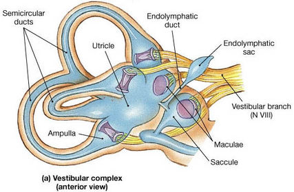
| Vestibular Labyrinth | The sensory system that provides the leading contribution about the sense of balance and spatial orientation -> for the purpose of coordinating movement with balance. Components: 1. Otolithic Organs (Utricule & Saccule). 2. Semicircular Canals. |
| Utricle & Saccule | Sense linear accelerations and gravity. Maintain head & body position. Initiate reflexes to keep head upright. |
| Semicircular Canals | 3 in each ear. Detects rotational movements. |
Want to create your own Flashcards for free with GoConqr? Learn more.