21788222
Description
Quiz by Charlotte Jakes, updated more than 1 year ago
|
|
Created by Charlotte Jakes
about 4 years ago
|
|
Question 1
Question
What is the upper boundary of the abdominal cavity?
Answer
-
Diaphragm
-
Costal margin
-
Parietal pleura
-
Superior thoracic aperture
Question 2
Question
What is the inferior boundary of the abdominal cavity?
Answer
-
The iliac crests
-
The pelvic inlet/brim
-
The pelvic outlet
-
The 12th ribs
Question 3
Question
Label these images to show the theoretical regions and quadrants of the abdomen.
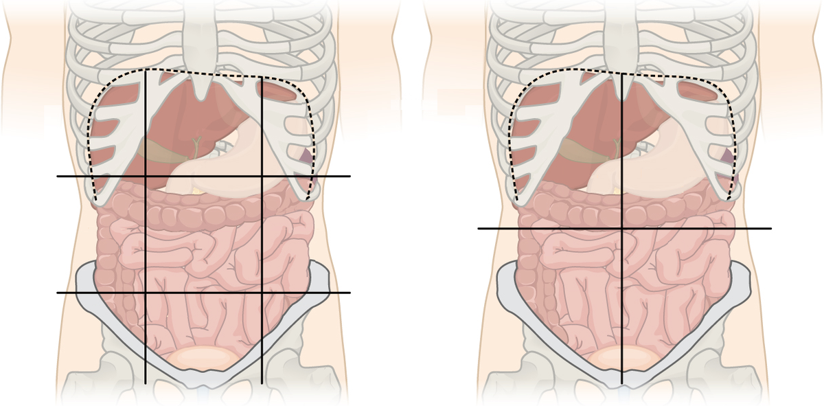
Answer
-
Right hypochondriac region
-
Epigastric region
-
Left hypochondriac region
-
Right lumbar region
-
Umbilical region
-
Left lumbar region
-
Right iliac region
-
Suprapubic region
-
Left iliac region
-
Right upper quadrant
-
Right lower quadrant
-
Left upper quadrant
-
Left lower quadrant
-
Transpyloric plane
-
Transtubercular plane
-
Midclavicular line
Question 4
Question
The transtubercular plane transverses the tubercles of what?
Answer
-
Iliac crests
-
Pubic bone
-
Ischium
-
Femurs
Question 5
Question
Where is the transpyloric plane?
Answer
-
Halfway between the jugular notch and pubic symphysis
-
1/3 of the way between the jugular notch and pubic symphysis
-
Halfway between the sternal angle and pubic symphysis
-
Halfway between the sternoclavicular joint and pubic symphysis
Question 6
Question
To find the level of the transpyloric plane, we measure a hands breadth below...
Answer
-
The sternal angle
-
The jugular notch
-
The junction between the body of the sternum and the xiphisternum
-
The costal margin
Question 7
Question
What is a raphe?
Answer
-
A seam or ridge marking the point of fusion of two embryologically different structures
-
A seam or ridge marking the point of fusion of two embryologically similar structures
-
A large sheet of fibrous tissue in place of a tendon
-
A membranous covering of organs within the peritoneal cavity
Question 8
Question
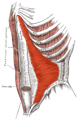
Drag and drop the correct answers to describe the anterior abdominal wall.
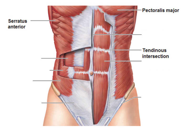
Answer
-
Linea alba
-
Rectus abdominis
-
Inguinal ligament
-
Aponeurosis
-
External oblique
-
Internal oblique
-
Transversus abdominis
Question 9
Question
Drag and drop the correct answers to describe the RECTUS ABDOMINIS muscle.
ORIGIN: [blank_start]pubic crest[blank_end]
INSERTION: [blank_start]xiphisternum[blank_end] and costal cartilages of ribs [blank_start]5-7[blank_end]
FUNCTION: compression of abdominal viscera, [blank_start]depression[blank_end] of ribs, stabilisation of [blank_start]pelvis[blank_end] during walking
INNERVATION: [blank_start]thoracoabdominal nerves (T7-T11)[blank_end]
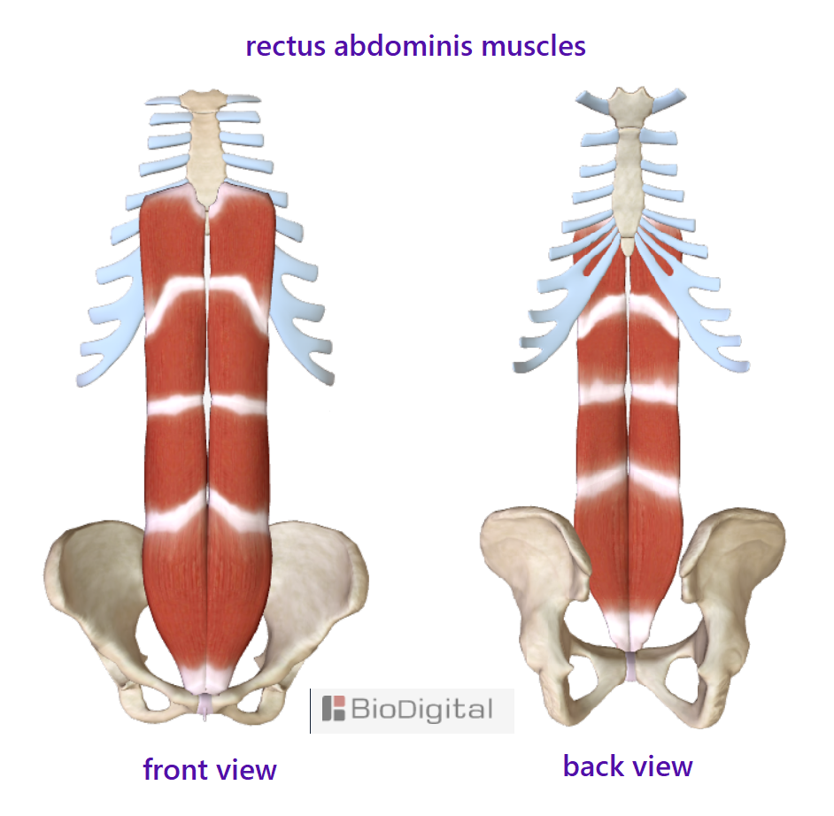
Answer
-
pubic crest
-
pubic tubercle
-
xiphisternum
-
sternal angle
-
5-7
-
6-8
-
10-12
-
depression
-
elevation
-
pelvis
-
ribs
-
thoracoabdominal nerves (T7-T11)
-
subcostal nerve (T12)
Question 10
Question
Drag and drop the correct answers to describe the EXTERNAL OBLIQUE muscle.
ORIGIN: ribs 5-12
INSERTION: iliac [blank_start]crest[blank_end] and pubic [blank_start]tubercle[blank_end]
FUNCTION: [blank_start]contralateral[blank_end] rotation of torso
INNERVATION: thoracoabdominal nerves [blank_start](T7-T11)[blank_end] and subcostal nerve [blank_start](T12)[blank_end]
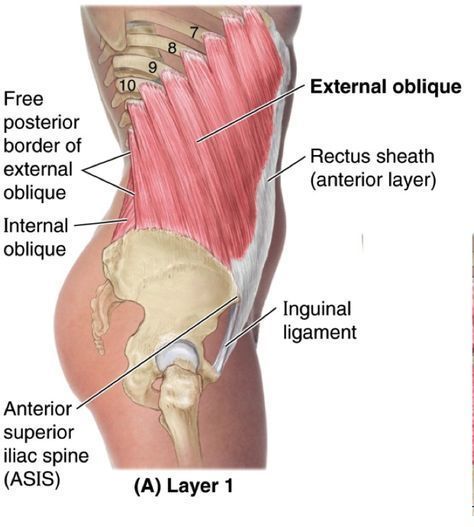
Answer
-
crest
-
fossa
-
tubercle
-
symphysis
-
contralateral
-
ipsilateral
-
(T7-T11)
-
(T9-T10)
-
(T12)
-
(C7)
Question 11
Question
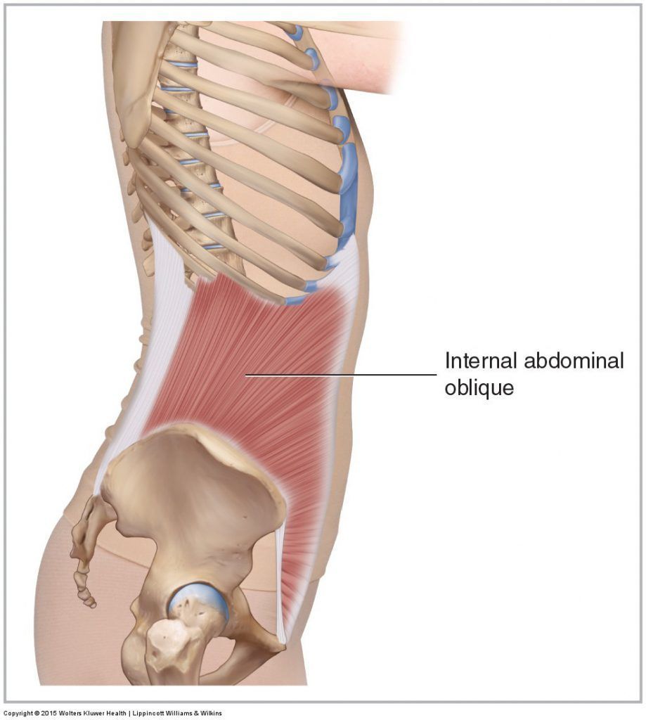
Drag and drop the correct answers to describe the INTERNAL OBLIQUE muscle.
ORIGIN: [blank_start]inguinal[blank_end] ligament, [blank_start]iliac[blank_end] crest and lumbodorsal [blank_start]fascia[blank_end]
INSERTION: ribs [blank_start]10-12[blank_end]
FUNCTION: compression of abdomen and [blank_start]ipsilateral[blank_end] rotation of torso
INNERVATION: thoracoabdominal nerves [blank_start](T7-T11)[blank_end], [blank_start]subcostal nerve[blank_end] (T12), [blank_start]iliohypogastric nerve[blank_end] (L1) and [blank_start]ilioinguinal nerve[blank_end] (L1)
Answer
-
inguinal
-
iliac
-
fascia
-
ligament
-
10-12
-
11-12
-
9-10
-
ipsilateral
-
contralateral
-
(T7-T11)
-
(T8-T10)
-
subcostal nerve
-
iliohypogastric nerve
-
ilioinguinal nerve
Question 12
Question
Drag and drop the correct answers to describe the TRANSVERSUS ABDOMINIS muscle.
ORIGIN: [blank_start]inguinal[blank_end] ligament, costal cartilages [blank_start]7-12[blank_end], iliac [blank_start]crest[blank_end] and thoracolumbar fascia
INSERTION: conjoint tendon, [blank_start]xiphisternum[blank_end], linea alba, [blank_start]pubic[blank_end] crest
FUNCTION: compression of abdomen
INNERVATION: [blank_start]thoracoabdominal nerves[blank_end] (T7-T11), subcostal nerve [blank_start](T12)[blank_end], ilioinguinal nerve (L1), [blank_start]iliohypogastric nerve[blank_end] (L1)
Answer
-
inguinal
-
7-12
-
8-10
-
5-7
-
crest
-
tubercle
-
xiphisternum
-
sternal angle
-
pubic
-
iliac
-
thoracoabdominal nerves
-
(T12)
-
(T11)
-
iliohypogastric nerve
Question 13
Question
Drag and drop the correct answers to describe this transverse section of the abdomen at the level of L3.
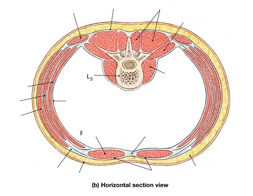
Answer
-
Psoas major
-
Quadratus lumborum
-
Erector spinae
-
Latissimus dorsi
-
Lumbodorsal fascia
-
Peritoneum
-
Transversus abdominis
-
Internal oblique
-
External oblique
-
Aponeurosis of external oblique
-
Skin
-
Rectus abdominis
-
Linea alba
-
Rectus sheath
-
Superficial fascia
Question 14
Question
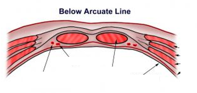
Below the arcuate line, which is true of the anterior abdominal wall?
Answer
-
The only layer deep to rectus abdominis is the transversalis fascia
-
There is no layer deep to rectus abdominis
-
The aponeurosis of the internal oblique splits where half lies deep to rectus abdominis and half lies superficial
-
The rectus abdominis muscle inserts and thus is no longer part of the abdominal wall
Question 15
Question
What is significant about the layers of the anterior abdominal wall below the arcuate line?
Answer
-
The inferior epigastric artery can penetrate the rectus sheath
-
The superior epigastric artery can penetrate the rectus sheath
-
This is a point of weakness where femoral hernias can occur
-
It occurs exactly at the umbilicus
Question 16
Question
Drag and drop the correct answers to describe the layers of the anterior abdominal wall ABOVE the arcuate line.
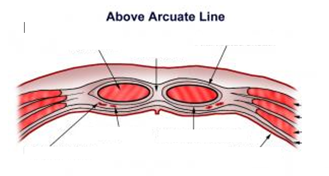
Answer
-
Rectus abdominis
-
Linea alba
-
Anterior rectus sheath
-
Inferior epigastric vein
-
Inferior epigastric artery
-
Posterior rectus sheath
-
Peritoneum
-
External oblique
-
Internal oblique
-
Transversus abdominis
-
Transversalis fascia
Question 17
Question
Drag and drop the correct answers to describe the layers of the anterior abdominal wall BELOW the arcuate line.
Answer
-
Inferior epigastric vein
-
Inferior epigastric artery
-
Rectus abdominis
-
External oblique
-
Internal oblique
-
Transversus abdominis
-
Transversalis fascia
-
Peritoneum
Question 18
Question
Label this image to show the anterior abdominal wall from a posterior view, in particular the arcuate line.
Answer
-
External oblique
-
Internal oblique
-
Transversus abdominis
-
Linea alba
-
Linea semilunaris
-
Arcuate line
-
Medial umbilical ligament
-
Median umbilical ligament
-
External iliac artery
-
Inferior epigastric artery
-
Superior epigastric artery
Question 19
Question
Where is the arcuate line?
Answer
-
Around halfway between the umbilicus and pubic crest
-
At the level of the umbilicus
-
At the transtubercular plane
-
2/3 of the way between the umbilicus and pubic crest
Question 20
Question
Drag and drop the correct answers to describe the layers of the anterior abdominal wall.
Answer
-
Skin
-
Camper's fascia
-
Scarpa's fascia
-
Deep fascia
-
External oblique
-
Internal oblique
-
Transversus abdominis
-
Extraperitoneal fat
-
Transversalis fascia
-
Parietal peritoneum
Question 21
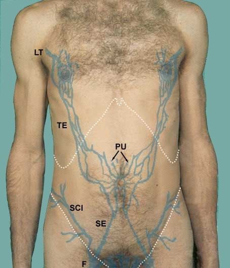
Question
Fill in the blanks to describe the veins that drain the anterior abdominal wall.
LT - [blank_start]lateral thoracic[blank_end] veins
TE - [blank_start]thoracoepigastric[blank_end] veins
PU - [blank_start]paraumbilical[blank_end] veins
SE - [blank_start]superficial epigastric[blank_end] veins
SCI - [blank_start]superficial circumflex iliac[blank_end] veins
F - [blank_start]femoral[blank_end] vein
Answer
-
lateral thoracic
-
thoracoepigastric
-
paraumbilical
-
superficial epigastric
-
superficial circumflex iliac
-
femoral
Question 22
Question
Drag and drop the correct answers to describe the blood supply of the anterior abdominal wall.
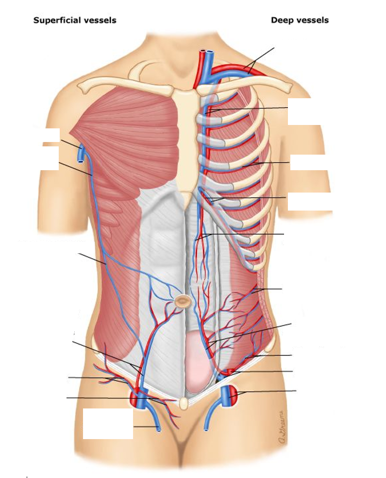
Answer
-
Internal thoracic vessels
-
Subclavian vessels
-
Intercostal vessels
-
Musculophrenic vessels
-
Superior epigastric vessels
-
SUbcostal vessels
-
Inferior epigastric vessels
-
Deep circumflex iliac vessels
-
External iliac vessles
-
Femoral vessels
-
Greater saphenous vein
-
Pudendal vessels
-
Superficial circumflex iliac vessels
-
Superficial epigastric vessels
-
Thoracoepigastric vein
-
Lateral thoracic vein
-
Axillary vein
Question 23
Question
Which spinal nerve supplies the dermatome overlying the umbilicus?
Answer
-
T10
-
T9
-
T11
-
T12
Question 24
Question
Which spinal nerves supply the dermatomes of the abdomen?
Answer
-
T7-L1
-
T7-T12
-
T10-L1
-
T1-L1
Question 25
Question
Drag and drop the correct answers to label the nerves of the anterior abdominal wall.
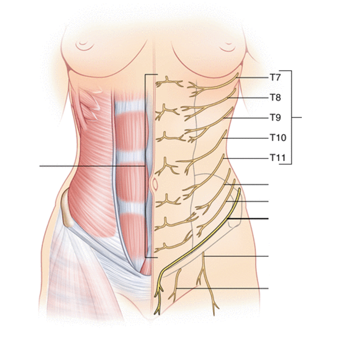
Answer
-
Thoracoabdominal nerves
-
Subcostal nerve (T12)
-
Iliohypogastric nerve (L1)
-
Ilioinguinal nerve (L1)
-
Femoral branch of genitofemoral nerve
-
Genital branch of genitofemoral nerve
-
Anterior cutaneous nerves
Question 26
Question
The superior epigastric artery branches off the internal thoracic artery.
Answer
- True
- False
Question 27
Question
What is the inferior epigastric artery a branch of?
Answer
-
External iliac artery
-
Internal iliac artery
-
Femoral artery
-
Internal thoracic artery
Want to create your own Quizzes for free with GoConqr? Learn more.