5606360

Question 1
Question
Where should the front edge of a bitewing radiograph be aligned?
Answer
-
Mesial edge of the upper first pre-molar
-
Distal edge of the upper first pre-molar
-
Mesial edge of the upper second pre-molar
-
Distal edge of the upper first molar
Question 2
Question
Which type of bitewing radiography is more commonly used?
Answer
-
Horizontal
-
Vertical
Question 3
Question
In a bitewing radiograph, the film should be positioned [blank_start]perpendicular[blank_end] to the occlusal surfaces of the teeth / central x-ray beam.
Answer
-
at 90°
-
at 45°
-
at 30°
Question 4
Question
When may a bitewing radiograph be prescribed?
Answer
-
Assessing occlusal and interproximal caries of multiple teeth
-
Assessing crowns of pre-molars and molars on one side of the mouth
-
Monitoring periodontal status
-
Assessing individual teeth and their apical tissues
Question 5
Question
What % of radiographs must be of excellent quality, with no errors in exposure, positioning or processing?
Answer
-
70%
-
80%
-
50%
-
30%
Question 6
Question
What colour of envelope is used to request a radiograph in the X-ray department?
Answer
-
White
-
Green
-
White/Purple
Question 7
Question
A _______ envelope is used to request a radiograph on clinics
Answer
-
Green
-
White
-
White/Purple
Question 8
Question
What size of film is used when taking anterior periapical radiographs?
Answer
-
Size 0
-
Size 2
-
Size 4
Question 9
Question
Size 4 film is used for what type of radiograph?
Answer
-
Occlusal
-
Posterior periapical
-
Bitewing
-
Anterior periapical
Question 10
Question
What size of film is used when taking posterior periapical radiographs?
Answer
-
Size 2
-
Size 4
-
Size 1
-
Size 0
Question 11
Question
Posterior periapicals and Bitewing radiographs are both taken using the same size of film.
Answer
- True
- False
Question 12
Question
The white side of the X-ray film faces [blank_start]toward[blank_end] the X-ray tube.
Answer
-
towards
-
away from
Question 13
Question
Radiographs should be prescribed to screen for disease.
Answer
- True
- False
Question 14
Question
Select the correct indications for use of a peri-apical radiograph
Answer
-
Periodontal status
-
Post-operative evaluation of implants
-
Apical infection
-
Endodontic treatment
-
Interproximal caries
-
Occlusal caries
Question 15
Question
Which type of X-ray film holder is used when using the bisecting angle technique?
Answer
-
Rinn Snaparay
-
Rinn anterior holder
-
Rinn endoray
-
Hawes-Kwikbite
Question 16
Question
The paralleling periapical technique is [blank_start]reproducible[blank_end] since the X-ray film and tube are held in place with a holder.
Answer
-
reproducible
-
not reproducible
Question 17
Question
In the paralleling periapical technique, the tooth and sensor are in contact.
Answer
- True
- False
Question 18
Question
In both the ideal and parallelling periapical technique, which two things are parallel?
Answer
-
Sensor
-
Long axis of the tooth
-
Occlusal surface of the tooth
-
Free gingival groove
Question 19
Question
In the ideal technique, the tooth and sensor are in contact.
Answer
- True
- False
Question 20
Question
The best way to describe the bisecting angle technique is...
Answer
-
Poorly reproducible
-
Easy to set up
-
Highly reproducible
-
A type of technique used when taking bitewing radiographs
Question 21
Question
The angle between which two features is bisected to reach the bisected angle?
Answer
-
Long axis of tooth
-
Long axis of sensor
-
Occlusal surface of tooth
-
Alveolar crest of bone
Question 22
Question
Where is the central X-ray beam directed in the bisecting angle technique?
Answer
-
Apex of the tooth
-
Long axis of the tooth
-
Pulpal horns
-
Pulp chamber of the tooth
Question 23
Question
What does ALARP stand for?
Answer
-
As low as reasonably practicable
-
As little as radiographically possible
-
As low as radiationally possible
-
Administer local anaesthetic for radiation protection
Question 24
Question
Poor contrast may be as a result of...
Answer
-
Poor processing
-
Wrong kV used
-
Exposure to daylight
-
Insufficient exposure
Question 25
Question
Shorter roots can be caused by [blank_start]too much[blank_end] vertical angulation.
Answer
-
too much
-
too little
Question 26
Question
An overexposed image is darker than an optimally exposed image.
Answer
- True
- False
Question 27
Question
Which side of the sensor faces the X-ray tube?
Answer
-
White side
-
White/Purple side
Question 28
Question
Which side of the sensor faces away from the X-ray tube?
Answer
-
White/Purple side
-
White side
Question 29
Question
For peri-apical radiographs, the embossed dot should aim towards the...
Answer
-
Crown of the teeth
-
Apex of the root
-
Pulp chamber
Question 30
Question
For bite wing radiographs, the embossed dot should aim towards the...
Answer
-
Upper teeth
-
Lower teeth
-
Apex of the lower teeth
Question 31
Question
What type of film is used for extra oral radiography?
Answer
-
Direct
-
Indirect
Question 32
Question
What type of film is used for intra oral radiography?
Answer
-
Direct
-
Indirect
Question 33
Question
Direct film is primarly responsive to
Answer
-
X-ray photons
-
Light photons
Question 34
Question
Indirect film is primarly sensitive to
Answer
-
Light photons
-
X-ray photons
Question 35
Question
Indirect film does not have embossed dot therefore L+R markers are used in the casette.
Answer
- True
- False
Question 36
Question
Which of these are components used in intensifying screens?
Answer
-
Calcium Tungstate
-
Lanthanum
-
Gadolinium
-
Silver halide
-
Yttrium
Question 37
Question
Intensifying screens reduce patient dose but consequently produce a lower resolution image.
Answer
- True
- False
Question 38
Question
If a direct film is placed the wrong way round, there is no way of telling that an error has been made.
Answer
- True
- False
Question 39
Question
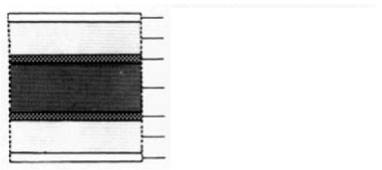
Label the image of the direct X-ray film
Answer
-
Protective gelatin layer
-
Emulsion (silver halide)
-
Adhesive
-
Plastic base
Question 40
Question
When X-ray photons strike the silver halide emulsion of the film, what happens to the silver halide emulsion?
Answer
-
It is sensitised
-
It is excited
-
It is neutralised
-
It is oxidised
Question 41
Question
When X-ray photons hit the silver halide emulsion, does this form the visible or latent image?
Answer
-
Latent image
-
Visible image
Question 42
Question
The embossed dot should face toward the operator and x-ray tube.
Answer
- True
- False
Question 43
Question
In a CCD X-rays hit the [blank_start]scintillation[blank_end] layer above the matrix/array and are converted to [blank_start]light[blank_end] photons. This creates a [blank_start]charge packet[blank_end] which represents the [blank_start]latent[blank_end] image. This is transmitted as an [blank_start]analogue[blank_end] voltage then converted by an analogue-digital converter to the digital image.
Answer
-
scintillation
-
light
-
charge packet
-
latent
-
analogue
Question 44
Question
Instead of film, what do PSPP systems use?
Answer
-
Barium fluorohalide phosphor
-
Silver halide
-
Gadolinium
-
Lanthanum
Question 45
Question
For a PSPP system, the barium [blank_start]fluorohalide[blank_end] phosphor layer absorbs the X-ray photons and stores them (X-rays not [blank_start]attenuated[blank_end] by patient). The image plate is then placed in a laser reader and this causes the stored X-rays to be released as light. A [blank_start]photomultiplier[blank_end] tube then detects this light energy and converts it into a voltage which is then interpreted by a computer and displayed as a digital image.
Answer
-
fluorohalide
-
attenuated
-
photomultiplier
Question 46
Question
Select the disadvantages of digital film
Answer
-
Loss of quality if printed
-
Image alteration possible
-
Expensive to initially implement
-
Higher dose
-
Slower processing
Question 47
Question
Select the advantages of digital film
Answer
-
Lower dose
-
Faster processing
-
Easy transfer
-
Inexpensive to implement
-
Less security required in software
Question 48
Question
Digital X-ray processing:
1. [blank_start]Analogue[blank_end] voltage converted to numerical [blank_start]digital[blank_end] signal
2. Each pixel given x,y coordinate and [blank_start]number[blank_end]
3. The computer then allocates the number a shade of [blank_start]grey[blank_end] scale
Answer
-
Analogue
-
Digital
-
digital
-
analogue
-
number
-
hash
-
grey
-
the visible spectrum
Question 49
Question
A number 0 given to a pixel represents a black shade on the grey scale. What does this mean about X-ray attenuation of the patient?
Answer
-
The patient has not attenuated any of the X-rays
-
The patient has fully attenuated the X-rays
Question 50
Question
A number 255 given to a pixel represents a white shade on the grey scale. What does this mean about X-ray attenuation of the patient?
Answer
-
All of the X-rays have been attenuated by the patient
-
Some of the X-rays have been attenuated by the patient
-
None of the X-rays have been attenuated by the patient
Question 51
Question
Which factors affect the final resolution and size of a digital X-ray image?
Answer
-
Number of pixels
-
Size of pixels
-
Number of shades available from grey scale
-
Type of pixel
-
X-ray attenuation by patient
Question 52
Question
Put in the correct order the processing of X-ray film
1. [blank_start]Development[blank_end]
2. [blank_start]Fixation[blank_end]
3. [blank_start]Washing[blank_end]
Answer
-
Development
-
Fixation
-
Washing
Question 53
Question
During development of X-ray film, sensitised silver halide ions are reduced to black metallic silver. What part of the image does this produce?
Answer
-
Black/Grey part
-
White part
Question 54
Question
Too long development of X-ray film leads to an image that is...
Answer
-
Too dark
-
Too pale
Question 55
Question
What is the name of the active ingredient in the developing solution used to develop X-ray film?
Answer
-
Hydroquinone
-
Camphoroquinone
-
Gamma-methacryloxypropyltrimethoxysilane
Question 56
Question
During fixation of the X-ray film image, what substance is used to remove unsensitised silver halide ions?
Answer
-
Ammonium thiosulphate
-
Copper sulphate
-
Sodium hexafluoride
Question 57
Question
The removal of unsensitised silver halide ions results in which parts of the image?
Answer
-
Transparent/White
-
Black/Grey
Question 58
Question
The time taken to clear unsensitised silver halide ions is known as the...
Answer
-
Clearing time
-
Washing time
-
Fixation time
-
Oxidation time
Question 59
Question
The X-ray film is fixed for ........ the clearing time
Answer
-
Double
-
Triple
-
Half
Question 60
Question
The fixation solution is alkaline and thus contamination with the alkaline developer solution is not a problem.
Answer
- True
- False
Question 61
Question
The X-ray film emulsion is ........... by aliminium chloride.
Answer
-
Hardened
-
Softened
-
Fixed
Question 62
Question
After fixation, X-rays films are washed to remove residual chemicals. What chemical is used to wash them?
Answer
-
Water
-
Ammonium thiosulphate
-
Hydrogen peroxide
-
Ethanol
Question 63
Question
In manual processing there is sometimes an extra washing stage - when is this completed?
Answer
-
Before development
-
Between development and fixation
-
Between fixation and washing
Want to create your own Quizzes for free with GoConqr? Learn more.