
Flussdiagramm
Integumentary System
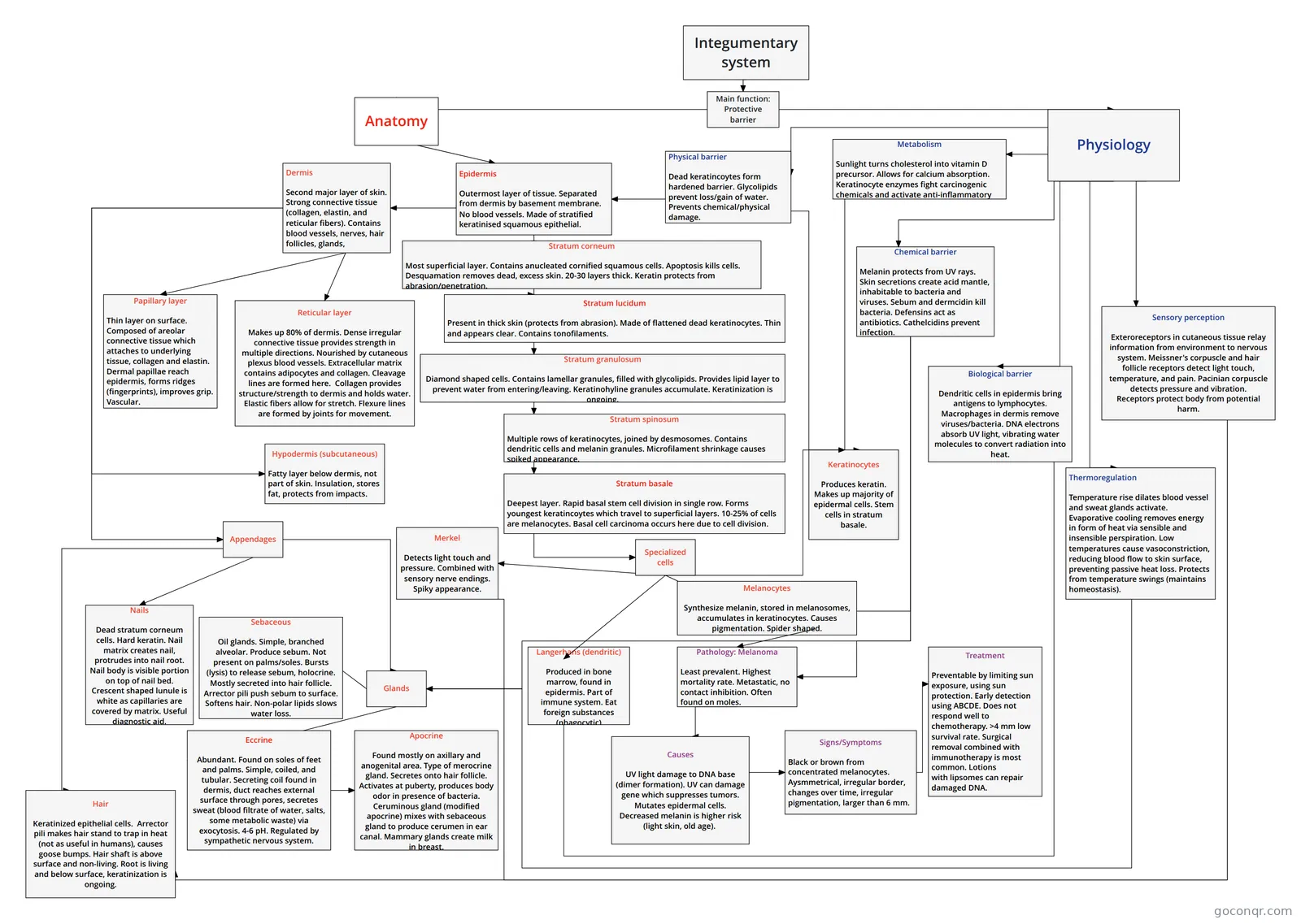
Flowchart: Integumentary system
Inhalt des Flussdiagramms
Anatomy
Physiology
Epidermis
Outermost layer of tissue. Separated from dermis by basement membrane. No blood vessels. Made of stratified keratinised squamous epithelial.
Dermis
Second major layer of skin. Strong connective tissue (collagen, elastin, and reticular fibers). Contains blood vessels, nerves, hair follicles, glands,
Stratum basale
Deepest layer. Rapid basal stem cell division in single row. Forms youngest keratincoytes which travel to superficial layers. 10-25% of cells are melanocytes. Basal cell carcinoma occurs here due to cell division.
Stratum granulosum
Diamond shaped cells. Contains lamellar granules, filled with glycolipids. Provides lipid layer to prevent water from entering/leaving. Keratinohyline granules accumulate. Keratinization is ongoing.
Stratum spinosum
Multiple rows of keratinocytes, joined by desmosomes. Contains dendritic cells and melanin granules. Microfilament shrinkage causes spiked appearance.
Stratum lucidum
Present in thick skin (protects from abrasion). Made of flattened dead keratinocytes. Thin and appears clear. Contains tonofilaments.
Stratum corneum
Most superficial layer. Contains anucleated cornified squamous cells. Apoptosis kills cells. Desquamation removes dead, excess skin. 20-30 layers thick. Keratin protects from abrasion/penetration.
Papillary layer
Thin layer on surface. Composed of areolar connective tissue which attaches to underlying tissue, collagen and elastin. Dermal papillae reach epidermis, forms ridges (fingerprints), improves grip. Vascular.
Appendages
Nails
Dead stratum corneum cells. Hard keratin. Nail matrix creates nail, protrudes into nail root. Nail body is visible portion on top of nail bed. Crescent shaped lunule is white as capillaries are covered by matrix. Useful diagnostic aid.
Glands
Physical barrier
Dead keratincoytes form hardened barrier. Glycolipids prevent loss/gain of water. Prevents chemical/physical damage.
Thermoregulation
Temperature rise dilates blood vessel and sweat glands activate. Evaporative cooling removes energy in form of heat via sensible and insensible perspiration. Low temperatures cause vasoconstriction, reducing blood flow to skin surface, preventing passive heat loss. Protects from temperature swings (maintains homeostasis).
Integumentary system
Main function: Protective barrier
Specialized cells
Melanocytes
Synthesize melanin, stored in melanosomes, accumulates in keratinocytes. Causes pigmentation. Spider shaped.
Merkel
Detects light touch and pressure. Combined with sensory nerve endings. Spiky appearance.
Langerhans (dendritic)
Produced in bone marrow, found in epidermis. Part of immune system. Eat foreign substances (phagocytic)
Keratinocytes
Produces keratin. Makes up majority of epidermal cells. Stem cells in stratum basale.
Hypodermis (subcutaneous)
Fatty layer below dermis, not part of skin. Insulation, stores fat, protects from impacts.
Sensory perception
Exteroreceptors in cutaneous tissue relay information from environment to nervous system. Meissner's corpuscle and hair follicle receptors detect light touch, temperature, and pain. Pacinian corpuscle detects pressure and vibration. Receptors protect body from potential harm.
Chemical barrier
Melanin protects from UV rays. Skin secretions create acid mantle, inhabitable to bacteria and viruses. Sebum and dermcidin kill bacteria. Defensins act as antibiotics. Cathelcidins prevent infection.
Reticular layer
Makes up 80% of dermis. Dense irregular connective tissue provides strength in multiple directions. Nourished by cutaneous plexus blood vessels. Extracellular matrix contains adipocytes and collagen. Cleavage lines are formed here. Collagen provides structure/strength to dermis and holds water. Elastic fibers allow for stretch. Flexure lines are formed by joints for movement.
Biological barrier
Dendritic cells in epidermis bring antigens to lymphocytes. Macrophages in dermis remove viruses/bacteria. DNA electrons absorb UV light, vibrating water molecules to convert radiation into heat.
Metabolism
Sunlight turns cholesterol into vitamin D precursor. Allows for calcium absorption. Keratinocyte enzymes fight carcinogenic chemicals and activate anti-inflammatory steroids.
Pathology: Melanoma
Least prevalent. Highest mortality rate. Metastatic, no contact inhibition. Often found on moles.
Causes
UV light damage to DNA base (dimer formation). UV can damage gene which suppresses tumors. Mutates epidermal cells. Decreased melanin is higher risk (light skin, old age).
Signs/Symptoms
Black or brown from concentrated melanocytes. Aysmmetrical, irregular border, changes over time, irregular pigmentation, larger than 6 mm.
Sebaceous
Oil glands. Simple, branched alveolar. Produce sebum. Not present on palms/soles. Bursts (lysis) to release sebum, holocrine. Mostly secreted into hair follicle. Arrector pili push sebum to surface. Softens hair. Non-polar lipids slows water loss.
Treatment
Preventable by limiting sun exposure, using sun protection. Early detection using ABCDE. Does not respond well to chemotherapy. >4 mm low survival rate. Surgical removal combined with immunotherapy is most common. Lotions with lipsomes can repair damaged DNA.
Eccrine
Abundant. Found on soles of feet and palms. Simple, coiled, and tubular. Secreting coil found in dermis, duct reaches external surface through pores, secretes sweat (blood filtrate of water, salts, some metabolic waste) via exocytosis. 4-6 pH. Regulated by sympathetic nervous system.
Apocrine
Found mostly on axillary and anogenital area. Type of merocrine gland. Secretes onto hair follicle. Activates at puberty, produces body odor in presence of bacteria. Ceruminous gland (modified apocrine) mixes with sebaceous gland to produce cerumen in ear canal. Mammary glands create milk in breast.
Hair
Keratinized epithelial cells. Arrector pili makes hair stand to trap in heat (not as useful in humans), causes goose bumps. Hair shaft is above surface and non-living. Root is living and below surface, keratinization is ongoing.
Informationen
- Beschreibung
- Flussdiagramm am Integumentary System, erstellt von Matt Wyatt am 12/09/2018.
- Erstellt von
- Matt Wyatt
- Aufrufe
- 1
- Bewertung
Mehr zu Skin Structure and Layers