
Karteikarten
Micro Lab Practical
Karten in diesem Stapel (27)
- 1Phase Contrast Microscope vs. Compound Light MicroscopePhase Contrast Microscope:
- 1 eyepiece
Compound Microscopes:
- 2 eyepieces
-Can see microbes
- Two lenses between the eye and the object
- Optical system magnifies the object, light source illuminates it - 2Calculation of Total MagnificationMagnification of Ocular Lens (10x) x Magnification of Objective Lens
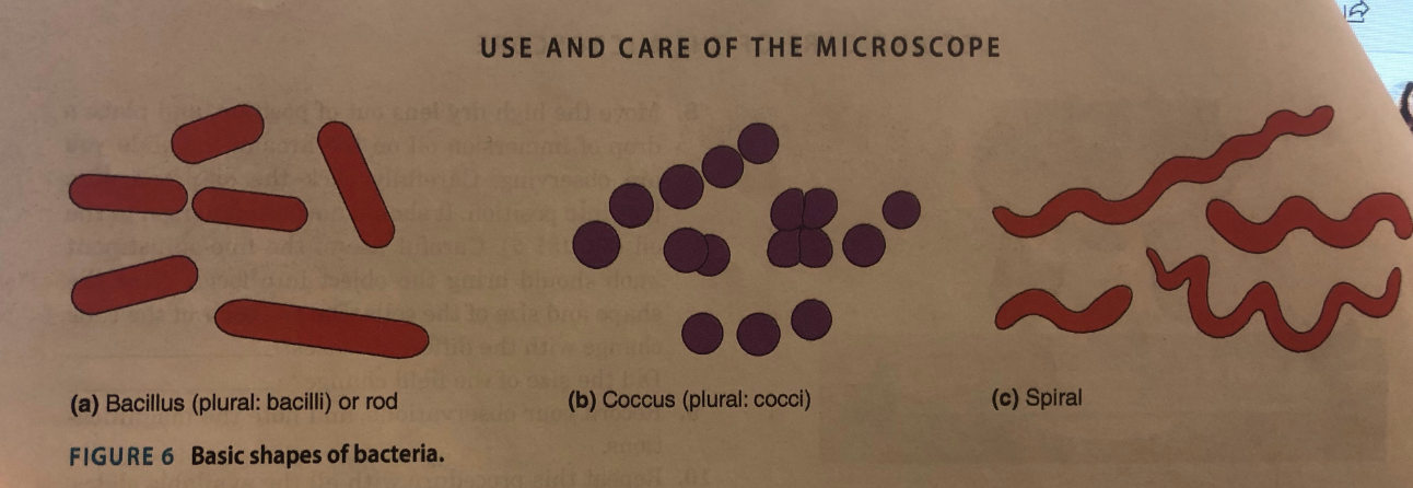
- 3Cell Morphology
- 4Negative Staining→ direct stains: stains bacteria
vs.
→ negative stains: stains background
Basic stain permeates the cell wall; adheres via weak ionic bonds to the bacterial cell, which is slightly negatively charged
Can be used to tell cell morphology, size, and arrangement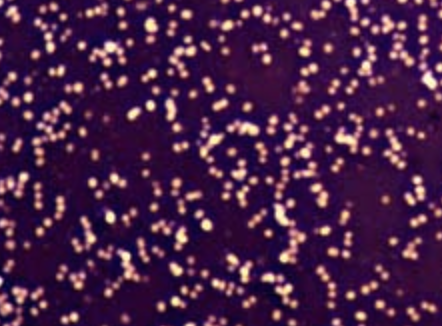
- 5Gram StainingPROCEDURE:
1. Crystal Violet (primary stain): stains all bacteria purple
2. Iodine (mordant): fixes crystal violet stain to Gram positives
3. Alcohol: washes out primary stain
4. Counterstain (safranin): dye that stains the decolorized bacteria red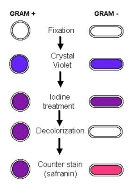
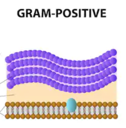
- 6Gram Positive- purple cocci
- thick layer of peptidoglycan, prevents stain from washing out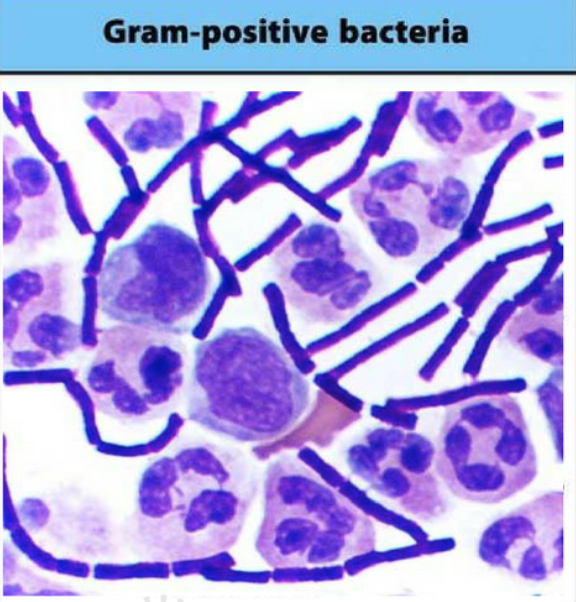
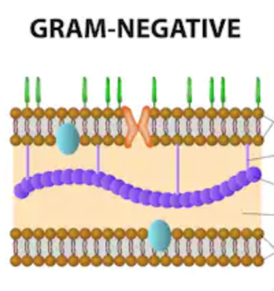
- 7Gram Negative- pink bacillus
- thin layer of peptidoglycan surrounded by an outer layer of phospholipid membrane
- decolorizing agent dissolves outer lipid layer and dye washes out through thin peptidoglycan layer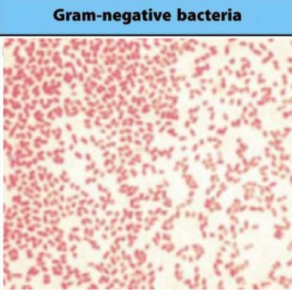
- 8Aseptic Technique1. Flame the loop/needle to sterilize
2. Remove cap of test tube with same hand that’s holding the loop/needle
3. Pass the mouth of the tube through the flame three times
4. Get loopful of culture → innoculate
5. Flame the mouth of tube again, replace cap
6. Flame loop/needle - 9Streak for Isolation1. Loop is used to streak the mixture sample many times over the surface of a solid culture medium
2. This causes the bacteria to fall off the loop one by one and distribute over the surface
3. Each cell/group = the same bacteria - 10Antimicrobial DrugsDisk-Diffusion Method: Petri plate w/ agar inoculated w/ microbe
→Paper disks w/ various antimicrobial agents placed on surface
Zone of Inhibition: area around disk that has successfully inhibited bacterial growth
→ compare ZoI to MIC index - 11Selective MediaUsed to isolate specific groups of bacteria
- 12Differential MediaUsed to differentiate closely related organisms within these groups
→ inhibit growth of one kind while permitting growth of another - 13Phenylethyl Alcohol Agar:- Selective: gram-positives
- Partially inhibits gram-negatives → still form visible colonies, but much smaller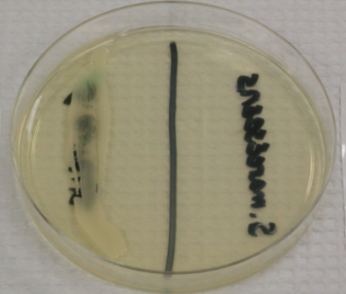
- 14Mannitol Salt Agar (MSA)High in salt; inhibits growth of most, but not all bacteria
Selective: Staphylococci
Differential:
Contains carb mannitol, which some staph. can ferment → which produces acid
Phenol red indicator → yellow zone if fermentation occurs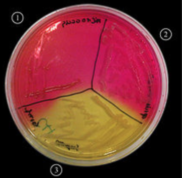
- 15Eosin-Methylene Blue Agar (EMB)Contain lactose and dyes
Differentiate: between enteric lactose fermenters and non-fermenters
→ Identify E. coli → Blue-black with green sheen caused by acid production from dye
Inhibits growth of gram-positives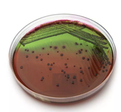
- 16CARBOHYDRATE CATABOLISM: OF Glucose TestType: OF deep
Procedure: needle/add mineral oil to one
Purpose: Determines if an organism is oxidative or fermentative
1.) Oxidative: Needs oxygen
2.) Fermentative: Does not require oxygen
POSITIVE: YELLOW
→ If both are yellow, ox. and ferm.
NEGATIVE: GREEN → BLUE: ammonia production → catabolizes peptone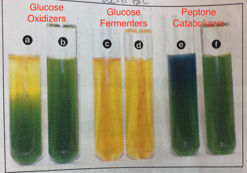
- 17FERMENTATION: Carbohydrate FermentationType: carb broth, peptone, Durham tube
Purpose: used to detect acid and gas from carb catabolism
POSITIVE: YELLOW
NEGATIVE: RED (or peptone growth)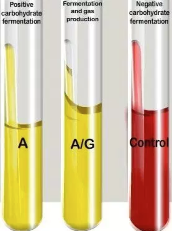
- 18FERMENTATION: MRVP TestType: Broth
Purpose: helps distinguish organism that produce acid
from those that produce acetoin → from glucose
MR Test: add methyl red
POSITIVE: remains red
NEGATIVE: turns yellow
VP Test: add potassium hydroxide
POSITIVE: upper part turns red
NEGATIVE: turns light brown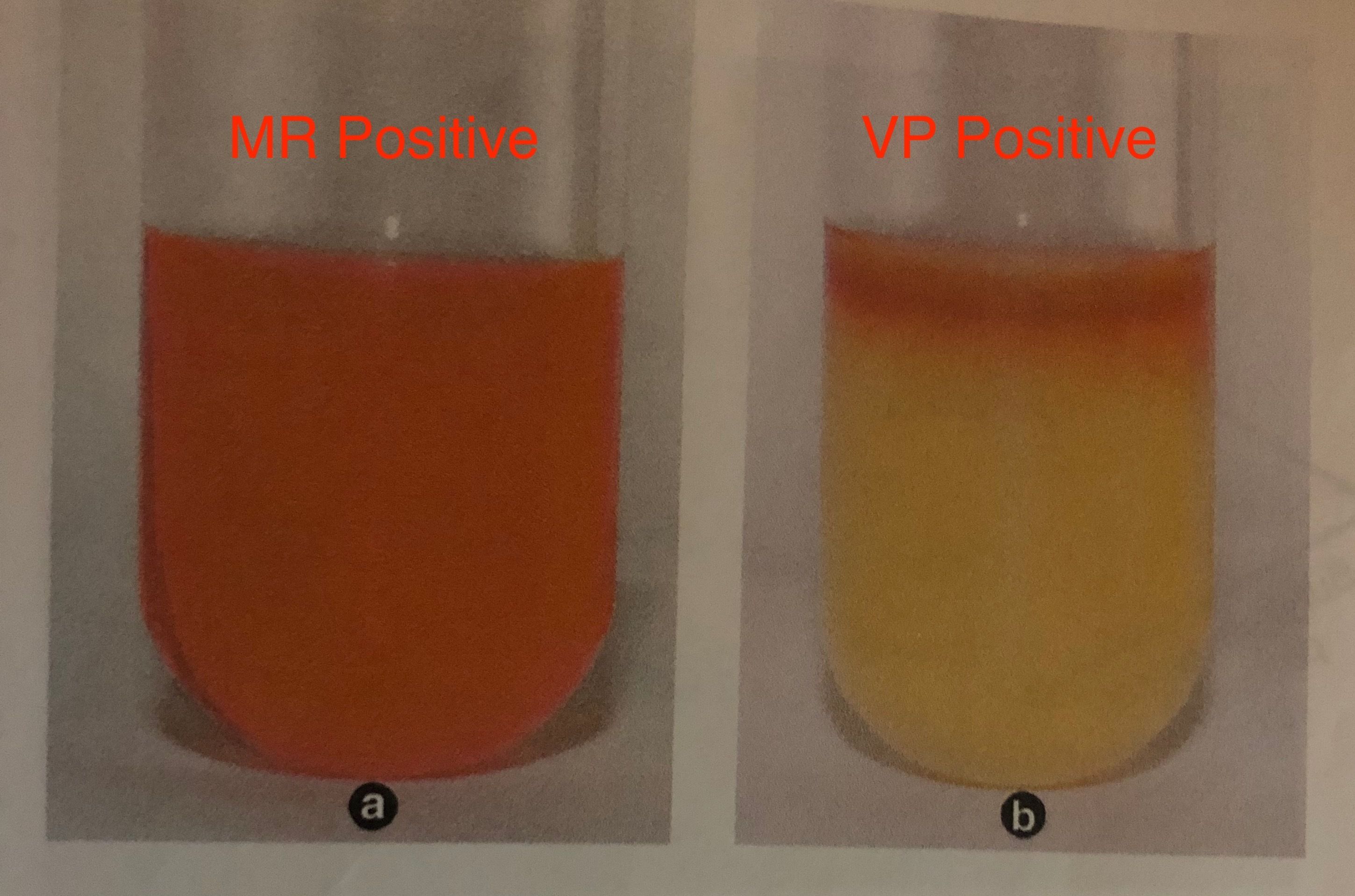
- 19FERMENTATION: Citrate TestType: Slant
Purpose: detects if bacterium can break down citrate to form pyruvate
POSITIVE: blue
NEGATIVE: green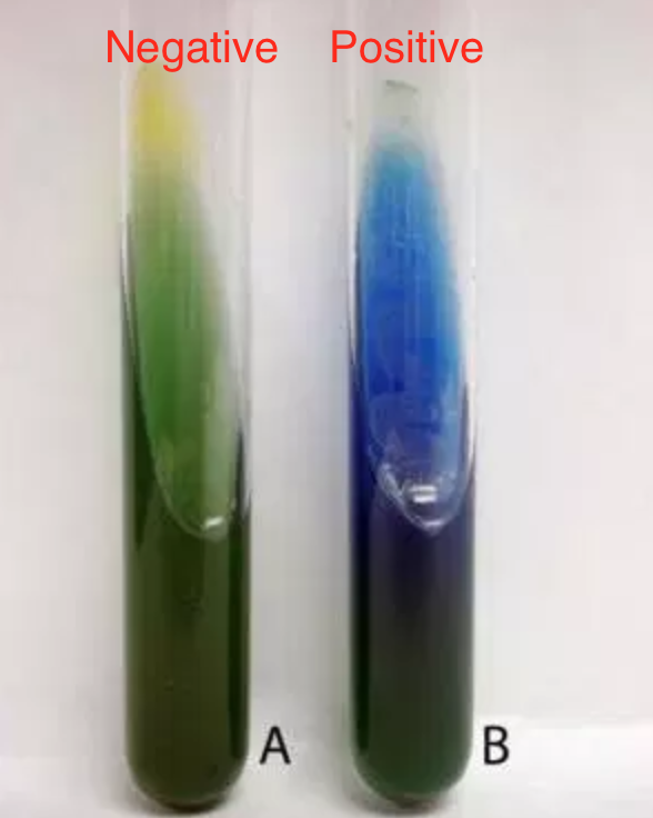
- 20PROTEIN CATABOLISM: Litmus MilkType: broth
Purpose: detects lactose fermentation
POSITIVE:
Lactose fermentation → acid production → Pink
Catabolism of amino acid → alkaline production → Purple
Reduces litmus → White
NEGATIVE: light purple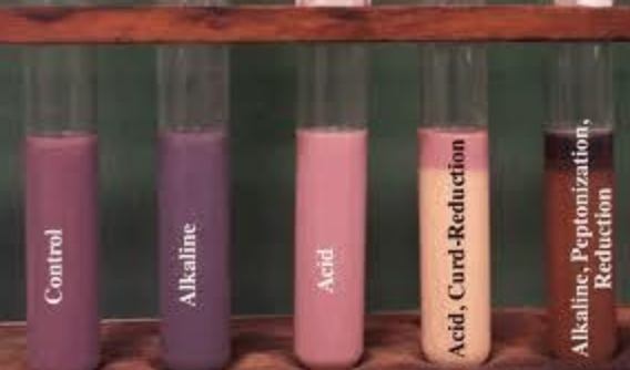
- 21PROTEIN CATABOLISM: UreaseType: Slant
Purpose: detects presence of urea
POSITIVE: pink → produces ammonia
NEGATIVE: yellow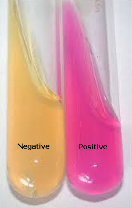
- 22PROTEIN CATABOLISM (PART 2): Phenylalanine DeaminationType: Slant
Purpose: detects deamination of phenylalanine
→ add ferric chloride
POSITIVE: dark green
NEGATIVE: clear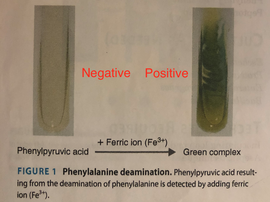
- 23PROTEIN CATABOLISM (PART 2): H2S ProductionType: peptone iron deep
Purpose: desulfhydrase enzyme removes H2S from amino acids
POSITIVE: black precipitate → H2S has been produced
NEGATIVE: no precipitate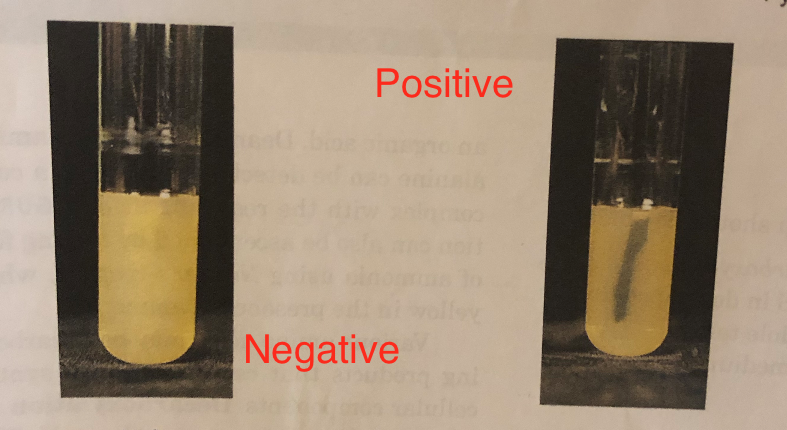
- 24PROTEIN CATABOLISM (PART 2): Indole TestType: tryptone broth
Purpose: detects indole production
→ Add Kovacs reagent
POSITIVE: red ring at top
NEGATIVE: clear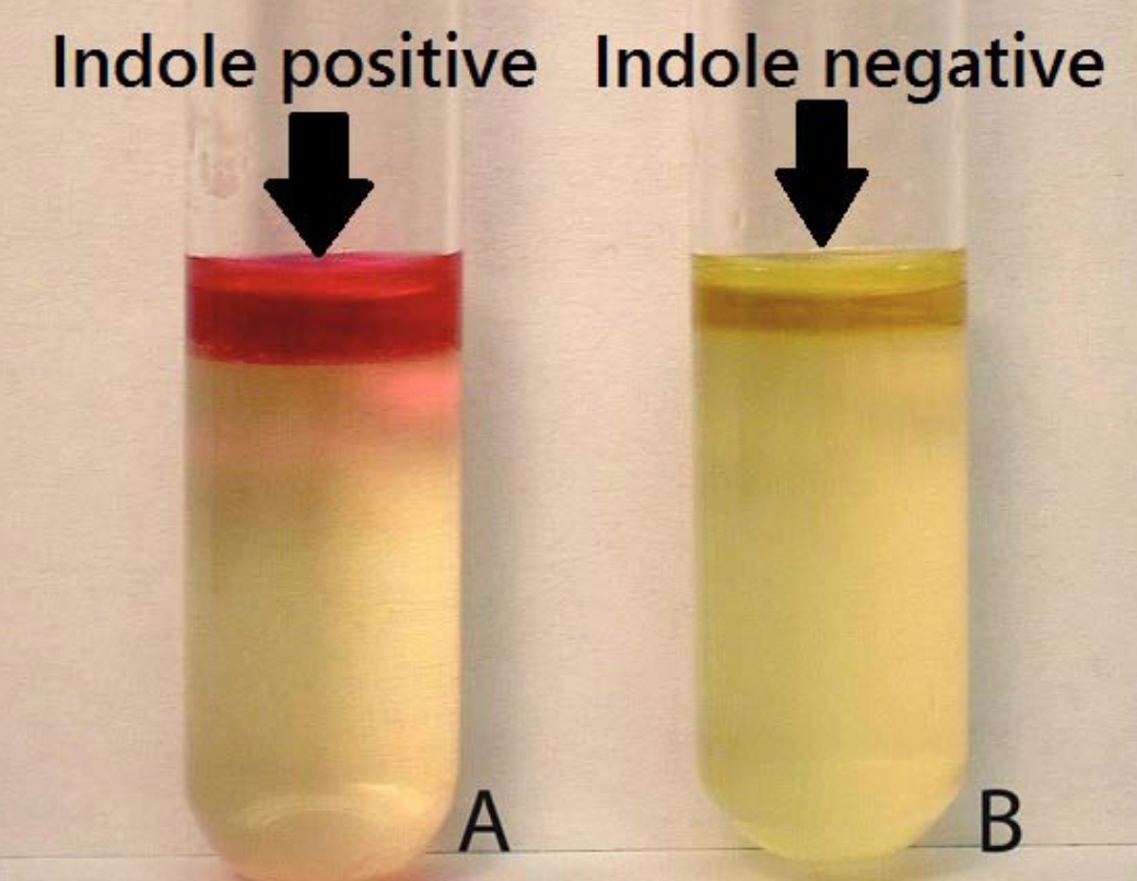
- 25RESPIRATION: Nitrate ReductionType: broth
Purpose: determines bacterium’s ability to reduce nitrates
POSITIVE: Gas production (no color change) → Reduced to N2
NEGATIVE: no color change or gas production
→Add Reagent A and B
→Positive: Red
→If Negative: add Zinc
→Positive: no change
→Negative: If it turns red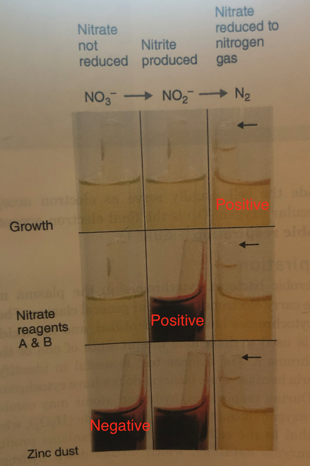
- 26RESPIRATION: Catalase TestPurpose: aerobic organisms produce enzyme catalase
POSITIVE: bubbles
NEGATIVE: no bubbles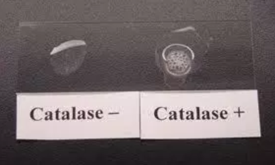
- 27CARBOHYDRATE CATABOLISM: Starch HydrolysisType: Plate
Purpose: presence of an exoenzyme is detected by looking for a change in the substrate outside a colony
POSITIVE: Clear space (contains Amylase)
NEGATIVE: Dark blue from iodine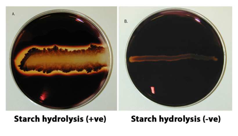
Informationen
- Beschreibung
- Karteikarten am Micro Lab Practical, erstellt von Maddie S am 06/12/2018.
- Erstellt von
- Maddie S
- Aufrufe
- 16
- Bewertung